Category:Hearing: Difference between revisions
From Embryology
mNo edit summary |
mNo edit summary |
||
| (2 intermediate revisions by the same user not shown) | |||
| Line 1: | Line 1: | ||
This {{Embryology}} category shows pages and media related to hearing and vestibular | This {{Embryology}} category shows pages and media related to sensory development of hearing and vestibular systems. | ||
Note that there are additional sub-categories for each part. | Note that there are additional sub-categories for each part: [[:Category:Outer Ear|Category:Outer Ear]], [[:Category:Middle Ear|Category:Middle Ear]], [[:Category:Inner Ear|Category:Inner Ear]] and [[:Category:Balance|Category:Balance]] (or vestibular). | ||
Latest revision as of 14:42, 4 October 2017
This Embryology category shows pages and media related to sensory development of hearing and vestibular systems.
Note that there are additional sub-categories for each part: Category:Outer Ear, Category:Middle Ear, Category:Inner Ear and Category:Balance (or vestibular).
Subcategories
This category has the following 6 subcategories, out of 6 total.
Pages in category 'Hearing'
The following 200 pages are in this category, out of 295 total.
(previous page) (next page)2
A
B
- Template:Balance
- Template:Bast TH.
- BGD Lecture - Face and Ear Development
- BGD Practical - Face and Ear Quiz
- BGD Practical - Face Quiz
- Template:BGDB Face
- BGDB Face and Ear - Abnormalities
- BGDB Face and Ear - Early Embryo
- BGDB Face and Ear - Fetal
- BGDB Face and Ear - Late Embryo
- BGDB Face and Ear - Postnatal
- BGDB Face and Ear - Trilaminar Embryo
- BGDB Practical - Face and Ear Development
- Talk:BGDB Practical - Face and Ear Development
- BGDB Practical - Face and Ear Development Interactive
- Template:BGDB Practical 6 - Abnormalities Interactive
- Template talk:BGDB Practical 6 - Abnormalities Interactive
- Template:BGDB Practical 6 - Early Embryo Interactive
- Template:BGDB Practical 6 - Fetal Interactive
- Template:BGDB Practical 6 - Late Embryo Interactive
- Template:BGDB Practical 6 - Postnatal Interactive
- Template:BGDB Practical 6 - Trilaminar Embryo Interactive
- Book - A textbook of histology, including microscopic technic (1910) Special Histology 9
- Book - Anatomical and physiological studies on the growth of the inner ear of the albino rat (1923)
- Book - Contributions to Embryology Carnegie Institution No.20
- Book - Contributions to Embryology Carnegie Institution No.20 part 1
- Book - Contributions to Embryology Carnegie Institution No.20 part 2
- Book - Contributions to Embryology Carnegie Institution No.20 part 3
- Book - Contributions to Embryology Carnegie Institution No.20 part 4
- Book - Contributions to Embryology Carnegie Institution No.20 part 5
- Book - Contributions to Embryology Carnegie Institution No.20 part 6
- Book - Contributions to Embryology Carnegie Institution No.20 part 7
- Book - Contributions to Embryology Carnegie Institution No.21
- Book - Contributions to Embryology Carnegie Institution No.69
- Book - Human Embryology and Morphology 4
- Book - Manual of Human Embryology 16
- Book - Manual of Human Embryology 16-1
- Book - Manual of Human Embryology 16-2
- Book - Manual of Human Embryology 16-3
- Book - Manual of Human Embryology 16-4
- Book - Text-Book of Embryology 18
C
E
H
- Template:Hardesty1915 table1
- Template:Hardesty1915 table2
- Template:Hardesty1915 table3
- Template:Hardesty1915 table4
- Template:Hardesty1915 table5
- Template:Hardesty1915 table6
- Template:Hearing
- Hearing - Inner Ear Development
- Hearing - Middle Ear Development
- Hearing - Neural Pathway
- Hearing - Outer Ear Development
- Template:Hearing abnormalities
- Template:Hearing EAM timeline
- Template:Hearing Embryonic Origins table1
- Template:Hearing Links
- Template:Hearing neural
- Template:Hearing terms
- Hearing test
- Template:Hearing test
- Template:Hearing timeline
- Template:Hearing timeline table
- Human Embryology and Morphology 16
- Template:Human Embryology Manual 2 16
- Human System Development
O
P
- Paper - 1880 The Platypus Cochlea
- Paper - 1906 Observations on the Labyrinth of Certain Animals
- Paper - 1917 The Typical Form of the Cochlea and Its Variations
- Paper - A model to illustrate the probable action of the tectorial membrane (1915)
- Paper - A note on the length of the basilar membrane in man and in various mammals (1940)
- Paper - Abnormal ossification of Meckel's cartilage
- Paper - Adult form of the human stapes in the light of its development
- Paper - Blood supply of the otic capsule of a 150 mm (C.R.) human fetus
- Paper - Comparative morphology of the ear 3
- Paper - Contribution to the structure and development of the vertebrate head
- Paper - Contribution to the structure and development of the vertebrate head 1
- Paper - Contribution to the structure and development of the vertebrate head 2
- Paper - Contribution to the structure and development of the vertebrate head 3
- Paper - Development of the aquaductus cochleae and the periotic (perilymphatic) duct
- Paper - Development of the aquaeductus cochleae and its contained periotic duct and cochlear vein in human embryos
- Paper - Development of the incus of the human ear - illustrated in atlas series
- Paper - Development of the malleus of the human ear - Illustrated in atlas series
- Paper - Development of the Otic Capsule 1
- Paper - Development of the otic capsule 2
- Paper - Development of the Otic Capsule 3
- Paper - Development of the Otic Capsule 4
- Paper - Development of the otic capsule of the human ear - illustrated in atlas series
- Paper - Development of the stapes of the human ear - illustrated in atlas series
- Paper - Experimental observations on the development of the amphibian ear vesicle (1909)
- Paper - Histogenesis of the otic capsule (1917)
- Paper - Major features in the developmental history of the human stapes (1940)
- Paper - Migration of the ear vesicle in the tadpole during normal development (1921)
- Paper - On the development of the external ear passages
- Paper - On the development of the membrana tectoria with reference to its structure and attachments
- Paper - On the development of the membranous labyrinth and the acoustic and facial nerves in the human embryo
- Paper - On the development of the retina and optic nerve, and of the membranous labyrinth and auditory nerve
- Paper - On the proportions, development and attachment of the tectorial membrane (1915)
- Paper - Ossification of the otic capsule in human fetuses
- Paper - Perichondrial ossification and the fate of the perichondrium with special reference to that of the otic capsule
- Paper - Postnatal growth and adult structure of the otic (endolymphatic) sac
- Paper - Some experiments on the developing ear vesicle of the tadpole with relation to equilibration
- Paper - Some factors in the development of the amphibian ear vesicle and further experiments on equilibration
- Paper - Some features of the auditory apparatus of a 16 mm human embryo
- Paper - Some uniform characteristics of the primate auricle (1922)
- Paper - Stapes, fissula ante fenestram and associated structures in man 1
- Paper - Stapes, fissula ante fenestram and associated structures in man 2
- Paper - Stapes, fissula ante fenestram and associated structures in man 3
- Paper - Stapes, fissula ante fenestram and associated structures in man 4
- Paper - Stapes, fissula ante fenestram and associated structures in man 5
- Paper - The comparison of auricular height determinations (1925)
- Paper - The cytological processes involved in the formation of the scalae of the internal ear
- Paper - The development and structure of the otic (endolymphatic) sac
- Paper - The development of the auditory nerve in vertebrates (1910)
- Paper - The development of the auditory ossicles and associated structures in man
- Paper - The development of the auditory ossicles, the otic capsule and the extracapsular tissues
- Paper - The development of the cochlear fenestra, fossula and secondary tympanic membrane
- Paper - The development of the ear-bones in the mouse
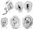
- Paper - The development of the external ear (1934)
- Paper - The development of the first branchial arch in man and the fate of Meckel's cartilage
- Paper - The development of the otic capsule in the region of surgical fenestration 1
- Paper - The development of the otic capsule in the region of surgical fenestration 2
- Paper - The development of the otic capsule in the region of the vestibular aqueduct
- Paper - The development of the pillar cells, tunnel space, and Nuel's spaces in the organ of Corti (1919)
- Paper - The Development of the Scala Tympani, Scala Vestibuli and Perioticular Cistern in the Human Embryo
- Paper - The development of the second branchial arch (Reichert's cartilage), facial canal and associated structures in man
- Paper - The developmental and adult anatomy of the air-cells in the petrous part of the temporal bone
- Paper - The developmental course of the human auditory vesicle
- Paper - The distal projection of the endolymphatic sac in human embryos
- Paper - The early development of the membranous labyrinth in mammalian embryos
- Paper - The early development of the otic vesicle in staged human embryos
- Paper - The early embryology of the auditory ossicles in man
- Paper - The early formations of the middle ear and eustachian tube - a criticism
- Paper - The early relation of the auditory vesicle to the ectoderm in human embryos
- Paper - The Factors Involved in the Excavation of the Cavities in the Cartilaginous Capsule of the Ear in the Human Embryo
- Paper - The fissula ante fenestram of the human otic capsule; aberrant form and contents
- Paper - The form and structure of the endolymphatic and associated ducts in the child
- Paper - The genesis and structure of the membrana tectoria and the crista spiralis of the cochlea (1918)
- Paper - The Origin of the Otic and Optic Primordia in Man
- Paper - The Typical Form of the Cochlea and its Variations
- Paper - The vascular drainage of the endolymphatic sac and its topographical relation to the transverse sinus in the human
- Paper - Vertebrate cephalogenesis 1 (1890)
- Paper - Vertebrate cephalogenesis 2 (1892)
- Template:Placode
- Template:Placodes
Media in category 'Hearing'
The following 200 files are in this category, out of 375 total.
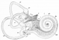
(previous page) (next page) Adult cochlea cartoon 01.jpg 986 × 800; 123 KB
 Adult cochlea nerve glia cartoon.jpg 1,000 × 725; 85 KB
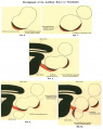
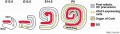
 Adult hearing embryonic origins c.jpg 400 × 270; 20 KB
 Adult hearing embryonic origins.jpg 1,000 × 675; 80 KB
 Anotia 01.jpg 796 × 660; 61 KB
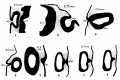 Anson1934 fig01-8.jpg 1,337 × 888; 126 KB
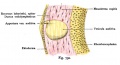
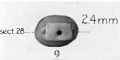 Anson1934 fig09.jpg 546 × 272; 16 KB
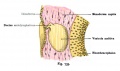
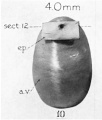 Anson1934 fig10.jpg 520 × 612; 31 KB
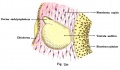
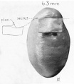 Anson1934 fig11.jpg 758 × 870; 52 KB
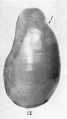 Anson1934 fig12.jpg 545 × 968; 44 KB
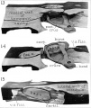
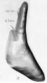 Anson1934 fig13.jpg 761 × 1,323; 74 KB
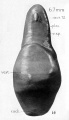 Anson1934 fig14.jpg 761 × 1,323; 81 KB
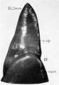 Anson1934 fig15.jpg 693 × 1,003; 62 KB
 Anson1934 fig16.jpg 691 × 985; 47 KB
 Anson1934 fig17.jpg 605 × 1,143; 57 KB
 Anson1934 fig18.jpg 418 × 1,161; 34 KB
 Anson1934 fig19.jpg 524 × 1,218; 48 KB
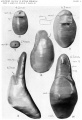 Anson1934 plate01.jpg 1,557 × 2,279; 288 KB
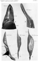 Anson1934 plate02.jpg 1,464 × 2,311; 259 KB
 AnsonKarabinMartin1939 fig01-06.jpg 1,280 × 1,650; 324 KB
 AnsonKarabinMartin1939 fig07-12.jpg 1,280 × 1,880; 444 KB
 AnsonKarabinMartin1939 fig13-15.jpg 1,280 × 1,519; 171 KB
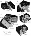 AnsonKarabinMartin1939 fig60-64.jpg 1,280 × 1,496; 130 KB
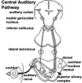 Auditory neural pathway.jpg 450 × 457; 46 KB
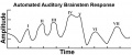 Automated Auditory Brainstem Response.jpg 1,239 × 521; 37 KB
 Bailey473.jpg 427 × 419; 40 KB
 Bailey474.jpg 1,321 × 869; 247 KB
 Bailey475.jpg 1,302 × 852; 213 KB
 Bailey476.jpg 688 × 648; 110 KB
 Bailey477.jpg 593 × 509; 57 KB
 Bast1931 plate01.jpg 1,280 × 871; 113 KB
 BeatonAnson1940 fig01-2.jpg 1,652 × 2,147; 652 KB
 Beckwith-Wiedemann syndrome ear lobe creases.jpg 257 × 414; 12 KB
 Beckwith-Wiedemann syndrome posterior helix pit.jpg 247 × 415; 10 KB
 BGDB PracManual 2011 Practical 6.pdf ; 426 KB
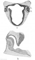
 CameronMilligan1910 fig01.jpg 750 × 695; 50 KB
 CameronMilligan1910 fig02.jpg 750 × 732; 46 KB
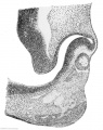
 CameronMilligan1910 fig05.jpg 750 × 800; 60 KB
 CameronMilligan1910 fig06-10.jpg 1,839 × 2,309; 208 KB
 CameronMilligan1910 fig11.jpg 800 × 551; 17 KB
 Cat inner ear MicroCT.jpg 1,159 × 1,300; 266 KB
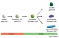 Cochlea glial lineage cartoon.jpg 1,000 × 651; 52 KB
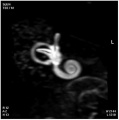 Cochlea MRI 01.jpg 380 × 382; 13 KB
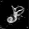 Cochlea MRI 02.jpg 381 × 380; 12 KB
 Cochlea stria vascularis cartoon 01.jpg 874 × 1,897; 338 KB
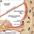 Cochlea stria vascularis cartoon 02.jpg 802 × 800; 138 KB
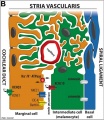 Cochlea stria vascularis cartoon 03.jpg 694 × 800; 125 KB
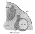
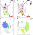 Cochlear and vestibular specification.jpg 1,000 × 1,063; 135 KB
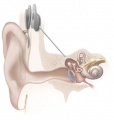 Cochlear implant.jpg 350 × 368; 31 KB
 Eustacian tube angle.jpg 609 × 458; 48 KB
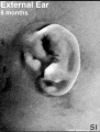 External Ear 5 months-icon.jpg 352 × 464; 63 KB
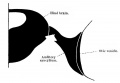
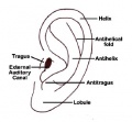 External ear anatomy.jpg 276 × 256; 13 KB
 External ear stages-14-23-adult a.jpg 800 × 524; 30 KB
 External ear stages-14-23-adult.jpg 1,000 × 655; 42 KB
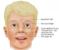 FASface.jpg 320 × 272; 8 KB
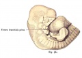
 Foster129.jpg 999 × 810; 114 KB
 Foster130.jpg 833 × 963; 103 KB
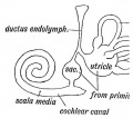
 Foster131.jpg 741 × 932; 87 KB
 Foster132.jpg 881 × 684; 82 KB
 Frazer1922 fig01.jpg 627 × 900; 102 KB
 Frazer1922 fig02.jpg 1,000 × 853; 182 KB
 Frazer1922 fig03.jpg 1,000 × 697; 165 KB
 Frazer1922 fig04.jpg 1,000 × 927; 136 KB
 Frazer1922 fig05.jpg 1,000 × 783; 94 KB
 Frazer1922 fig06.jpg 1,000 × 728; 116 KB
 Gray0043.jpg 800 × 496; 50 KB
 Gray0698.jpg 500 × 518; 47 KB
 Gray0898.jpg 400 × 295; 26 KB
 Gray0899.jpg 300 × 180; 9 KB
 Gray0900.jpg 647 × 800; 121 KB
 Gray0902.jpg 500 × 361; 40 KB
 Gray0903.jpg 600 × 389; 42 KB
 Gray0904.jpg 226 × 350; 26 KB
 Gray0905.jpg 430 × 275; 19 KB
 Gray0906.jpg 438 × 600; 81 KB
 Gray0907.jpg 679 × 600; 110 KB
 Gray0908.jpg 500 × 359; 30 KB
 Gray0909.jpg 600 × 437; 56 KB
 Gray0910.jpg 400 × 548; 78 KB
 Gray0911.jpg 651 × 400; 73 KB
 Gray0912.jpg 600 × 540; 87 KB
 Gray0913.jpg 671 × 600; 98 KB
 Gray0914.jpg 708 × 500; 102 KB
 Gray0915.jpg 720 × 600; 94 KB
 Gray0916.jpg 600 × 367; 28 KB
 Gray0917.jpg 600 × 379; 44 KB
 Gray0918.jpg 500 × 283; 17 KB
 Gray0919.jpg 500 × 731; 93 KB
 Gray0920.jpg 600 × 438; 69 KB
 Gray0921.jpg 640 × 500; 95 KB
 Gray0922.jpg 600 × 556; 71 KB
 Gray0923.jpg 691 × 500; 106 KB
 Gray0924.jpg 500 × 384; 26 KB
 Gray0925.jpg 693 × 500; 84 KB
 Gray0926.jpg 719 × 500; 90 KB
 Gray0927.jpg 542 × 500; 82 KB
 Gray0928.jpg 600 × 408; 56 KB
 Gray0929.jpg 700 × 287; 45 KB
 Gray0930.jpg 712 × 400; 65 KB
 Gray0931.jpg 600 × 334; 53 KB
 Gray0932.jpg 659 × 450; 111 KB
 Gray0933.jpg 500 × 360; 54 KB
 Hearing cartoon.jpg 800 × 553; 43 KB
 Hearing sound localization circuits brainstem.jpg 800 × 378; 45 KB
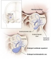 Hearing-vestibular sac abnormality.jpg 432 × 493; 45 KB
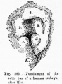 Hertwig285.jpg 400 × 544; 46 KB
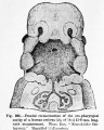 Hertwig286.jpg 642 × 800; 126 KB
 Human cochlea fetal development cartoon.jpg 592 × 1,200; 96 KB
 Human cochlea stria vascularis 01.jpg 1,854 × 1,806; 754 KB
 Human CS13 otic vesicle 01.jpg 1,028 × 774; 112 KB
 Human CS13-15 otic vesicle 01.jpg 1,574 × 1,779; 364 KB
 Human fetal cochlea 01.jpg 1,270 × 532; 266 KB
 Human fetal cochlea 02.jpg 1,270 × 532; 271 KB
 Human inner ear MicroCT.jpg 2,131 × 3,111; 1,001 KB
 Incomplete cochlea CT.jpg 550 × 217; 24 KB
 Inner ear development cartoon 01.jpg 714 × 800; 94 KB
 Inner ear haircells.jpg 800 × 671; 121 KB
 Keibel Mall 2 126.jpg 1,000 × 598; 66 KB
 Keibel Mall 2 158.jpg 563 × 700; 47 KB
 Keibel Mall 2 159.jpg 679 × 699; 47 KB
 Keibel Mall 2 160.jpg 501 × 700; 35 KB
 Keibel Mall 2 161.jpg 621 × 700; 46 KB
 Keibel Mall 2 162.jpg 579 × 800; 69 KB
 Keibel Mall 2 163.jpg 675 × 500; 51 KB
 Keibel Mall 2 164.jpg 610 × 500; 44 KB
 Keibel Mall 2 165.jpg 1,037 × 900; 166 KB
 Keibel Mall 2 166.jpg 969 × 1,000; 144 KB
 Keibel Mall 2 167.jpg 1,000 × 496; 86 KB
 Keibel Mall 2 168.jpg 583 × 1,000; 65 KB
 Keibel Mall 2 169.jpg 634 × 800; 97 KB
 Keibel Mall 2 170.jpg 1,000 × 793; 65 KB
 Keibel Mall 2 171.jpg 1,000 × 696; 84 KB
 Keibel Mall 2 172.jpg 822 × 800; 82 KB
 Keibel Mall 2 173.jpg 818 × 800; 142 KB
 Keibel Mall 2 174.jpg 607 × 600; 56 KB
 Keibel Mall 2 175.jpg 496 × 597; 40 KB
 Keibel Mall 2 176.jpg 992 × 800; 134 KB
 Keibel Mall 2 177.jpg 965 × 389; 48 KB
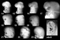
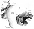
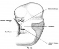
 Keith1902 fig035.jpg 800 × 633; 125 KB
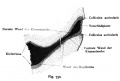
 Keith1902 fig036a.jpg 678 × 639; 93 KB
 Keith1902 fig036b.jpg 678 × 639; 70 KB
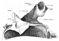
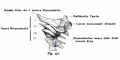
 Keith1902 fig037.jpg 800 × 668; 62 KB
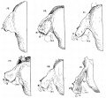
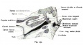
 Keith1902 fig038.jpg 800 × 641; 56 KB
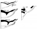
 Keith1902 fig039.jpg 751 × 600; 114 KB
 Keith1902 fig040.jpg 1,000 × 524; 85 KB
 Keith1902 fig041.jpg 555 × 400; 34 KB
 Keith1902 fig042.jpg 404 × 400; 21 KB
 Keith1902 fig043.jpg 505 × 400; 38 KB
 Keith1902 fig044.jpg 640 × 564; 54 KB
 Keith1902 fig045.jpg 613 × 400; 33 KB
 Keith1902 fig046.jpg 862 × 800; 116 KB
 Kollmann731.jpg 386 × 242; 21 KB
 Kollmann732.jpg 658 × 355; 41 KB
 Kollmann733.jpg 646 × 384; 40 KB
 Kollmann734.jpg 701 × 406; 46 KB
 Kollmann735.jpg 600 × 435; 48 KB
 Kollmann736-737.jpg 826 × 467; 56 KB
 Kollmann738.jpg 505 × 503; 34 KB
 Kollmann739.jpg 781 × 667; 79 KB
 Kollmann740.jpg 706 × 426; 43 KB
 Kollmann741.jpg 706 × 417; 51 KB
 Kollmann742.jpg 749 × 540; 67 KB
 Kollmann743.jpg 789 × 729; 94 KB
 Kollmann744.jpg 693 × 468; 56 KB
 Kollmann745.jpg 588 × 338; 43 KB
 Kollmann746.jpg 802 × 441; 110 KB
 Kollmann747.jpg 753 × 388; 44 KB
 Kollmann748.jpg 704 × 479; 46 KB
 Kollmann749.jpg 774 × 641; 66 KB
 Kollmann750.jpg 640 × 433; 37 KB
 Kollmann751.jpg 691 × 348; 34 KB
 Kollmann752.jpg 751 × 415; 67 KB
 Kollmann753.jpg 734 × 380; 41 KB
 Kollmann754.jpg 802 × 577; 71 KB
 Kollmann755.jpg 814 × 457; 59 KB
 Kollmann756.jpg 820 × 489; 67 KB
 Kollmann757.jpg 807 × 697; 98 KB
 Kollmann758.jpg 780 × 406; 45 KB
 Kollmann759.jpg 827 × 520; 72 KB
 Kollmann760.jpg 719 × 641; 85 KB
 Kollmann761.jpg 641 × 455; 45 KB
 Kollmann762-765.jpg 824 × 763; 66 KB
 Kollmann766-769.jpg 694 × 311; 25 KB
 Lewis1906 fig438.jpg 1,007 × 877; 149 KB
 Lewis1920 fig16.jpg 1,000 × 743; 101 KB
 Macklin-plate03.jpg 2,331 × 3,061; 1,010 KB
 Max Brödel-cochlea drawing1934.jpg 531 × 395; 69 KB
 Microtia.jpg 600 × 450; 27 KB
 Minot1897 429.jpg 1,200 × 805; 189 KB
 Minot1897 430.jpg 863 × 800; 150 KB
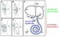
 Mouse - inner ear cartoon.jpg 800 × 498; 77 KB
 Mouse cochlea development cartoon.jpg 1,000 × 280; 53 KB
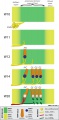
 Mouse cochlea gene expression.jpg 1,000 × 346; 75 KB
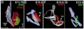
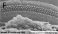
 Mouse E18.5 cochlea sem01.jpg 902 × 774; 150 KB
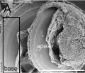
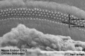
 Mouse E18.5 cochlea sem02.jpg 901 × 489; 75 KB
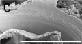
 Mouse E18.5 cochlea sem03.jpg 905 × 1,048; 183 KB
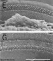
 Mouse E18.5 cochlea sem04.jpg 905 × 535; 85 KB
 Mouse E18.5 cochlea sem05.jpg 807 × 533; 78 KB
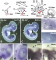
 Mouse inner ear development 01.jpg 600 × 373; 64 KB
 Mouse inner ear.jpg 478 × 508; 47 KB


