Paper - Histogenesis of the otic capsule (1917)
| Embryology - 27 Apr 2024 |
|---|
| Google Translate - select your language from the list shown below (this will open a new external page) |
|
العربية | català | 中文 | 中國傳統的 | français | Deutsche | עִברִית | हिंदी | bahasa Indonesia | italiano | 日本語 | 한국어 | မြန်မာ | Pilipino | Polskie | português | ਪੰਜਾਬੀ ਦੇ | Română | русский | Español | Swahili | Svensk | ไทย | Türkçe | اردو | ייִדיש | Tiếng Việt These external translations are automated and may not be accurate. (More? About Translations) |
Streeter GL. Histogenesis of the otic capsule. (1917) Anat. Rec. 11: 417-418.
| Historic Disclaimer - information about historic embryology pages |
|---|
| Pages where the terms "Historic" (textbooks, papers, people, recommendations) appear on this site, and sections within pages where this disclaimer appears, indicate that the content and scientific understanding are specific to the time of publication. This means that while some scientific descriptions are still accurate, the terminology and interpretation of the developmental mechanisms reflect the understanding at the time of original publication and those of the preceding periods, these terms, interpretations and recommendations may not reflect our current scientific understanding. (More? Embryology History | Historic Embryology Papers) |
Histogenesis of the Otic Capsule
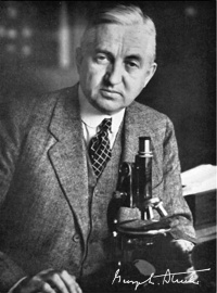
G. L. Streeter
Carnegie Laboratory of Embryology, Baltimore.
(Lantern.)
Presented at the 33rd Meeting of the American Association of Anatomists in New York (Dec 27-29, 1916).
The cartilaginous capsule of the ear is a favorable place for studying the histological features of the growth of cartilage and its associated tissues, particularly because of two reasons; in the first place, there are, on account of the intricacy of form of the labyrinth, many kinds of cartilaginous changes found there that are necessary to accommodate its growth, including the deposit of new tissue and the removal of old tissue; and in the second place the topography is so well marked by known landmarks that all of these changes as well as the location and direction of growth can be easily followed. From such a study one is forced to conclude that the tissues of the otic capsule are capable both of differentiation and dedifferentiation throughout a considerable period of their development. This progressive and retrogressive adaptability of the cartilaginous tissue makes possible those changes that are necessary in the growth arid alteration in form of the labyrinth.
In the earlier stages the precartilage tissue abuts directly against the epithelial labyrinth. Subsequently the periotic reticulum, beginning along the central borders of the canals, becomes established and spreads at the expense of the temporary precartilage thereby forming a crescentic shaped area of reticulum entirely inclosing the membranous canal. The invasion of the reticulum into the surrounding area of precartilage is brought about, at least in the later stages, by a dedifferentiation of the latter into the former.
At the same time that the precartilage is reverting into reticulum the inner margin of the cartilage that surrounds the canal is dedifferentiated into precartilage, so that a new and more peripheral area of precartilage becomes established as the old area disappears. In this way the margin of the true cartilage gradually recedes from the epithelial canal.
In human embryos 30 mm long the cartilaginous labyrinth has attained approximately the adult form. Its subsequent development consists primarily of an increase in size to accommodate the growing membranous labyrinth. However, if one compares the superior canal of an 80 mm. fetus with that of a 30 mm fetus it will be found that the canal has doubled its diameter and has trebled its length and furthermore its linear curvature corresponds to an arc with a longer radius. In reality therefore the developing cartilaginous labyrinth is continuously undergoing considerable changes both in size and form. The enlargement of the cartilaginous canals and their alterations in form and position involves both the excavation of cartilage and also the laying down of new cartilage, the excavation being accomplished by its dedifferentiation into precartilage and reticulum, and the new cartilage being built up, through a precartilage stage, from the periotic reticular tissue. Throughout the entire period of growth of the cartilagenous canals the elements of this continual transformation exist along their margin. The margin during this period is in a state of temporary equilibrium and is capable of advancing or receding as the conditions determine.
The first and relatively the major part of the hollowing-out of the cartilagkous canals is complete before the perichondrium makes its appearance. The perichondrium is formed as a membrane-like condensation of the periotic reticulum which can be first recognized in fetuses of about 70 mm CR length. In its histogenesisit is analogous to the niembrana propria of the epithelial canals.
Cite this page: Hill, M.A. (2024, April 27) Embryology Paper - Histogenesis of the otic capsule (1917). Retrieved from https://embryology.med.unsw.edu.au/embryology/index.php/Paper_-_Histogenesis_of_the_otic_capsule_(1917)
- © Dr Mark Hill 2024, UNSW Embryology ISBN: 978 0 7334 2609 4 - UNSW CRICOS Provider Code No. 00098G


