Category:Second Trimester
This Embryology category shows content related to the second trimester of human development.
- Links: Second Trimester | Third Trimester | Fetal Development
| Links: human timeline | first trimester timeline | second trimester timeline | third trimester timeline | ||
| Event | ||
| Clinical second trimester |  Week 12 - CRL 85 mm, femur length 15 mm, biparietal diameter 25 mm Week 12 - CRL 85 mm, femur length 15 mm, biparietal diameter 25 mm
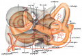
Hearing Week 12-16 - Capsule adjacent to membranous labrynth undegoes vacuolization to form a cavity (perilymphatic space) around membranous labrynth and fills with perilymph
Respiratory Month 3-6 - lungs appear glandular, end month 6 alveolar cells type 2 appear and begin to secrete surfactant Tongue Week 12 - first differentiated epithelial cells (Type II and III) Genital female genital canal (80 days) formed with absorption of the median septum | |
| tongue Week 12 to 13 - maximum synapses between cells and afferent nerve fibers
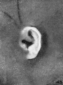
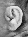
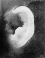
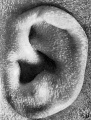
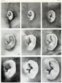
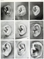
hearing outer ear Week 13 - Meatal plug disc-like, innermost surface in contact with the primordial malleus, contributes to the formation of the tympanic membrane. | ||
| tongue Week 14 to 15 - taste pores develop, mucous
ovary 100 days - primary follicles present nail toenails appear Head Development facial skeleton remodelling begins Hearing - Inner Ear Development Week 14 GA 16 - neural-crest-derived melanocytes, now intermediate cells of the stria vascularis, tightly integrate with Na+ /K+ -ATPase-positive marginal cells, which started to express KCNQ1 in their apical membrane.[1] | ||
| Pancreas glucagon detectable in fetal plasma.
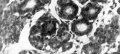
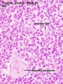
spleen Week 15 -alpha-smooth muscle actin (alpha-SMA)-positive reticulum cells scattered around the arterioles.[2] | ||
| 14 cm | 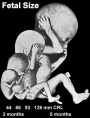 Hearing Week 16-24 - Centres of ossification appear in remaining cartilage of otic capsule form petrous portion of temporal bone. Continues to ossify to form mastoid process of temporal bone. Hearing Week 16-24 - Centres of ossification appear in remaining cartilage of otic capsule form petrous portion of temporal bone. Continues to ossify to form mastoid process of temporal bone.
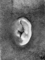
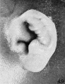
pituitary adenohypophysis fully differentiated respiratory Week 16 to 25 lung histology - canalicular Hearing - Outer Ear Development Week 16.5 - External auditory meatus is fully patent throughout its length, lumen is still narrow and curved. Hearing - Inner Ear Development Week 16 GA 18 - cells in the outer sulcus express KCNJ10 and gap junction proteins GJB2/CX26 and GJB6/CX30, but these are not expressed in the spiral ligament.[1] gap junction cartoon neural - Cerebrum development of the periinsular sulci (week 16-17, GA 18-19 weeks)[3]
primary follicles begin to form in the ovary and are characterized by an oocyte glandular urethra forms and skin folds present | |
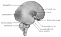
 Neural - Brain development histology week 17 Neural - Brain development histology week 17
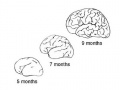
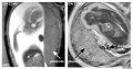
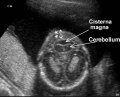
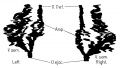
Cerebellum Magnetic Resonance Imaging (MRI) can study the developing cerebellum from 17 to 18 weeks (GA 19 to 20 weeks). tooth Week 17 - First papilla of the permanent dentition appear (first molar) immediately behind the second milk molar, milk teeth are well advanced (Fetus 180 mm). | ||
 tongue Week 18 - substance P detected in dermal papillae, not in taste bud primordia tongue Week 18 - substance P detected in dermal papillae, not in taste bud primordia
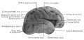
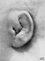
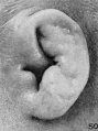
integumentary vernix caseosa covers skin spleen Week 18 - alpha-SMA-positive reticulum cells increase in number and began to form a reticular framework. An accumulation of T and B lymphocytes occurred within the framework, and a primitive white pulp was observed around the arterioles.[2] Hearing - Outer Ear Development week 18 - External auditory meatus is already fully expanded to its complete form. neural - Cerebrum central sulci and opercularization of the insula (week 18-20, GA 20-22 weeks)[3] | ||
| neural week 19 neuronal migration ends and the radial glial cells that aided the migration now become transformed into astrocytes and astrocytic precursors.[4] | ||
| pituitary week 20 to 24 growth hormone levels peak, then decline
integumentary lanugo, skin hair integumentary 5 months - Hair growth initiated at base of cord, lateral outgrowths form associated sebaceous glands; Other cords elongate and coil to form sweat glands; Cords in mammary region branch as they elongate to form mammary glands. touch pacinian corpuscle begin to develop[5] | ||
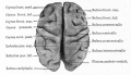
 Neural brain cortical sulcation - sylvian fissure, interhemispheric fissure, callosal sulcus, parietooccipital fissure, and hippocampic fissures present[6] Neural brain cortical sulcation - sylvian fissure, interhemispheric fissure, callosal sulcus, parietooccipital fissure, and hippocampic fissures present[6]
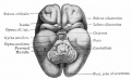
spleen - Week 22 - antigenic diversity of the reticular framework was observed, and T and B lymphocytes were segregated in the framework. T lymphocytes were sorted into the alpha-smooth muscle actin-positive reticular framework, and the periarteriolar lymphoid sheath (PALS) was formed around the arteriole. B lymphocytes aggregated in eccentric portions to the PALS and formed the lymph follicle (LF). The reticular framework of the LF was alpha-SMA-negative. [2] neural - Cerebrum covering of the posterior insula (week 22-24, GA 24-26 weeks)[3] | ||
| respiratory Week 24 to 40 lung histology - terminal sac
spleen Week 24 - marginal zone appeared in the alpha-smooth muscle actin-positive reticular framework around the white pulp.[2] tooth Week 24 - Permanent incisors and canines appear. Earliest potential survival expected if born ovary follicles can consist of growing oocytes surrounded by several layers of granulosa cells | ||
| respiratory end month 6 alveolar cells type 2 appear and begin to secrete surfactant
neural - Cerebrum closure of the laeteral sulcus (Sylvian fissure or lateral fissure) (week 25-26, GA 27-28 weeks)[3] | ||
| touch pacinian corpuscle well developed[5] | ||
- ↑ 1.0 1.1 Locher H, de Groot JC, van Iperen L, Huisman MA, Frijns JH & Chuva de Sousa Lopes SM. (2015). Development of the stria vascularis and potassium regulation in the human fetal cochlea: Insights into hereditary sensorineural hearing loss. Dev Neurobiol , 75, 1219-40. PMID: 25663387 DOI.
- ↑ 2.0 2.1 2.2 2.3 Satoh T, Sakurai E, Tada H & Masuda T. (2009). Ontogeny of reticular framework of white pulp and marginal zone in human spleen: immunohistochemical studies of fetal spleens from the 17th to 40th week of gestation. Cell Tissue Res. , 336, 287-97. PMID: 19255788 DOI.
- ↑ 3.0 3.1 3.2 3.3 Afif A, Bouvier R, Buenerd A, Trouillas J & Mertens P. (2007). Development of the human fetal insular cortex: study of the gyration from 13 to 28 gestational weeks. Brain Struct Funct , 212, 335-46. PMID: 17962979 DOI.
- ↑ Kadhim HJ, Gadisseux JF & Evrard P. (1988). Topographical and cytological evolution of the glial phase during prenatal development of the human brain: histochemical and electron microscopic study. J. Neuropathol. Exp. Neurol. , 47, 166-88. PMID: 3339373
- ↑ 5.0 5.1 Hewer EE. The development of nerve endings in the human foetus. (1935) J Anat. 69(3):369-79. PMID 17104543
- ↑ Garel C, Chantrel E, Brisse H, Elmaleh M, Luton D, Oury JF, Sebag G & Hassan M. (2001). Fetal cerebral cortex: normal gestational landmarks identified using prenatal MR imaging. AJNR Am J Neuroradiol , 22, 184-9. PMID: 11158907
Subcategories
This category has the following 11 subcategories, out of 11 total.
Pages in category 'Second Trimester'
The following 50 pages are in this category, out of 50 total.
C
- Template:CE1449
- Template:CE1561
- Template:CE1702
- Template:CE1708
- Template:CE1716
- Template:CE1724
- Template:CE1742
- Template:CE1782
- Template:CE1811
- Template:CE1840a
- Template:CE1845
- Template:CE1858
- Template:CE1980
- Template:CE2003
- Template:CE2066
- Template:CE2075
- Template:CE2079
- Template:CE2095
- Template:CE2118
- Template:CE2144
- Template:CE2163
- Template:CE2170
- Template:CE218
- Template:CE2185
- Template:CE2274
- Template:CE2328
- Template:CE642
- Template:CE9526
- Template:CE981
F
P
S
Media in category 'Second Trimester'
The following 76 files are in this category, out of 76 total.
 Bailey094.jpg 376 × 689; 27 KB
 Bailey330.jpg 680 × 539; 109 KB
 Bailey353.jpg 759 × 458; 60 KB
 Bailey443.jpg 666 × 539; 49 KB
 Bailey444.jpg 777 × 590; 127 KB
 Bailey445.jpg 900 × 655; 89 KB
 Bailey446.jpg 709 × 437; 40 KB
 Bailey448.jpg 722 × 416; 55 KB
 Bailey449.jpg 777 × 374; 45 KB
 Bailey450.jpg 680 × 419; 45 KB
 Bast1931 plate01.jpg 1,280 × 871; 113 KB
 Boyd1950 fig10.jpg 1,000 × 453; 114 KB
 Dev anat 01.jpg 500 × 375; 25 KB
 Fawcett1911 fig06.jpg 1,000 × 680; 161 KB
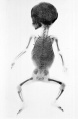 Fetal 5 month x-ray.jpg 1,500 × 2,298; 308 KB
 Fetal gonad retinoid receptor expression 01.jpg 1,004 × 1,000; 226 KB
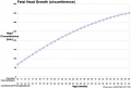 Fetal head growth circumference graph01.jpg 905 × 613; 58 KB
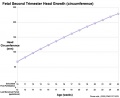 Fetal head growth circumference graph02.jpg 800 × 650; 44 KB
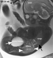 Fetal kidney MRI 01.jpg 797 × 880; 68 KB
 Fetal length change.jpg 972 × 648; 72 KB
 Fetal ovary meiosis 01.jpg 1,280 × 410; 132 KB
 Fetal ovary meiosis 02.jpg 496 × 600; 77 KB
 Fetal ovary meiosis 03.jpg 652 × 400; 64 KB
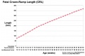
 Fetal palate growth graph.jpg 681 × 757; 77 KB
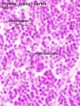
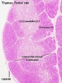 Fetal thymus.jpg 450 × 600; 122 KB
 Galletti1770 week 16.jpg 450 × 450; 32 KB
 Gillilan1959-fig03.jpg 1,042 × 1,366; 317 KB
 Gray0043.jpg 800 × 496; 50 KB
 Greater-omentum.jpg 537 × 419; 48 KB
 HansonAnson1962 fig04.jpg 1,280 × 598; 201 KB
 HansonAnson1962 fig05.jpg 1,280 × 604; 225 KB
 Herring1908b fig06.jpg 1,280 × 1,234; 283 KB
 Herring1908b fig07.jpg 1,280 × 974; 290 KB
 Human 15 weeks - terminal nerve and vomeronasal organ nerves.jpg 940 × 403; 306 KB
 Human fetal gonad retinoid receptor expression.jpg 1,004 × 1,000; 447 KB
 Keibel Mall 2 302.jpg 1,278 × 803; 149 KB
 Keibel Mall 2 583.jpg 1,280 × 1,044; 259 KB
 Keibel Mall 2 584.jpg 464 × 800; 68 KB
 Keibel Mall 2 585.jpg 1,280 × 1,144; 277 KB
 Keith1902 fig103.jpg 1,000 × 723; 139 KB
 Model male androsterone synthesis.jpg 740 × 518; 92 KB
 Placenta MRI 02.jpg 1,200 × 631; 91 KB
 Placental volume - second trimester.jpg 600 × 441; 59 KB
 Ramsey1972 fig04-16c.jpg 760 × 704; 47 KB
 Second Trimester Cerebellum.jpeg 475 × 385; 26 KB
 Streeter1922-fig42.jpg 581 × 778; 88 KB
 Streeter1922-fig43.jpg 583 × 780; 94 KB
 Streeter1922-fig44.jpg 589 × 780; 101 KB
 Streeter1922-fig45.jpg 581 × 781; 91 KB
 Streeter1922-fig46.jpg 592 × 779; 95 KB
 Streeter1922-fig47.jpg 581 × 777; 95 KB
 Streeter1922-fig48.jpg 591 × 767; 86 KB
 Streeter1922-fig49.jpg 592 × 770; 97 KB
 Streeter1922-fig50.jpg 582 × 781; 97 KB
 Streeter1922-fig51.jpg 579 × 766; 78 KB
 Streeter1922-fig52.jpg 586 × 774; 83 KB
 Streeter1922-fig53.jpg 592 × 778; 79 KB
 Streeter1922-fig54.jpg 574 × 780; 88 KB
 Streeter1922-fig55.jpg 586 × 779; 80 KB
 Streeter1922-fig56.jpg 592 × 783; 92 KB
 Streeter1922-fig57.jpg 577 × 775; 88 KB
 Streeter1922-fig58.jpg 586 × 772; 92 KB
 Streeter1922-fig59.jpg 591 × 778; 104 KB
 Streeter1922-plate05.jpg 900 × 1,200; 315 KB
 Streeter1922-plate06.jpg 904 × 1,200; 291 KB
 Thomson1899 fig03.jpg 1,209 × 1,678; 82 KB
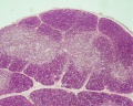
 Thymus - young 01.jpg 450 × 600; 93 KB
 Thymus - young 02.jpg 450 × 600; 91 KB
 Thymus histology 01.jpg 1,280 × 1,024; 723 KB
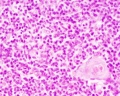 Thymus histology 02.jpg 1,280 × 1,024; 287 KB
 Thymus histology 03.jpg 1,280 × 1,024; 325 KB
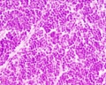
 Thymus histology 05.jpg 513 × 385; 41 KB
 Ultrasound - fetal abdominal circumference.jpg 672 × 512; 23 KB
 Watson1918 fig06.jpg 1,000 × 518; 47 KB
 Watson1918 fig07.jpg 1,000 × 570; 59 KB
 Watson1918 fig08.jpg 1,000 × 565; 71 KB


