ANAT2241 Integumentary System

| ANAT2241 This practical support page content is not part of the virtual science practical class and provides additional information for student self-directed learning purposes. All practical class pages are located on Moodle - ANAT2241 |
General Objective
To know the structure and function of skin and its appendages (derivatives).
Specific Objectives
- To know the microscopic structure of the epidermis and dermis.
- To know the histological differences between hairy (thin) and glabrous (thick) skin.
- To know the formation and histology of skin appendages: eccrine and apocrine sweat glands, sebaceous glands, hairs, nails and specialised glands as listed below.
- To know the histological features of Pacinian and Meissner corpuscles and free nerve endings.
Learning activities
Examine the following virtual slides, and in course manual identify draw and label the following structures and note their function.
Virtual Slides: Integumentary System (skin)
Histology

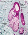
Skin overview (Stain - Haematoxylin Eosin) |

Skin overview (trichrome stain) |

Epidermis (thin skin) (Stain - Haematoxylin Eosin) |

Epidermis (thick skin) |

Epidermis and Dermis (Stain - Haematoxylin Eosin) |

Dermis elastic fibres ((Stain - van Gieson) and Silver Stain) |
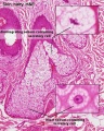
Pigmentation

Melanin in Basal Cell Layer
Glands and Hair

|

|

|
| Merocrine secretion (sweat gland) | Apocrine secretion (axilla) | Holocrine secretion (hair follicle) |
Sebaceous gland histology
Sebaceous gland histology
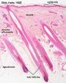
Hair follicles
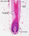
Hair follicle
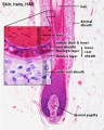
Hair follicle
Hair histology


Sensory
| Touch | Pressure |
|---|---|

|

|
| Meissner corpuscle a sensory touch cellular structure located in the dermis superficial region. | Pacinian corpuscle a sensory pressure cellular structure located in the dermis deep region. |
|
|
- Links: touch
Lip

vermilion border |
|
Nail

|
|
- Integument Histology Links: Adult Skin | Epidermis and Dermis | Thin Skin Epidermis | Thick Skin Epidermis | Elastic Fibres | Basal Cell Melanin | Foundations Practical Support | Integumentary System Development | Histology Stains
Course Links
- Histology Glossary: A | B | C | D | E | F | G | H | I | J | K | L | M | N | O | P | Q | R | S | T | U | V | W | X | Y | Z | ANAT2241 Support | Histology | Histology Stains | Embryology Glossary
| Common Histology Stains | ||||||||||||||||||||||||||||||||||||||||||||||||||||||||||||||||||||||||||||||||||||||||||||||||||||||||||||||||||||||||||||||||||||||||||||||||
|---|---|---|---|---|---|---|---|---|---|---|---|---|---|---|---|---|---|---|---|---|---|---|---|---|---|---|---|---|---|---|---|---|---|---|---|---|---|---|---|---|---|---|---|---|---|---|---|---|---|---|---|---|---|---|---|---|---|---|---|---|---|---|---|---|---|---|---|---|---|---|---|---|---|---|---|---|---|---|---|---|---|---|---|---|---|---|---|---|---|---|---|---|---|---|---|---|---|---|---|---|---|---|---|---|---|---|---|---|---|---|---|---|---|---|---|---|---|---|---|---|---|---|---|---|---|---|---|---|---|---|---|---|---|---|---|---|---|---|---|---|---|---|---|---|
| ||||||||||||||||||||||||||||||||||||||||||||||||||||||||||||||||||||||||||||||||||||||||||||||||||||||||||||||||||||||||||||||||||||||||||||||||
| ||||||||||||||||||||||||||||||||||||||||||||||||||||||||||||||||||||||||||||||||||||||||||||||||||||||||||||||||||||||||||||||||||||||||||||||||
Practical Support
- Pages can be accessed from any internet connected computer.
ANAT2241 Support Links: The Virtual Microscope | Covering and Lining Epithelia | Glandular Epithelia | CT Components | CT Types | Bone, Bone Formation and Joints | Muscle | Nervous | Blood | Eye | Cardiovascular | Respiratory | Integumentary | Gastrointestinal | Gastrointestinal Organs | Lymphatic and Immune | Endocrine | Urinary | Female Reproductive | Male Reproductive | Histology Stains | Histology Drawings | Practicals Health and Safety 2013 | Moodle - 2019
ANAT2241 This practical support page content is not part of the science practical class and provides only background information for student self-directed learning purposes.
Cite this page: Hill, M.A. (2024, May 4) Embryology ANAT2241 Integumentary System. Retrieved from https://embryology.med.unsw.edu.au/embryology/index.php/ANAT2241_Integumentary_System
- © Dr Mark Hill 2024, UNSW Embryology ISBN: 978 0 7334 2609 4 - UNSW CRICOS Provider Code No. 00098G