2018 Group Project 5: Difference between revisions
From Embryology
| Line 30: | Line 30: | ||
Link on current research for DRG {{#pmid:22375099|PMID22375099}} | Link on current research for DRG {{#pmid:22375099|PMID22375099}} | ||
===image | ===image of rat DRG=== | ||
[[file:rat L5 DRG.jpg|400px]] | [[file:rat L5 DRG.jpg|400px]] | ||
Revision as of 18:49, 27 August 2018
| Projects 2018: 1 Adrenal Medulla | 3 Melanocytes | 4 Cardiac | 5 Dorsal Root Ganglion |
Project Pages are currently being updated (notice removed when completed)
Dorsal Root Ganglion
Introduction
Dorsal Root Ganglion is a cluster of neurone found in the dorsal root of the spinal nerve. The cells found in the ganglion develops from the neural crest migration at about 4 weeks post-conception (pc).
History
Embryonic Origins
Developmental Process
Neural Crest Migration in Formation of the DRG
Trunk neural crest cells migrate via a ventromedial pathway on the neural tube during the fourth week of development. Depending on where these cells cease their migration will determine the structure into which they develop.[1]
Adult Function
Tissue / Organ Structure
Molecular Mechanisms / Factors / Genes
CXCR4 Chemokine Receptor
Abnormalities / Abnormal Development
Animal Models
Zebrafish Model
Current Research (Labs)
Link on current research for DRG [4]
image of rat DRG
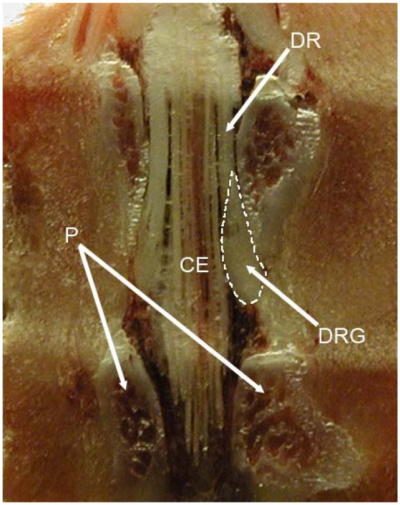

DRG L5 of rat [4]
Glossary
Reference List
- ↑ 1.0 1.1 Kasemeier-Kulesa JC, Kulesa PM & Lefcort F. (2005). Imaging neural crest cell dynamics during formation of dorsal root ganglia and sympathetic ganglia. Development , 132, 235-45. PMID: 15590743 DOI.
- ↑ 2.0 2.1 Kasemeier-Kulesa JC, McLennan R, Romine MH, Kulesa PM & Lefcort F. (2010). CXCR4 controls ventral migration of sympathetic precursor cells. J. Neurosci. , 30, 13078-88. PMID: 20881125 DOI.
- ↑ Honjo Y & Eisen JS. (2005). Slow muscle regulates the pattern of trunk neural crest migration in zebrafish. Development , 132, 4461-70. PMID: 16162652 DOI.
- ↑ 4.0 4.1 Sapunar D, Kostic S, Banozic A & Puljak L. (2012). Dorsal root ganglion - a potential new therapeutic target for neuropathic pain. J Pain Res , 5, 31-8. PMID: 22375099 DOI.
- ↑ George L, Dunkel H, Hunnicutt BJ, Filla M, Little C, Lansford R & Lefcort F. (2016). In vivo time-lapse imaging reveals extensive neural crest and endothelial cell interactions during neural crest migration and formation of the dorsal root and sympathetic ganglia. Dev. Biol. , 413, 70-85. PMID: 26988118 DOI.
- ↑ George L, Kasemeier-Kulesa J, Nelson BR, Koyano-Nakagawa N & Lefcort F. (2010). Patterned assembly and neurogenesis in the chick dorsal root ganglion. J. Comp. Neurol. , 518, 405-22. PMID: 20017208 DOI.
- ↑ Ogawa R, Fujita K & Ito K. (2017). Mouse embryonic dorsal root ganglia contain pluripotent stem cells that show features similar to embryonic stem cells and induced pluripotent stem cells. Biol Open , 6, 602-618. PMID: 28373172 DOI.