Category:Respiratory
From Embryology
This page lists UNSW Embryology content related to respiratory development. Note the section of notes include the musculoskeletal diaphragm development.
Subcategories
This category has the following 2 subcategories, out of 2 total.
Pages in category 'Respiratory'
The following 148 pages are in this category, out of 148 total.
2
A
B
- Book - A History of Science 9
- Book - Contributions to Embryology Carnegie Institution No.12
- Book - Contributions to Embryology Carnegie Institution No.38
- Book - Human Embryology and Morphology 2
- Book - Manual of Human Embryology 17-10
- Book - Manual of Human Embryology 17-9
- Book - Text-Book of Embryology 13
C
E
F
H
L
M
P
- Paper - A case of atresia of the esophagus combined with traoheoesophageal fistula in a 9 mm human embryo, and its embryological explanation
- Paper - A further communication on the formation of the nasal cavities (1912)
- Paper - A histological investigation of the development and structure of the human lung
- Paper - A preliminary communication on the formation of the nasal cavities (1911)
- Paper - Conditions of foetal respiration in the goat (1934)
- Paper - Congenital defects in the diaphragm (1940)
- Paper - Cor biloculare, with a note on the development of the pulmonary veins (1937)
- Paper - Development of olfactory and related structures in staged human embryos
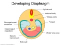
- Paper - Development of the human diaphragm and pleural sacs
- Paper - Development of the larynx (1910)
- Paper - Early developmental stages of the human lung
- Paper - Malformation of the diaphragm in a dog (1924)
- Paper - Normal development of the trachea and esophagus in man
- Paper - Note on a case of defective development of the diaphragm, accompanied by stenosis of the anal canal (1918)
- Paper - On the genesis of air cells in the conchae nasales (1910)
- Paper - On the origin of the pulmonary arteries in mammals
- Paper - On the origin of the pulmonary arteries in mammals 2
- Paper - On the prenatal and neonatal lung (1913)
- Paper - Origin of the pulmonary vessels in the chick (1922)
- Paper - Studies on the development of the human larynx (1911)
- Paper - The Course of the Phrenic Nerve in the Embryo
- Paper - The development of nerve-endings in the respiratory muscles of the sheep (1940)
- Paper - The development of the bronchopulmonary segments in human embryos of horizons XVII to XIX
- Paper - The development of the lungs
- Paper - The Development of the Nose and of the Pharynx and its Derivatives in Man
- Paper - The development of the terminal air passages of the human lung
- Paper - The embryology of the bird's lung 2 (1916)
- Paper - The functional history of the coelom and the diaphragm (1913)
- Paper - The genesis and development of the nasolacrimal passages in man
- Paper - The lateral wall of the cavum nasi in man, with especial reference to the various developmental stages
- Paper - The lung of a human foetus of 170 mm C.R. length
- Paper - The right lung of a human foetus of 152 millimeters CRL
- Paper - The sinus maxillarus and its relations in the embryo, child, and adult man
- Paper - The terminals of the human bronchiole (1922)
- Paper - True congenital diverticulum of the trachea in a subject showing also right aortic arch (1929)
- Paper - Two anomalies in the construction of the diaphragm (1924)
- Template:Pseudoglandular stage
R
- Template:Ref-AddisonHow1913
- Template:Ref-Amin1914
- Template:Ref-BarcroftElliottFlexner1934
- Template:Ref-BarnardDay1937
- Template:Ref-Bremer1902
- Template:Ref-Bremer1909
- Template:Ref-Cooper1938
- Template:Ref-Dickson1940
- Template:Ref-Flint1906
- Template:Ref-Frazer1910
- Template:Ref-Frazer1911a
- Template:Ref-Frazer1911b
- Template:Ref-Frazer1912
- Template:Ref-Goodrich1918
- Template:Ref-Grosser1912
- Template:Ref-GrosserLewisMcMurrich1912
- Template:Ref-Guinane1924
- Template:Ref-Huntington1919
- Template:Ref-LiebowMiller1940
- Template:Ref-LocyLarsell1916b
- Template:Ref-Mall1901c
- Template:Ref-Miller1919
- Template:Ref-Palmer1936a
- Template:Ref-Palmer1936b
- Template:Ref-Schaeffer1910c
- Template:Ref-Stibbe1929
- Template:Ref-Wells1954
- Template:Ref-WellsBoyden1954
- Template:Ref-Willson1922
- Template:Respiratory
- Template:Respiratory abnormalities
- Template:Respiratory Links
- Template:Respiratory Postnatal Timeline table
- Respiratory Quiz
- Template:Respiratory Species Comparison collapsetable
- Template:Respiratory Species Comparison table
- Respiratory System - Abnormalities
- Respiratory System - Carnegie Stage 13
- Respiratory System - Carnegie Stage 22
- Respiratory System - Diaphragm
- Respiratory System - Histology
- Respiratory System - Molecular
- Respiratory System - Postnatal
- Respiratory System - Upper Respiratory Tract
- Respiratory System Development
- Template:Respiratory terms
S
Media in category 'Respiratory'
The following 200 files are in this category, out of 275 total.
(previous page) (next page)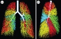 3D model of the air way tree.jpg 1,280 × 819; 215 KB
 Agenesis of left lung.jpg 600 × 424; 28 KB
 Alveolar-sac-01.jpg 720 × 478; 73 KB
 Amin1914 fig01.jpg 1,000 × 652; 120 KB
 Amin1914 fig02.jpg 1,000 × 681; 152 KB
 Amin1914 fig03.jpg 1,000 × 689; 134 KB
 Amin1914 fig04.jpg 1,000 × 727; 139 KB
 Amin1914 fig05.jpg 1,000 × 646; 91 KB
 Bailey282.jpg 876 × 597; 114 KB
 Bailey283.jpg 604 × 424; 56 KB
 Bailey284.jpg 885 × 353; 42 KB
 Bailey285.jpg 588 × 332; 52 KB
 Bailey286.jpg 842 × 481; 85 KB
 Bailey287.jpg 785 × 281; 48 KB
 Bailey288.jpg 857 × 400; 90 KB
 Bailey289.jpg 917 × 533; 130 KB
 Bailey290.jpg 935 × 635; 154 KB
 Bailey298.jpg 342 × 713; 56 KB
 Bailey471.jpg 711 × 368; 43 KB
 Bailey472.jpg 797 × 518; 105 KB
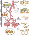
 Birth lymphatics lung inflation cartoon.jpg 845 × 510; 85 KB
 Branching.jpg 634 × 407; 70 KB
 Bronchi lungs.jpg 690 × 444; 41 KB
 Bronchial epithelial bridge.jpg 397 × 482; 28 KB
 Buell-plate01.jpg 1,221 × 1,500; 236 KB
 Buell-plate02.jpg 1,124 × 1,500; 287 KB
 Buell01.jpg 713 × 800; 70 KB
 Buell02.jpg 977 × 800; 86 KB
 Buell03.jpg 790 × 800; 73 KB
 Buell04.jpg 913 × 800; 69 KB
 Buell05.jpg 1,176 × 800; 96 KB
 Buell06.jpg 1,037 × 800; 76 KB
 Buell07.jpg 1,200 × 878; 195 KB
 Buell08.jpg 1,037 × 1,000; 176 KB
 Buell09.jpg 724 × 1,000; 83 KB
 Buell10.jpg 665 × 1,000; 113 KB
 Cilium cartoon.jpg 800 × 682; 114 KB
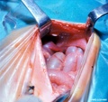
 Congenital diaphragmatic hernia 01.jpg 1,000 × 494; 108 KB
 Congenital diaphragmatic hernia 02.jpg 578 × 800; 48 KB
 Congenital diaphragmatic hernia 03.jpg 611 × 800; 82 KB
 Congenital diaphragmatic hernia 04.jpg 637 × 600; 66 KB
 Congenital lobar emphysema.jpg 1,204 × 1,130; 71 KB
 CPAMXCT.jpg 1,559 × 838; 157 KB
 Diaphragm components.jpg 600 × 450; 41 KB
 Dickie1914 fig07.jpg 407 × 577; 74 KB
 Fetal lung histology 01.jpg 1,280 × 1,024; 339 KB
 Fetal lung histology 02.jpg 450 × 600; 74 KB
 Fetal lung histology.jpg 450 × 600; 83 KB
 Fetal rabbit neuroepithelial body 01.jpg 793 × 1,200; 98 KB
 Fgf signalling.jpg 400 × 219; 46 KB
 Flint1906 plate01.jpg 1,280 × 843; 149 KB
 Flint1906 textfig01.jpg 700 × 473; 33 KB
 Frazer1910 fig01.jpg 650 × 740; 91 KB
 Frazer1910 fig02.jpg 750 × 804; 144 KB
 Frazer1910 fig03.jpg 1,000 × 704; 130 KB
 Frazer1910 fig04.jpg 1,000 × 637; 129 KB
 Frazer1910 fig05.jpg 900 × 825; 169 KB
 Frazer1910 fig06.jpg 1,400 × 1,251; 319 KB
 Frazer1910 fig06a.jpg 863 × 742; 129 KB
 Frazer1910 fig06b.jpg 565 × 584; 52 KB
 Frazer1910 fig06c.jpg 739 × 520; 82 KB
 Frazer1910 fig06d.jpg 678 × 679; 61 KB
 Frazer1910 fig07.jpg 1,300 × 1,368; 279 KB
 Frazer1910 fig07a.jpg 666 × 747; 118 KB
 Frazer1910 fig07b.jpg 700 × 716; 83 KB
 Frazer1910 fig07c.jpg 400 × 495; 10 KB
 Frazer1910 fig07d.jpg 1,036 × 634; 72 KB
 Frazer1910 fig08.jpg 1,300 × 708; 202 KB
 Frazer1910 fig08a.jpg 463 × 708; 72 KB
 Frazer1910 fig08b.jpg 660 × 708; 86 KB
 Frazer1910 fig08c.jpg 281 × 708; 48 KB
 Frazer1910 fig09.jpg 1,000 × 582; 80 KB
 Frazer1910 fig10.jpg 750 × 702; 60 KB
 Frazer1910 fig11.jpg 755 × 702; 70 KB
 Frazer1910 fig12.jpg 1,300 × 450; 69 KB
 Frazer1910 fig13.jpg 700 × 1,248; 83 KB
 Frazer1910 fig14.jpg 380 × 302; 19 KB
 Frazer1910 fig15.jpg 720 × 620; 65 KB
 Frazer1910 fig16.jpg 700 × 745; 89 KB
 Frazer1910 fig17.jpg 900 × 794; 76 KB
 Frazer1910 fig18.jpg 540 × 490; 43 KB
 Frazer1910 fig19.jpg 1,300 × 446; 62 KB
 Gray0051.jpg 600 × 423; 91 KB
 Gray0391.jpg 600 × 530; 79 KB
 Gray0622.jpg 737 × 700; 110 KB
 Gray0804.jpg 550 × 700; 75 KB
 Gray0806.jpg 600 × 771; 173 KB
 Gray0859.jpg 694 × 600; 114 KB
 Gray0947.jpg 600 × 398; 56 KB
 Gray0948.jpg 400 × 203; 21 KB
 Gray0949.jpg 400 × 276; 28 KB
 Gray0950 arytenoid cartilage.jpg 600 × 500; 39 KB
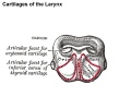 Gray0950 cricoid cartilage.jpg 600 × 500; 42 KB
 Gray0950 epiglottis cartilage.jpg 600 × 500; 24 KB
 Gray0950 thyroid cartilage.jpg 600 × 500; 54 KB
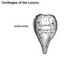
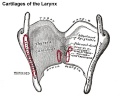
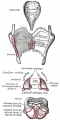 Gray0950.jpg 500 × 1,000; 106 KB
 Gray0951.jpg 600 × 724; 100 KB
 Gray0952.jpg 588 × 800; 102 KB
 Gray0953.jpg 588 × 800; 106 KB
 Gray0954.jpg 497 × 800; 94 KB
 Gray0955.jpg 616 × 600; 91 KB
 Gray0956.jpg 508 × 400; 49 KB
 Gray0957.jpg 421 × 700; 74 KB
 Gray0958.jpg 508 × 700; 85 KB
 Gray0959.jpg 471 × 700; 91 KB
 Gray0960.jpg 530 × 650; 99 KB
 Gray0961.jpg 600 × 769; 66 KB
 Gray0962.jpg 801 × 669; 191 KB
 Gray0963.jpg 600 × 300; 48 KB
 Gray0964.jpg 536 × 800; 139 KB
 Gray0965.jpg 600 × 750; 130 KB
 Gray0966.jpg 531 × 800; 132 KB
 Gray0968.jpg 700 × 550; 96 KB
 Gray0971.jpg 800 × 583; 166 KB
 Gray0974.jpg 617 × 800; 125 KB
 Gray0975.jpg 800 × 799; 130 KB
 Gray0976.jpg 600 × 599; 146 KB
 Gray0983.jpg 800 × 517; 38 KB
 Gray0984.jpg 800 × 477; 58 KB
 Gray0987.jpg 1,000 × 579; 114 KB
 Gray0987a.jpg 554 × 579; 50 KB
 Gray0987b.jpg 554 × 579; 67 KB
 Gray0988.jpg 397 × 800; 48 KB
 Gray0989.jpg 700 × 685; 107 KB
 Gray804.gif 471 × 600; 30 KB
 Head arches cartoon.jpg 394 × 402; 29 KB
 Historic-lungs.jpg 600 × 493; 79 KB
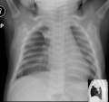
 Human congenital diaphragmatic hernia.jpg 800 × 626; 86 KB
 Human developing lung protein 01.jpg 657 × 1,000; 241 KB
 Human developing lung protein 02.jpg 800 × 302; 82 KB
 Human lung inter-alveolar septum em01.jpg 785 × 589; 168 KB
 Human lung pseudoglandular.jpg 672 × 1,000; 121 KB
 Human lung stages 01.jpg 787 × 534; 138 KB
 Human right lung 7-8 weeks.jpg 709 × 897; 789 KB
 Hyaline cartilage 03.jpg 500 × 626; 92 KB
 Hyaline cartilage 04.jpg 500 × 626; 101 KB
 Keibel Mall 2 266.jpg 1,000 × 754; 90 KB
 Keibel Mall 2 314.jpg 760 × 1,000; 103 KB
 Keibel Mall 2 315.jpg 604 × 800; 61 KB
 Keibel Mall 2 316.jpg 899 × 1,000; 148 KB
 Keibel Mall 2 317.jpg 805 × 1,000; 95 KB
 Keibel Mall 2 318.jpg 1,077 × 1,000; 111 KB
 Keibel Mall 2 319.jpg 811 × 800; 82 KB
 Keibel Mall 2 320.jpg 973 × 800; 110 KB
 Keibel Mall 2 321.jpg 904 × 800; 76 KB
 Keibel Mall 2 322.jpg 729 × 800; 47 KB
 Keibel Mall 2 323.jpg 718 × 800; 54 KB
 Keibel Mall 2 324.jpg 1,280 × 930; 125 KB
 Keibel Mall 2 325.jpg 1,280 × 882; 225 KB
 Keibel Mall 2 326.jpg 1,280 × 730; 207 KB
 Keibel Mall 2 327.jpg 1,100 × 676; 114 KB
 Keibel Mall 2 328.jpg 1,100 × 534; 96 KB
 Keibel Mall 2 329.jpg 930 × 582; 74 KB
 Keibel Mall 2 330.jpg 1,280 × 1,009; 342 KB
 Keibel Mall 2 331.jpg 690 × 966; 62 KB
 Keibel Mall 2 332.jpg 703 × 900; 48 KB
 Keibel Mall 2 333.jpg 811 × 900; 88 KB
 Keibel Mall 2 334.jpg 1,000 × 332; 50 KB
 Keibel Mall 2 335.jpg 793 × 800; 61 KB
 Keibel Mall 2 336.jpg 777 × 800; 34 KB
 Keibel Mall 2 337.jpg 847 × 660; 53 KB
 Keibel Mall 2 338.jpg 1,111 × 800; 101 KB
 Keibel Mall 2 339.jpg 842 × 800; 74 KB
 Keibel Mall 2 340.jpg 986 × 800; 99 KB
 Keibel Mall 2 341.jpg 865 × 800; 65 KB
 Keibel Mall 2 342.jpg 800 × 668; 41 KB
 Keibel Mall 2 343-344.jpg 1,200 × 510; 115 KB
 Keibel Mall 2 343.jpg 942 × 800; 99 KB
 Keibel Mall 2 344.jpg 942 × 800; 111 KB
 Keibel Mall 2 345.jpg 1,028 × 838; 144 KB
 Keibel Mall 2 346.jpg 1,200 × 801; 224 KB
 Keibel Mall 2 347.jpg 1,200 × 741; 110 KB
 Keibel Mall 2 348.jpg 902 × 791; 92 KB
 Keibel Mall 2 349-350.jpg 1,200 × 566; 87 KB
 Keibel Mall 2 349.jpg 597 × 566; 44 KB
 Keibel Mall 2 350.jpg 604 × 566; 44 KB
 Keibel Mall 2 351-352.jpg 1,000 × 551; 55 KB
 Keibel Mall 2 351.jpg 468 × 551; 28 KB
 Keibel Mall 2 352.jpg 452 × 551; 28 KB
 Keibel Mall 2 353.jpg 671 × 788; 37 KB
 Keibel Mall 423.jpg 766 × 550; 38 KB
 Keith1902 fig007.jpg 946 × 700; 80 KB
 Keith1902 fig015b.jpg 1,000 × 719; 97 KB
 Keith1902 fig019.jpg 1,000 × 562; 119 KB
 Kollmann407-409.jpg 729 × 1,000; 134 KB
 Kollmann417.jpg 778 × 583; 123 KB
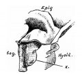
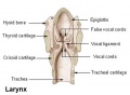 Larynx.jpg 473 × 345; 16 KB
 Lung alveoli development cartoon.jpg 500 × 652; 38 KB
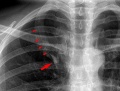 Lung Azygos Lobe 02.jpg 600 × 457; 114 KB
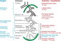
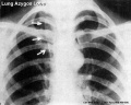 Lung Azygos Lobe.jpg 800 × 637; 100 KB
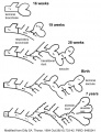
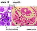 Lung development stage13-22.jpg 450 × 377; 57 KB
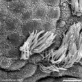 Lung epithelium sem11.jpg 1,000 × 999; 240 KB
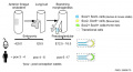
 Lung Fgf10 expression cartoon.jpg 1,280 × 821; 96 KB
 Lung human and mouse Sox expression.jpg 1,280 × 692; 62 KB
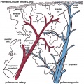 Lung primary lobule 01.jpg 800 × 799; 140 KB
 Lung secondary lobule 01.jpg 617 × 800; 146 KB
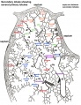
 Lung subdivisions cartoon.jpg 600 × 717; 148 KB
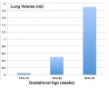 Lung volume graph 01.jpg 800 × 644; 47 KB
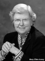 Mary Ellen Avery.jpg 588 × 784; 27 KB
 MAS.jpg 640 × 513; 30 KB


