Musculoskeletal System - Limb Abnormalities
Introduction
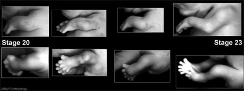
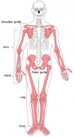
Musculoskeletal and limb abnormalities are one of the largest groups of congenital abnormalities. The upper and lower limbs have a large number of different genetic and environment derived abnormalities, some of which can be surgically repaired, while others may indicate other syndromes or karyotype anomalies (trisomy etc) by association (More? aneuploidy effect). Many of the hand and foot abnormalities are described as "dactyly" from the Greek (daktulos) for finger or digit.
A 1994 review of data from Hungary (1975-1984)[1] showed in this population an overall 1 in 1,816 prevalence of limb deficiency at birth.
A key historic area of limb abnormalities were those associated with limb reduction (and other defects) with the "morning sickness" prescription drug Thalidomide and its significant teratogenic effects (More? Abnormal Development - Thalidomide).
--Mark Hill 09:25, 14 April 2010 (EST) Page Template only - content from original UNSW Embryology site currently being edited and updated.
Some Recent Findings
Sun M, Ma F, Zeng X, Liu Q, Zhao XL, Wu FX, Wu GP, Zhang ZF, Gu B, Zhao YF, Tian SH, Lin B, Kong XY, Zhang XL, Yang W, Lo W, Zhang X. Triphalangeal thumb-polysyndactyly syndrome and syndactyly type IV are caused by genomic duplications involving the long-range, limb-specific SHH enhancer. J Med Genet. 2008 Apr 16;
"Both Triphalangeal thumb-polysyndactyly syndrome (TPTPS) and syndactyly type IV (SD4) are due to duplications involving ZPA regulatory sequence (ZRS), the limb-specific Sonic hedgehog (SHH) enhancer. Point mutations in the ZRS and duplications encompassing the ZRS cause distinctive limb phenotypes."
Dauwerse JG, de Vries BB, Wouters CH, Bakker E, Rappold G, Mortier GR, Breuning MH, Peters DJ. A t(4;6)(q12;p23) translocation disrupts a membrane-associated O-acetyl transferase gene (MBOAT1) in a patient with a novel brachydactyly-syndactyly syndrome. Eur J Hum Genet. 2007 Apr 18;
"One of the genes on chromosome 6, the membrane-associated O-acetyl transferase gene 1 (MBOAT1), was disrupted by the breakpoint. ...Identification of the transferred acyl group and the target may reveal the signaling pathways altered in this novel brachydactyly-syndactyly syndrome."
Nissim S, Allard P, Bandyopadhyay A, Harfe BD, Tabin CJ. Characterization of a novel ectodermal signaling center regulating Tbx2 and Shh in the vertebrate limb. Dev Biol. 2007 Apr 1;304(1):9-21
"The data presented here identify the non-AER border of dorsal-ventral ectoderm as a new signaling center in limb development that localizes the ZPA to the limb margin. This finding explains the tight restriction of Shh expression to the posterior margin throughout limb outgrowth as well as the tight restriction of Shh expression to the anterior margin in many mutants exhibiting preaxial polydactyly."
Textbooks
- The Developing Human: Clinically Oriented Embryology (8th Edition) by Keith L. Moore and T.V.N Persaud - Moore & Persaud Chapter 15 the skeletal system
- Larsen’s Human Embryology by GC. Schoenwolf, SB. Bleyl, PR. Brauer and PH. Francis-West - Chapter 11 Limb Dev (bone not well covered in this textbook)
- Before we Are Born (5th ed.) Moore and Persaud Chapter 16,17: p379-397, 399-405
- Essentials of Human Embryology Larson Chapter 11 p207-228
Limb Abnormality Classification
There have been a number of different classifications applied to limb abnormalities (Classic Classification, Frantz Classification, International Society for Prosthetics and Orthotics (ISPO) Classification System).
The current preferred classification system is the [#limbclassificationref ISPO Classification] which divides all limb deformities into either transverse (no distal remaining portions) or longitudinal (has distal portions).
The original classical classification of limb deficiencies was:
Amelia complete absence of a limb.
Meromelia the partial absence of a limb.
Hemimelia the absence of half a limb.
Phocomelia a flipper-like appendage attached to the trunk.
Acheiria a missing hand or foot.
Adactyly the absence of metacarpal or metatarsal (More? [#handclassification Hand Abnormality Classification]).
Aphalangia an absent digit, finger or toe (More? [#handclassification Hand Abnormality Classification]).
Day HJ. The ISO/ISPO classification of congenital limb deficiency. Prosthet Orthot Int. 1991 Aug;15(2):67-9.
Hand Abnormality Classification
There is now a [#handclassificationref international classification for congenital hand anomalies] based on an extension of an earlier classification system.
Some of these abnormalities can be initially detected prenatally by ultrasound and may be associated with other syndromes or karyotype anomalies.
Groups:
I. Failure of formation; transverse (A), or longitudinal (B) (radial and ulnar deficiencies, symbrachydactyly)
II. Failure of differentiation
III. Polydactyly (513 anomalies, Madelung deformity, the Kirner deformity and congenital trigger fingers and trigger thumbs, Triphalangeal thumbs)
IV. Overgrowth
V. Undergrowth
VI. Amniotic band syndrome (amniotic bands)
VII. Generalized skeletal syndromes.
Failure of finger ray induction (cleft hand (IC), central polydactyly (III) and (bony) syndactyly (II)
Unclassifiable
De Smet L; IFSSH. International Federation for Societies for Surgery of the Hand JSSH. Japanese Society for Surgery of the Hand. Classification for congenital anomalies of the hand: the IFSSH classification and the JSSH modification. Genet Couns. 2002;13(3):331-8.
alignment abnormalities (clenched hand, camptodactyly, clinodactyly, hypokinesia, clubhand, phocomelia), thumb anomalies, abnormal size (macrodactyly, trident hand), abnormal echogenicity (abnormal calcifications), abnormal number (polydactyly, syndactyly, ectrodactyly), and constriction band sequence.
Syndactyly
| Fusion of fingers or toes (Greek, syn = together, dactyly = digit) which may be single or multiple and may affect: skin only, skin and soft tissues or skin, soft tissues and bone. The condition is unimportant in toes but disabling in fingers and requires operative separation and is frequently inherited as an autosomal dominant. The presence of this additional "webbing" reflects preservation of the developmental tissues that in normal development are removed by programmed cell death (apotosis).
Syndactyly occuring in cattle is known as "mulefoot" an autosomal recessive trait and has been associated with mutations in the low density lipoprotein receptor-related protein 4 gene (LRP4). When shortening of the syndactyal digits also occurs it is then described as brachysyndactyly (Greek, brachys = short, syn = together, dactyly = digit). (More? [#Brachydactyly Brachydactyly]) |
File:Syndactylysm.jpg |
OMIM: [../OMIMfind/skmus/OMIM-185900.htm Syndactyly I] | [../OMIMfind/skmus/OMIM-186000.htm Syndactyly II] | [../OMIMfind/skmus/OMIM-186100.htm Syndactyly III] | [../OMIMfind/skmus/OMIM-186200.htm Syndactyly IV] | [../OMIMfind/skmus/OMIM-186300.htm Syndactyly V] | [../OMIMfind/skmus/OMIM-syndactyly_list.htm Syndactyly List (1999)]
Ota S, Zhou ZQ, Keene DR, Knoepfler P, Hurlin PJ. Activities of N-Myc in the developing limb link control of skeletal size with digit separation. Development. 2007 Apr;134(8):1583-92.
Search PubMed Now: Syndactyly | Syndactylia |
Polydactyly
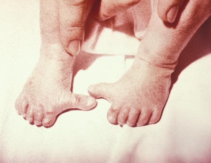
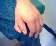 Polydactylia (Image: CDC Imagebank)
Presence of additional toes or fingers (Greek, poly = many, dactyly = digit) also called polydactylia or polydactylism. The condition is often treated surgically in the infant. Polydactyly can also be associated with a number of different syndromes including Greig cephalopolysyndactyly syndrome (GCPS).
There are also several forms of polydactyly including: preaxial polydactyly type-IV (PPD-IV) and postaxial polydactyly.
Preaxial polydactyly (PPD) has been shown as a defect in Sonic Hedgehog (Shh) expression. SHH is normally expressed specifically in the zone of polarizing activity (ZPA) located posteriorly in the limb bud and is expressed in an additional ectopic site (at the anterior margin) in a mouse model of this disorder.
This expression appears to be due to point mutations in the limb-specific regulatory element of the SHH gene.
File:Cat6toes sm.jpg The author Ernest Hemmingway in the 1930's had a six-toed cat (Snowball) showing a form of polydactyly and cats with a similar condition today (image) are now called "Hemmingway cats".
Search PubMed Now: Polydactyly | Polydactylia |
Polysyndactyly
Developmental abnormality where there is a combination of additional digits (polydactyly) that are fused together (syndactyly) and is known as polysyndactyly.
References:
Netscher DT, Baumholtz MA. Treatment of congenital upper extremity problems. Plast Reconstr Surg. 2007 Apr 15;119(5):101e-129e.
Search PubMed Now: Polysyndactyly
Brachydactyly
Middle phalanges of both hands and feet are very short (Greek, brachys = short, dactyly = digit) in length or absent.
Condition can also be associated with endocrine abnormality, pseudohypoparathyroidism (end-organ unresponsiveness to parathyroid hormone) leading to short stature, round facies, brachydactyly, and short fourth or fifth metacarpals.
References:
Dauwerse JG, de Vries BB, Wouters CH, Bakker E, Rappold G, Mortier GR, Breuning MH, Peters DJ. A t(4;6)(q12;p23) translocation disrupts a membrane-associated O-acetyl transferase gene (MBOAT1) in a patient with a novel brachydactyly-syndactyly syndrome. Eur J Hum Genet. 2007 Apr 18;
"One of the genes on chromosome 6, the membrane-associated O-acetyl transferase gene 1 (MBOAT1), was disrupted by the breakpoint. ...Identification of the transferred acyl group and the target may reveal the signaling pathways altered in this novel brachydactyly-syndactyly syndrome."
Search PubMed Now: Brachydactyly
Symbrachydactyly
Symbrachydactyly describes a combination of [#Syndactyly syndactyly] accompanied by brachydactyly.
References:
Gulgonen A, Gudemez E. Reconstruction of the first web space in symbrachydactyly using the reverse radial forearm flap. J Hand Surg [Am]. 2007 Feb;32(2):162-7.
Search PubMed Now: Symbrachydactyly
Ectrodactyly
| Ectrodactyly or split hand/foot malformation (SHFM) or cleft hand, central ray deficiency (previously called "lobster-claw malformation"). Abnormality is a deep median cleft of either the hand and/or foot due to the embryonic absence of the central rays.
Highly variable malformation (genetic heterogeneous, 5+ loci mapped) occuring in isolation or in association with other systematic anomalies including congenital heart defects. |
File:Clefthand-apical-defect sm.jpg |
| During limb development at the time of had or foot formation, the median apical ectodermal ridge (AER) fails to be maintained leading to an absence of its developmental regional signaling. Can have isolated vascular supply related cases and occur as part of a syndrome.
Search PubMed Now: Ectrodactyly | Split hand/foot malformation | |
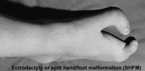
|
Triphalangeal Thumb (TPT)
| Developmental abnormality with three phalanges instead of two, forming a long, finger-like thumb.
The isolated triphalangeal thumb anomaly has also been mapped to chromosome region 7q36 and has been identified as caused by point mutations in the ZPA regulatory sequence (ZRS) which is a long-range cis-regulator for the SHH gene (Sun M etal., 2008). |
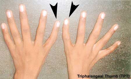 [images/skmus/Polydactylia.jpg Triphalangeal Thumb (Image modified from: Indian Pediatrics)] [images/skmus/Polydactylia.jpg Triphalangeal Thumb (Image modified from: Indian Pediatrics)] |
References: Sun M, Ma F, Zeng X, Liu Q, Zhao XL, Wu FX, Wu GP, Zhang ZF, Gu B, Zhao YF, Tian SH, Lin B, Kong XY, Zhang XL, Yang W, Lo W, Zhang X. Triphalangeal thumb-polysyndactyly syndrome and syndactyly type IV are caused by genomic duplications involving the long-range, limb-specific SHH enhancer. J Med Genet. 2008 Apr 16;
Search PubMed Now: Triphalangeal Thumb
Links: OMIM - Sall4 | OMIM - Tbx5 | Indian Pediatrics - Indian Pediatrics 2005;42:1246-1247
Talipes Equinovarus
File:Talipes equinovarus sm.jpg
Abnormality of the lower limb which begins in the embryonic period (first trimester of pregnancy) resulting in the foot is then turned inward and downward at birth, described as "club foot".
Occurs in approximately 1 in 1,000 births, postnatally it affects how children walk on their toes with the foot pointed downward like a horse (Latin, talipes = ankle bone, pes = foot, equinus = horse).
Can also occur in asociation with other syndromes. For example, camptomelic dysplasia, an extremely rare (2 per million live births) lethal congenital bony dysplasia which can be detected by ultrasound (25 weeks) visible as anterior bowing of long bones.
Search PubMed Now: talipes equinovarus
Links: Medline Plus - Clubfoot | The Clubfoot Club |
Amniotic band syndrome (amniotic bands)
Amniotic constriction bands are relatively rare and is caused by damage to the amnion, producing fiber-like bands that trap periperal structures (arms, legs, fingers, or toes) reducing local blood supply leading to abnormal development.
Abnormalities range from a permanent band or indentation around the structure (arm, leg, finger, or toe), digital webbing, too all or part of the limb missing.
Search PubMed Now: Amniotic band syndrome | amniotic bands |
Limb Reduction
Genetic
There are many different genetic abnormalities and syndromes that have an associated limb reduction.
| File:Trisomy21-hand.jpg | Trisomy 21 - (Down Syndrome) features short and broad hands, clinodactyly (curving of the fifth finger, little finger) with a single flexion crease (20%), hyperextensible finger joints, space between the great toe (big) and the second toe is increased, and acquired hip dislocation (6%). |
Diastrophic dysplasia - an autosomal recessive disorder, due to mutations in the DTD sulphate transporter gene (chromosome 5q32–q33). Leads to severe short-limbed dwarfism, progressive spinal and joint problems and can be detected by ultrasound (16 and 19 weeks of gestation).
Environment
Thalilomide was the most celebrated limb reducing insult (teratogen) in humans which also produced a range of other deformities depending on developmental time and concentration of the drug exposure.
Many additional substances (teratogens) have been found capable of producing limb reduction defects in experimental animals but few have been related to humans.
Limb reduction defects may also be either direct or indirect, for example with loss of blood supply to part of the limb or abnormal innervation at the spinal or cerebral level. There are a number of as yet undefined mechanisms involved.
Limb reduction defects may be apical (congenital amputation) or pre- or post-axial (absence of radius and lateral digits; ulnar and medial digits).
Questions:
What area is missing in the reduced limb shown above?
What will be the relative growth rates of the right and left humeri in this child?
Links: [../Defect/page5i.htm Abnormal Development - Thalidomide] | The Swedish Thalidomide Society |
Search PubMed Now: Congenital Limb Reduction | Thalidomide |
Nail Abnormalities
Covered in developmental notes on [skin2.htm Integumentary Development Abnormalities], [skin12.htm Nail Development] and [skin.htm Integumentary Development].
Congenital hyponychia
Anonychia
Nail-patella syndrome
Ectodermal dysplasias
Brachydactylies
Search PubMed Now: Congenital hyponychia | Anonychia |
Objectives
- Identify the components of a somite and the adult derivatives of each component.
- Give examples of sites of (a) endochondral and (b) intramembranous ossification and to compare these two processes.
- Identify the general times (a) of formation of primary and (b) of formation of secondary ossification centres, and (c) of fusion of such centres with each other.
- Briefly summarise the development of the limbs.
- Describe the developmental abnormalities responsible for the following malformations: selected growth plate disorders; congenital dislocation of the hip; scoliosis; arthrogryposis; and limb reduction deformities.
Computer Activities
Development Overview
Below is a very brief overview using simple figures of 3 aspects of early musculoskeletal development. More detailed overviews are shown on other notes pages Mesoderm and Somite, Vertebral Column, Limb in combination with serial sections and Carnegie images.
Mesoderm Development

|
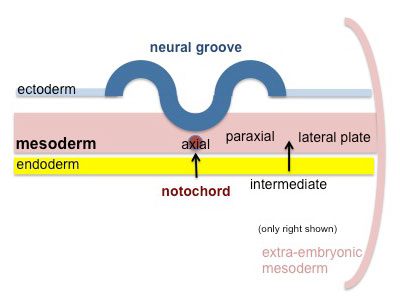
Cells migrate through the primitive streak to form mesodermal layer. Extraembryonic mesoderm lies adjacent to the trilaminar embryo totally enclosing the amnion, yolk sac and forming the connecting stalk. |
|
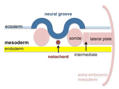
Paraxial mesoderm accumulates under the neural plate with thinner mesoderm laterally. This forms 2 thickened streaks running the length of the embryonic disc along the rostrocaudal axis. In humans, during the 3rd week, this mesoderm begins to segment. The neural plate folds to form a neural groove and folds. |
|
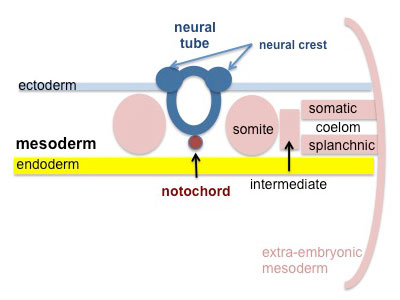
Segmentation of the paraxial mesoderm into somites continues caudally at 1 somite/90minutes and a cavity (intraembryonic coelom) forms in the lateral plate mesoderm separating somatic and splanchnic mesoderm.
Note intraembryonic coelomic cavity communicates with extraembryonic coelom through portals (holes) initially on lateral margin of embryonic disc. |
|
Somites continue to form. The neural groove fuses dorsally to form a tube at the level of the 4th somite and "zips up cranially and caudally and the neural crest migrates into the mesoderm. |
Somite Development

|
Mesoderm beside the notochord (axial mesoderm, blue) thickens, forming the paraxial mesoderm as a pair of strips along the rostro-caudal axis. |
|
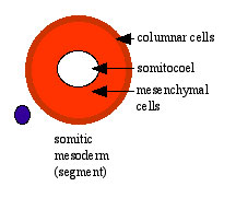
Paraxial mesoderm towards the rostral end, begins to segment forming the first somite. Somites are then sequentially added caudally. The somitocoel, is a cavity forming in early somites, which is lost as the somite matures. |
|
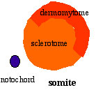
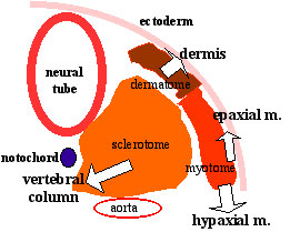
Cells in the somite differentiate medially to form the sclerotome (forms vertebral column) and dorsolaterally to form the dermomyotome. |
|
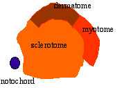
The dermomyotome then forms the dermotome (forms dermis) and myotome (forms muscle).
Neural crest cells migrate beside and through somite. |
|
The myotome differentiates to form 2 components dorsally the epimere and ventrally the hypomere, which in turn form epaxial and hypaxial muscles respectively. The bulk of the trunk and limb muscle coming from the Hypaxial mesoderm. Different structures will be contributed depending upon the somite level. |
Limb Axis Formation
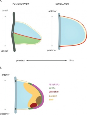
Four Concepts - much of the work has been carried out using the chicken and more recently the mouse model of development.
- Limb Initiation
- Proximodistal Axis
- Dorsoventral Axis
- Anteroposterior Axis
Limb Initiation
- Fibroblast growth factor (FGF) coated beads can induce additional limb
- FGF10 , FGF8 (lateral plate intermediate mesoderm) prior to bud formation
- FGF8 (limb ectoderm) FGFR2
- FGF can respecify Hox gene expression (Hox9- limb position)
- Hox could then activate FGF expression
Note that during the embryonic period there is a rostrocaudal (anterior posterior) timing difference between the upper and lower limb development
- this means that developmental changes in the upper limb can precede similar changes in the lower limb (2-5 day difference in timing)
Limb Identity
Forelimb and hindlimb (mouse) identity appears to be regulated by T-box (Tbx) genes, which are a family of transcription factors.
- hindlimb Tbx4 is expressed.
- forelimb Tbx5 is expressed.
- Tbx2 and Tbx3 are expressed in both limbs.
Related Research - PMID: 12490567 | Development 2003 Figures | Scanning electron micrographs of E9 Limb bud wild-type and Tbx5del/del A model for early stages of limb bud growth | PMID: 12736217 | Development 2003 Figures
Body Axes
- Anteroposterior - (Rostrocaudal, Craniocaudal, Cephalocaudal) from the head end to opposite end of body or tail.
- Dorsoventral - from the spinal column (back) to belly (front).
- Proximodistal - from the tip of an appendage (distal) to where it joins the body (proximal).
Proximodistal Axis
- Apical Ectodermal Ridge (AER) formed by Wnt7a
- then AER secretes FGF2, 4, 8
- stimulates proliferation and outgrowth
apical ectodermal ridge | AER and vascular channel
Dorsoventral Axis
- Somites - provides dorsal signal to mesenchyme which dorsalizes ectoderm
- Ectoderm - then in turn signals back (Wnt7a) to mesenchyme to pattern limb
Wnt7a
- name was derived from 'wingless' and 'int’
- Wnt gene first defined as a protooncogene, int1
- Humans have at least 4 Wnt genes
- Wnt7a gene is at 3p25 encoding a 349aa secreted glycoprotein
- patterning switch with different roles in different tissues
- mechanism of Wnt and receptor distribution still being determined (free diffusion, restricted diffusion and active transport)
One WNT receptor is Frizzled (FZD)
- Frizzled gene family encodes a 7 transmembrane receptor
Fibroblast growth factors (FGF)
- Family of at least 17 secreted proteins
- bind membrane tyrosine kinase receptors
- Patterning switch with many different roles in different tissues
- FGF8 = androgen-induced growth factor, AIGF
FGF receptors
- comprise a family of at least 4 related but individually distinct tyrosine kinase receptors (FGFR1- 4) similar protein structure
- 3 immunoglobulin-like domains in extracellular region
- single membrane spanning segment
- cytoplasmic tyrosine kinase domain
Anteroposterior Axis
- Zone of polarizing activity (ZPA)
- a mesenchymal posterior region of limb
- secretes sonic hedgehog (SHH)
- apical ectodermal ridge (AER), which has a role in patterning the structures that form within the limb
- majority of cell division (mitosis) occurs just deep to AER in a region known as the progress zone
- A second region at the base of the limbbud beside the body, the zone of polarizing activity (ZPA) has a similar patterning role to the AER, but in determining another axis of the limb
Wing as Limb Model
- chicken wing easy to manipulate
- removal, addition and rotation of limb regions
- grafting additional AER, ZPA
- implanting growth factor secreting structures
UNSW Embryology - Axes Formation - Limb | Signal Factors - Wnt
References
- ↑ <pubmed>8172251</pubmed>
- ↑ <pubmed>20644713</pubmed>| PMC2903596 | PLoS
Reviews
- How to make a zone of polarizing activity: insights into limb development via the abnormality preaxial polydactyly. Hill RE. Dev Growth Differ. 2007 Aug;49(6):439-48. Review. PMID: 17661738
Articles
Search PubMed
Search Pubmed: Limb Development | apical ectodermal ridge | zone polarizing activity]
Additional Images
Terms
Glossary Links
- Glossary: A | B | C | D | E | F | G | H | I | J | K | L | M | N | O | P | Q | R | S | T | U | V | W | X | Y | Z | Numbers | Symbols | Term Link
Cite this page: Hill, M.A. (2024, May 23) Embryology Musculoskeletal System - Limb Abnormalities. Retrieved from https://embryology.med.unsw.edu.au/embryology/index.php/Musculoskeletal_System_-_Limb_Abnormalities
- © Dr Mark Hill 2024, UNSW Embryology ISBN: 978 0 7334 2609 4 - UNSW CRICOS Provider Code No. 00098G
