Cell Division - Mitosis
| Embryology - 10 Jun 2024 |
|---|
| Google Translate - select your language from the list shown below (this will open a new external page) |
|
العربية | català | 中文 | 中國傳統的 | français | Deutsche | עִברִית | हिंदी | bahasa Indonesia | italiano | 日本語 | 한국어 | မြန်မာ | Pilipino | Polskie | português | ਪੰਜਾਬੀ ਦੇ | Română | русский | Español | Swahili | Svensk | ไทย | Türkçe | اردو | ייִדיש | Tiếng Việt These external translations are automated and may not be accurate. (More? About Translations) |
Introduction

Normal cell division in all cells, except germ cells, occurs by 2 mechanical processes that initially divide the nucleus then the cell cytoplasm. This process produces two (daughter) cells that should be genetically identical to the parent cell.
Germ cells, oocyte and spermatozoa, undergo meiotic cell division.
- Mitosis segregation of chromosomes and formation of 2 nuclei
- Cytokinesis splitting of the cell as a whole into 2 daughter cells
- Recent Nobel Prizes- 2001 Cell Cycle, 2002 Cell Death
| Mitosis of the single zygote produces how many cells in the adult? |
|---|
 |
| Cell Division Links: meiosis | mitosis | Lecture - Cell Division and Fertilization | spermatozoa | oocyte | fertilization | zygote | Genetics |
Some Recent Findings
|
| More recent papers |
|---|
This table allows an automated computer search of the external PubMed database using the listed "Search term" text link.
More? References | Discussion Page | Journal Searches | 2019 References | 2020 References Search term: Mitosis <pubmed limit=5>Mitosis</pubmed> |
Movies
| <html5media height="420" width="400">File:Mitosis 01.mp4</html5media> |
Mitosis This movie shows a cell dividing by mitosis with a fluorescently labelled protein that is located at the kinetochores and along the axes of the chromosome arms. This allows you to see the chromosomes and the linking region (kinetochore) between chromosome pairs and the mitotic spindle microtubules.[4]
|
|
|
|
|
|
Cell Changes
- Nucleus
- Chromosome condensation
- Nuclear envelope breakdown
- Cytoplasm
- Cytoskeleton reorganization
- Spindle formation (MT) Contractile ring (MF)
- Organelle redistribution
- Mitosis Energy
- Cell division uses up a lot of energy, so cells ensure they have enough resources to complete the job before committing to it.
Mitosis Phases
- Based on light microscopy of living cells light and electron microscopy of fixed and stained cells
- 5 Phases - prophase, prometaphase, metaphase, anaphase, and telophase
- Cytokinesis 6th stage overlaps the end of mitosis
MBC The stages of mitosis and cytokinesis in an animal cell
Interphase
- not a mitotic phase (discussed in cell cycle)
- Chromosomes dispersed in nucleus
- Gene expression
- Cytoskeleton and cell organelles - Distributed and functioning
- Mitochondria undergo independent proliferation/division
Chromosome Changes

Prophase

- Chromosome DNA has been earlier duplicated (S Phase)
- Chromosomes begin condensing
- Chromosome pairs (chromatids) held together at centromere
- Microtubules disassemble
- Mitotic spindle begins to form
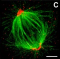
Spindle Apparatus
- 3 sets of microtubules - (+) ends point away from centrosome at each pole.
- astral microtubules - anchor the pole end in position
- kinetochore microtubules - connected to chromosomes
- polar microtubules - form the structure of the spindle apparatus
Spindle Apparatus EM | Spindle Apparatus | MBC Movie- Microtubule dynamics during mitosis
At end of prophase nuclear envelope breaks down
Prometaphase
- Microtubules now enter nuclear region
- Nuclear envelope forms vesicles around mitotic spindle
- Kinetochores form on centromere attach to some MTs of spindle
Dynamic instability and the capture of chromosomes
Centromeric attachment of microtubules
At end of prometaphase chromosomes move to metaphase plate
Metaphase

- Kinetochore MTs align chromosomes in one midpoint plane.
- Astrin is a spindle-associated protein required for chromosome alignment at the metaphase plate.[6]
Proposed alternative mechanisms for chromosome congression
Metaphase ends when sister kinetochores separate
Anaphase

- Separation of sister Kinetochores
- shortening of Kinetochore microtubules pulls chromosome to spindle pole.
- Katanin is a microtubule-severing complex involved with this stage of microtubule dynamics.[7]
Anaphase ends as nuclear envelope (membrane) begins to reform.
Telophase

- Chromosomes arrive at spindle poles
- Kinetochore MTs lost
- Condensed chromosomes begin expanding
- Continues through cytokinesis
Links: Figure 19-41 Microtubule dynamics during mitosis | Figure 19-34. The stages of mitosis and cytokinesis in an animal cell | Cytokinetic abscission: cellular dynamics at the midbody
Cleavage of Zygote
Mouse zygote mitosis[8]

|

|
| First metaphase | First anaphase |
Cleavage of the zygote forms 2 blastomeres and is cleavage with no cytoplasm synthesis.
- special "embryonic" cell cycle S phases and M phases alternate without any intervening G1 or G2 phases (MSMSMSMS, adult MG1SG2) therefore individual cell volume decreases
Cell division within these cells is initially synchronous (at the same time), then becomes asynchronously (at different times).
- slow- centre cells, larger fast- peripheral cells
- Links: Zygote | Cell Division - Mitosis | Movie - Early Cell Division | Movie - Week 1 Cell Cleavage | Carnegie stage 1
Cytokinesis
- Division of cytoplasmic contents
- Contractile ring forms at midpoint under membrane
- Microfilament ring - contracts forming cleavage furrow
- myosin II is the motor
- Eventually fully divides cytoplasm
Links: Cytokinesis | Cytokinesis in Plants
Mitotic Spindle

Spindle assembly motors[9]
Microtubule (MT)-bound motors promote bipolar spindle formation, whereas chromosome-associated motors drive proper kinetochore orientation and chromosome movement to the equator.
| Box 1 | Box 2 | Box 3 | Box 4 | Box 5 |
|---|---|---|---|---|
| Motor-dependent mechanisms establish bipolarity as Eg5 (kinesin-5) motors slide antiparallel microtubules apart with their minus ends leading and their plus ends directed toward the spindle equator. | Minus end–directed motors such as dynein move microtubules poleward with their minus ends leading, thereby incorporating K-fibers into the spindle and focusing spindle poles. | Kinetochore-associated dynein transports chromosomes along astral microtubules toward the spindle poles from the periphery. | Plus end–directed chromokinesins (kinesin-4 and -10) eject chromosome arms outward. | CENP-E (kinesin-7) transports unattached kinetochores toward the equator along spindle microtubules. MTOC, microtubule organizing centre. |
Cell Organelles
Mitochondria
- Divide independently of cell mitosis
- distributed into daughter cells
Peroxisomes
- localise at spindle poles

Peroxisome (red) location at Interphase (a) and during Mitosis (b and c)[10]
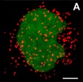
Interphase
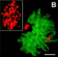
Mitosis
Mitosis


Endoplasmic Reticulum
- Associated with nuclear membrane.
Golgi
- 2 processes - disassembly and reassembly[11]
- Golgi stack undergoes a continuous fragmentation process
- fragments are distributed into daughter cells
- are reassembled into new Golgi stacks
Disassembly
- Unstacking - mediated by two mitotic kinases (cdc2 and plk)
- Vesiculation - mediated by COPI budding machinery ARF1 and the coatomer complex
Reassembly
- Fusion - formation of single cisternae by membrane fusion
- Restacking - requires dephosphorylation of Golgi stacking proteins by protein phosphatase PP2A
References
- ↑ <pubmed>23829164</pubmed>
- ↑ <pubmed>28457629</pubmed>
- ↑ <pubmed>22555603</pubmed>
- ↑ <pubmed>12105179</pubmed>
- ↑ Russan NM. Let's Build a Spindle. ASCB Image & Video Library. 2008;CYT-190. Available at: http://cellimages.ascb.org/u?/p4041coll12,521
- ↑ <pubmed>21402792</pubmed>
- ↑ <pubmed>17452528</pubmed>
- ↑ <pubmed>21321204</pubmed>| PMC2132672 | PNAS
- ↑ <pubmed>26668328</pubmed>
- ↑ <pubmed>19194514</pubmed>| PMC2633614 | PLoS One.
- ↑ <pubmed>18156178</pubmed>
Reviews
Articles
Search Pubmed
Search Pubmed: mitosis
NCBI - Policies and Guidelines | PubMed | Help:Reference Tutorial
Additional Images
Centrosome cartoon
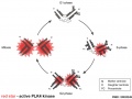
Polo-like kinase 4 centriole duplication activity
Chromosome telomeres


Terms
| Cell Division Terms (expand to view) | ||
|---|---|---|
meiosis | mitosis
| ||
|
External Links
External Links Notice - The dynamic nature of the internet may mean that some of these listed links may no longer function. If the link no longer works search the web with the link text or name. Links to any external commercial sites are provided for information purposes only and should never be considered an endorsement. UNSW Embryology is provided as an educational resource with no clinical information or commercial affiliation.
- Nature - Cell Division Milestones
Glossary Links
- Glossary: A | B | C | D | E | F | G | H | I | J | K | L | M | N | O | P | Q | R | S | T | U | V | W | X | Y | Z | Numbers | Symbols | Term Link
Cite this page: Hill, M.A. (2024, June 10) Embryology Cell Division - Mitosis. Retrieved from https://embryology.med.unsw.edu.au/embryology/index.php/Cell_Division_-_Mitosis
- © Dr Mark Hill 2024, UNSW Embryology ISBN: 978 0 7334 2609 4 - UNSW CRICOS Provider Code No. 00098G