Heart Abnormalities
Shown below are images showing the major Congenital Heart Disease (CHD). More details about the abnormality and the associated epidemiology and disease classification (ICD) can be seen by opening the table beside the image. Within the tables for the major CHDs, there are linked pages just about that abnormality.
| About - The International Classification of Diseases (ICD)
|
The International Classification of Diseases (ICD) World Health Organization's classification used worldwide as the standard diagnostic tool for epidemiology, health management and clinical purposes. This includes the analysis of the general health situation of population groups. It is used to monitor the incidence and prevalence of diseases and other health problems. Within this classification "congenital malformations, deformations and chromosomal abnormalities" are (Q00-Q99) but excludes "inborn errors of metabolism" (E70-E90).
- Links: ICD - XVII Congenital Malformations
|
CHD is a feature of many genetic abnormalities, the most relevant statistically would be Trisomy 21, but there are others that can be identified from the genetic links below.
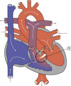
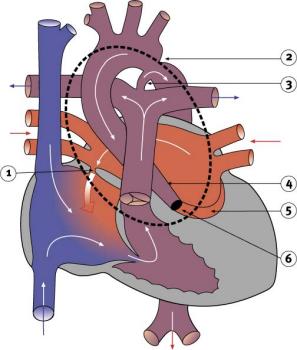
Ventricular Septal Defect
|
| About - Ventricular Septal Defect
|
The Ventricular Septal Defect (VSD)
- Usually occurs in the membranous (perimembranous) rather than muscular interventricular septum, and is more frequent in males that females.
- Perimembranous defects are located close to the aortic and tricuspid valves and adjacent to atrioventricular conduction bundle.
- Growth failure of the membranous interventricular septum or endocardial cushions, resulting in a lack of closure of the interventricular foramen.
- 30-50% close spontaneously; large VSDs result in dyspnoea and cardiac failure in infancy.
- Epidemiology - 25% of CHD; more frequent in males.
- ICD-10 Q21.0 Ventricular septal defect
- Links: Ventricular Septal Defects | Search PubMed
|
|
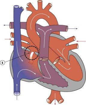
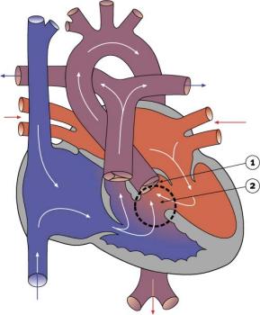
Atrial Septal Defects
|
| About - Atrial Septal Defect
|
The Atrial Septal Defect (ASD)
- Are a group of common (1% of cardiac) congenital anomolies defects occuring in a number of different forms and more often in females.
- patent foramen ovale- allows a continuation of the atrial shunting of blood
- in 25% of people a probe patent (allowing a probe to be passed from one atria to the other) foramen ovale exists.
- ostium secundum defect
- endocardial cushion defect involving ostium primum
- sinus venosus defect - contributes about 10% of all ASDs and occurs mainly in a common and less common form. Common ("usual type") - in upper atrial septum which is contiguous with the superior vena cava. Less common - at junction of the right atrium and inferior vena cava.
- common atrium
ICD-10 Q21.1 Atrial septal defect Coronary sinus defect Patent or persistent: foramen ovale ostium secundum defect (type II) Sinus venosus defect
Treatment: The surgical repair requires a cardiopulmonary bypass and is recommended in most cases of ostium secundum ASD, even though there is a significant risk involved. Ostium primum defects tend to present earlier and are often associated with endocardial cushion defects and defective mitral or tricuspid valves. In such cases, valve replacement may be necessary and the extended operation has a considerable chance of mortality.
- Increasingly closure by a transcatheter device closure has been applied.
- Repair of atrial septal defects on the perfused beating heart (atrial septal defect size 2 cm - 4.5 cm) [1]
- Links: Atrial Septal Defects | OMIM: Atrial Septal Defect | Search PubMed | Medline Plus - ASD Repair Video
|
|
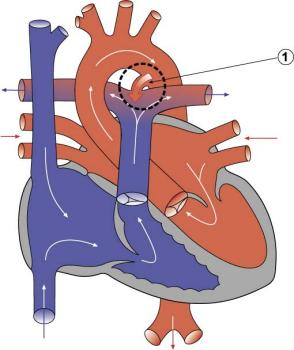
Patent Ductus Arteriosus
|
| About - Patent Ductus Arteriosus
|
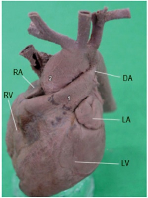
* Patent ductus arteriosus (PDA), or Patent arterial duct (PAD), occurs commonly in preterm infants, and at approximately 1 in 2000 full term infants.
- more common in females (to male ratio is 2:1).
- Can also be associated with specific genetic defects, trisomy 21 and trisomy 18, and the Rubinstein-Taybi and CHARGE syndromes.
- The opening is asymptomatic when the duct is small and can close spontaneously (by day three in 60% of normal term neonates).
- The remainder are ligated simply and with little risk, with transcatheter closure of the duct generally indicated in older children.
- The operation is always recommended even in the absence of cardiac failure and can often be deferred until early childhood.
- ICD-10 Q25.0 Patent ductus arteriosus Patent ductus Botallo Persistent ductus arteriosus
Patent ductus arteriosus classification
- Links: Patent Ductus Arteriosus |Search PubMed
|
|
Tetralogy of Fallot
|
| About - Tetralogy of Fallot
|
- Named after Etienne-Louis Arthur Fallot (1888) who described it as "la maladie blue" and is a common developmental cardiac defect.
- The syndrome consists of a number of a number of cardiac defects possibly stemming from abnormal neural crest migration.
- ICD-10 Q21.3 Tetralogy of Fallot Ventricular septal defect with pulmonary stenosis or atresia, dextroposition of aorta and hypertrophy of right ventricle.
- Links: Gene expression in cardiac tissues from infants with idiopathic conotruncal defects | Search PubMed
|
|
Hypoplastic Left Heart
|
| About - Hypoplastic Left Heart
|
- Characterized by hypoplasia (underdevelopment or absence) of the left ventricle obstructive valvular and vascular lesion of the left side of the heart.
- ICD-10 Q23.4 Hypoplastic left heart syndrome Atresia, or marked hypoplasia of aortic orifice or valve, with hypoplasia of ascending aorta and defective develop-ment of left ventricle (with mitral valve stenosis or atresia).
- Links: Search PubMed
|
|
Double Outlet Right Ventricle
|
| About - Double Outlet Right Ventricle
|
- De-oxygenated blood enters the aorta from the right ventricle and is returned to the body.
- ICD-10 Q20.1 Double outlet right ventricle Taussig-Bing syndrome
- Links: Search PubMed
|
|
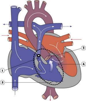
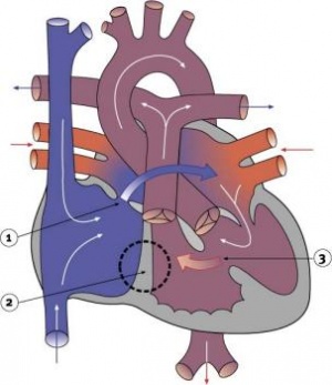
Tricuspid Atresia
|
| About - Tricuspid Atresia
|
- Blood is shunted through an atrial septal defect to the left atrium and through the ventricular septal defect to the pulmonary artery.
- The shaded arrows in the cartoon indicate mixing of the blood.
- ICD-10 Q22.4 Congenital tricuspid stenosis Tricuspid atresia
- Fontan Procedure - a surgical procedure developed by Fontan and Baudet (1971) to restore a circulation in patients with tricuspid atresia.
- Links: Search PubMed | Fontan procedure
|
|
Dextrocardia
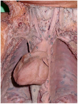
Dextrocardia anatomical heart position[2]
|
Dextrocardia (postnatal 1 year old)[2]
|
| About - Dextrocardia
|
- Initial malrotation of the heart tube bending left instead of right. Results in heart and greater vessels reversed.
- Can also occur with situs invertus, where viscera are transposed LR.
- Anatomical left-right normal asymmetry is called situs solitus. The alternative heterotaxy can be either randomization (situs ambiguus) or a complete reversal (situs inversus) of normal organ position.
- Links: Chicken Abnormal Heart Movie | Search PubMed
|
Abnormalities of Conducting System
Also variously called the cardiac conduction system (CCS), cardiac pacemaking and conduction system (CPCS), or atrioventricular conduction system (AVCS). Recently animal models (CCS-lacZ transgenic mouse) have helped identify key processes in the development of this specialized conduction system.
"Known arrhythmogenic areas including Bachmann's bundle, the pulmonary veins, and sinus venosus derived internodal structures, demonstrate lacZ expression." (Jongbloed et al, 2004)
Long QT Syndrome
Congenital long QT syndrome (LQTS) is a group of rare genetic disorders with prolonged ventricular repolarization and a risk of ventricular tachyarrhythmias. Cause is mutations in genes encoding either cardiac ion channels or channel interacting proteins.
Search NCBI Bookshelf: Congenital long-QT syndrome
- Links: Search PubMed
Glossary Links
- Glossary: A | B | C | D | E | F | G | H | I | J | K | L | M | N | O | P | Q | R | S | T | U | V | W | X | Y | Z | Numbers | Symbols | Term Link
Cite this page: Hill, M.A. (2024, June 10) Embryology ANAT2341 Lab 11 - Heart Abnormalities. Retrieved from https://embryology.med.unsw.edu.au/embryology/index.php/ANAT2341_Lab_11_-_Heart_Abnormalities
- What Links Here?
- © Dr Mark Hill 2024, UNSW Embryology ISBN: 978 0 7334 2609 4 - UNSW CRICOS Provider Code No. 00098G


