Category:Hearing: Difference between revisions
From Embryology
mNo edit summary |
mNo edit summary |
||
| Line 1: | Line 1: | ||
This {{Embryology}} category shows pages and media related to hearing and vestibular development. | This {{Embryology}} category shows pages and media related to hearing and vestibular development. | ||
Note that there are additional sub-categories for each part | Note that there are additional sub-categories for each part: [[:Category:Outer Ear|Category:Outer Ear]] [[:Category:Middle Ear|Category:Middle Ear]] [[:Category:Inner Ear|Category:Inner Ear]] | ||
Revision as of 12:00, 12 January 2017
This Embryology category shows pages and media related to hearing and vestibular development.
Note that there are additional sub-categories for each part: Category:Outer Ear Category:Middle Ear Category:Inner Ear
Subcategories
This category has the following 6 subcategories, out of 6 total.
Pages in category 'Hearing'
The following 200 pages are in this category, out of 295 total.
(previous page) (next page)2
A
B
- Template:Balance
- Template:Bast TH.
- BGD Lecture - Face and Ear Development
- BGD Practical - Face and Ear Quiz
- BGD Practical - Face Quiz
- Template:BGDB Face
- BGDB Face and Ear - Abnormalities
- BGDB Face and Ear - Early Embryo
- BGDB Face and Ear - Fetal
- BGDB Face and Ear - Late Embryo
- BGDB Face and Ear - Postnatal
- BGDB Face and Ear - Trilaminar Embryo
- BGDB Practical - Face and Ear Development
- Talk:BGDB Practical - Face and Ear Development
- BGDB Practical - Face and Ear Development Interactive
- Template:BGDB Practical 6 - Abnormalities Interactive
- Template talk:BGDB Practical 6 - Abnormalities Interactive
- Template:BGDB Practical 6 - Early Embryo Interactive
- Template:BGDB Practical 6 - Fetal Interactive
- Template:BGDB Practical 6 - Late Embryo Interactive
- Template:BGDB Practical 6 - Postnatal Interactive
- Template:BGDB Practical 6 - Trilaminar Embryo Interactive
- Book - A textbook of histology, including microscopic technic (1910) Special Histology 9
- Book - Anatomical and physiological studies on the growth of the inner ear of the albino rat (1923)
- Book - Contributions to Embryology Carnegie Institution No.20
- Book - Contributions to Embryology Carnegie Institution No.20 part 1
- Book - Contributions to Embryology Carnegie Institution No.20 part 2
- Book - Contributions to Embryology Carnegie Institution No.20 part 3
- Book - Contributions to Embryology Carnegie Institution No.20 part 4
- Book - Contributions to Embryology Carnegie Institution No.20 part 5
- Book - Contributions to Embryology Carnegie Institution No.20 part 6
- Book - Contributions to Embryology Carnegie Institution No.20 part 7
- Book - Contributions to Embryology Carnegie Institution No.21
- Book - Contributions to Embryology Carnegie Institution No.69
- Book - Human Embryology and Morphology 4
- Book - Manual of Human Embryology 16
- Book - Manual of Human Embryology 16-1
- Book - Manual of Human Embryology 16-2
- Book - Manual of Human Embryology 16-3
- Book - Manual of Human Embryology 16-4
- Book - Text-Book of Embryology 18
C
E
H
- Template:Hardesty1915 table1
- Template:Hardesty1915 table2
- Template:Hardesty1915 table3
- Template:Hardesty1915 table4
- Template:Hardesty1915 table5
- Template:Hardesty1915 table6
- Template:Hearing
- Hearing - Inner Ear Development
- Hearing - Middle Ear Development
- Hearing - Neural Pathway
- Hearing - Outer Ear Development
- Template:Hearing abnormalities
- Template:Hearing EAM timeline
- Template:Hearing Embryonic Origins table1
- Template:Hearing Links
- Template:Hearing neural
- Template:Hearing terms
- Hearing test
- Template:Hearing test
- Template:Hearing timeline
- Template:Hearing timeline table
- Human Embryology and Morphology 16
- Template:Human Embryology Manual 2 16
- Human System Development
O
P
- Paper - 1880 The Platypus Cochlea
- Paper - 1906 Observations on the Labyrinth of Certain Animals
- Paper - 1917 The Typical Form of the Cochlea and Its Variations
- Paper - A model to illustrate the probable action of the tectorial membrane (1915)
- Paper - A note on the length of the basilar membrane in man and in various mammals (1940)
- Paper - Abnormal ossification of Meckel's cartilage
- Paper - Adult form of the human stapes in the light of its development
- Paper - Blood supply of the otic capsule of a 150 mm (C.R.) human fetus
- Paper - Comparative morphology of the ear 3
- Paper - Contribution to the structure and development of the vertebrate head
- Paper - Contribution to the structure and development of the vertebrate head 1
- Paper - Contribution to the structure and development of the vertebrate head 2
- Paper - Contribution to the structure and development of the vertebrate head 3
- Paper - Development of the aquaductus cochleae and the periotic (perilymphatic) duct
- Paper - Development of the aquaeductus cochleae and its contained periotic duct and cochlear vein in human embryos
- Paper - Development of the incus of the human ear - illustrated in atlas series
- Paper - Development of the malleus of the human ear - Illustrated in atlas series
- Paper - Development of the Otic Capsule 1
- Paper - Development of the otic capsule 2
- Paper - Development of the Otic Capsule 3
- Paper - Development of the Otic Capsule 4
- Paper - Development of the otic capsule of the human ear - illustrated in atlas series
- Paper - Development of the stapes of the human ear - illustrated in atlas series
- Paper - Experimental observations on the development of the amphibian ear vesicle (1909)
- Paper - Histogenesis of the otic capsule (1917)
- Paper - Major features in the developmental history of the human stapes (1940)
- Paper - Migration of the ear vesicle in the tadpole during normal development (1921)
- Paper - On the development of the external ear passages
- Paper - On the development of the membrana tectoria with reference to its structure and attachments
- Paper - On the development of the membranous labyrinth and the acoustic and facial nerves in the human embryo
- Paper - On the development of the retina and optic nerve, and of the membranous labyrinth and auditory nerve
- Paper - On the proportions, development and attachment of the tectorial membrane (1915)
- Paper - Ossification of the otic capsule in human fetuses
- Paper - Perichondrial ossification and the fate of the perichondrium with special reference to that of the otic capsule
- Paper - Postnatal growth and adult structure of the otic (endolymphatic) sac
- Paper - Some experiments on the developing ear vesicle of the tadpole with relation to equilibration
- Paper - Some factors in the development of the amphibian ear vesicle and further experiments on equilibration
- Paper - Some features of the auditory apparatus of a 16 mm human embryo
- Paper - Some uniform characteristics of the primate auricle (1922)
- Paper - Stapes, fissula ante fenestram and associated structures in man 1
- Paper - Stapes, fissula ante fenestram and associated structures in man 2
- Paper - Stapes, fissula ante fenestram and associated structures in man 3
- Paper - Stapes, fissula ante fenestram and associated structures in man 4
- Paper - Stapes, fissula ante fenestram and associated structures in man 5
- Paper - The comparison of auricular height determinations (1925)
- Paper - The cytological processes involved in the formation of the scalae of the internal ear
- Paper - The development and structure of the otic (endolymphatic) sac
- Paper - The development of the auditory nerve in vertebrates (1910)
- Paper - The development of the auditory ossicles and associated structures in man
- Paper - The development of the auditory ossicles, the otic capsule and the extracapsular tissues
- Paper - The development of the cochlear fenestra, fossula and secondary tympanic membrane
- Paper - The development of the ear-bones in the mouse
- Paper - The development of the external ear (1934)
- Paper - The development of the first branchial arch in man and the fate of Meckel's cartilage
- Paper - The development of the otic capsule in the region of surgical fenestration 1
- Paper - The development of the otic capsule in the region of surgical fenestration 2
- Paper - The development of the otic capsule in the region of the vestibular aqueduct
- Paper - The development of the pillar cells, tunnel space, and Nuel's spaces in the organ of Corti (1919)
- Paper - The Development of the Scala Tympani, Scala Vestibuli and Perioticular Cistern in the Human Embryo
- Paper - The development of the second branchial arch (Reichert's cartilage), facial canal and associated structures in man
- Paper - The developmental and adult anatomy of the air-cells in the petrous part of the temporal bone
- Paper - The developmental course of the human auditory vesicle
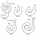
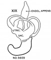
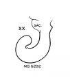
- Paper - The distal projection of the endolymphatic sac in human embryos
- Paper - The early development of the membranous labyrinth in mammalian embryos
- Paper - The early development of the otic vesicle in staged human embryos
- Paper - The early embryology of the auditory ossicles in man
- Paper - The early formations of the middle ear and eustachian tube - a criticism
- Paper - The early relation of the auditory vesicle to the ectoderm in human embryos
- Paper - The Factors Involved in the Excavation of the Cavities in the Cartilaginous Capsule of the Ear in the Human Embryo
- Paper - The fissula ante fenestram of the human otic capsule; aberrant form and contents
- Paper - The form and structure of the endolymphatic and associated ducts in the child
- Paper - The genesis and structure of the membrana tectoria and the crista spiralis of the cochlea (1918)
- Paper - The Origin of the Otic and Optic Primordia in Man
- Paper - The Typical Form of the Cochlea and its Variations
- Paper - The vascular drainage of the endolymphatic sac and its topographical relation to the transverse sinus in the human
- Paper - Vertebrate cephalogenesis 1 (1890)
- Paper - Vertebrate cephalogenesis 2 (1892)
- Template:Placode
- Template:Placodes
Media in category 'Hearing'
The following 175 files are in this category, out of 375 total.
(previous page) (next page) Mouse organ of corti 01.jpg 1,280 × 1,024; 339 KB
 Mouse organ of corti 02.jpg 1,280 × 1,024; 320 KB
 Mouse organ of corti 03.jpg 1,280 × 1,024; 207 KB
 Mouse organ of corti 04.jpg 1,280 × 1,024; 202 KB
 Mouse organ of corti 05.jpg 1,280 × 1,024; 171 KB
 Mouse organ of corti NeuroD1.jpg 1,779 × 2,383; 1.11 MB
 Mouse otic placode gene expression 01.jpg 358 × 677; 86 KB
 Mouse otic placode gene expression 02.jpg 500 × 486; 100 KB
 Mouse- early Pax8 and Pax2 expression.jpg 1,000 × 1,250; 191 KB
 Neural domain.jpg 452 × 778; 54 KB
 Newborn hearing test.jpg 200 × 153; 6 KB
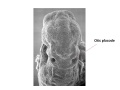
 Otic placode embryo.jpg 500 × 461; 23 KB
 Otic placode label 1.jpg 960 × 720; 53 KB
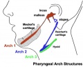 Pharyngeal arch cartilages.jpg 400 × 324; 26 KB
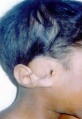
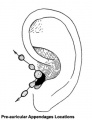 Pre-auricular appendages locations.jpg 306 × 400; 17 KB
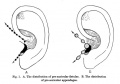 Pre-auricular fistulae and appendage locations.jpg 647 × 454; 36 KB
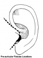 Pre-auricular fistulae locations.jpg 296 × 398; 16 KB
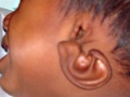
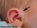 Preauricular sinus.jpg 600 × 450; 38 KB
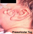 Preauricular tag 01.jpg 395 × 408; 28 KB
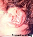 Preauricular tag 02.jpg 388 × 439; 50 KB
 Rugh 125.jpg 856 × 500; 82 KB
 Rugh 126.jpg 986 × 800; 166 KB
 Rugh 127.jpg 1,000 × 564; 110 KB
 Rugh 128.jpg 826 × 800; 145 KB
 Rugh 167.jpg 770 × 800; 153 KB
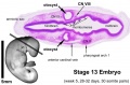
 Stage 13 image 001.jpg 1,000 × 357; 44 KB
 Stage 13 image 002.jpg 1,000 × 359; 52 KB
 Stage 13 image 003.jpg 1,000 × 436; 71 KB
 Stage 13 image 004.jpg 1,000 × 386; 65 KB
 Stage 13 image 051.jpg 1,000 × 382; 55 KB
 Stage 13 image 052.jpg 1,000 × 476; 81 KB
 Stage 13 image 053.jpg 1,000 × 423; 76 KB
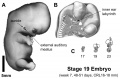 Stage 19 ear.jpg 1,200 × 786; 116 KB
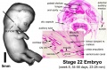
 Stage 22 image 159.jpg 1,000 × 665; 118 KB
 Stage 22 image 161.jpg 1,000 × 667; 154 KB
 Stage 22 image 162.jpg 1,000 × 657; 176 KB
 Stage 22 image 218.jpg 1,200 × 730; 308 KB
 Stage 22 image 219.jpg 1,250 × 892; 295 KB
 Stage11 sem20.jpg 668 × 1,000; 132 KB
 Stage11 sem20a.jpg 534 × 800; 92 KB
 Stage11 sem20b.jpg 401 × 600; 58 KB
 Stage11 sem20c.jpg 267 × 400; 29 KB
 Stage12 sem6.jpg 1,620 × 1,612; 190 KB
 Stage12 sem6a.jpg 1,000 × 995; 100 KB
 Stage12 sem6b.jpg 800 × 796; 74 KB
 Stage12 sem6c.jpg 600 × 597; 49 KB
 Stage13 otocyst.jpg 1,000 × 655; 55 KB
 Stage22 ear.jpg 1,000 × 655; 80 KB
 Streeter001.jpg 423 × 800; 79 KB
 Streeter002-3.jpg 607 × 800; 106 KB
 Streeter004.jpg 487 × 800; 92 KB
 Streeter005-13.jpg 657 × 800; 178 KB
 Streeter014-19.jpg 640 × 800; 199 KB
 Streeter020-25.jpg 636 × 800; 105 KB
 Streeter026.jpg 774 × 1,000; 45 KB
 Streeter027.jpg 774 × 1,000; 51 KB
 Streeter028-30.jpg 748 × 1,000; 134 KB
 Streeter028.jpg 774 × 1,000; 69 KB
 Streeter029.jpg 774 × 1,000; 74 KB
 Streeter030.jpg 774 × 1,000; 78 KB
 Streeter031.jpg 774 × 1,000; 79 KB
 Streeter1906 fig01.jpg 1,193 × 1,318; 167 KB
 Streeter1906 fig02.jpg 1,527 × 2,059; 244 KB
 Streeter1906 fig03.jpg 1,472 × 1,943; 285 KB
 Streeter1906 fig04.jpg 2,237 × 1,113; 404 KB
 Streeter1906 fig05.jpg 1,487 × 2,108; 554 KB
 Streeter1906 fig06.jpg 1,089 × 833; 240 KB
 Streeter1906 fig07.jpg 1,254 × 591; 75 KB
 Streeter1906 fig08.jpg 874 × 617; 147 KB
 Streeter1906 plate01.jpg 2,708 × 1,786; 701 KB
 Streeter1906 plate02.jpg 2,783 × 1,819; 499 KB
 Streeter1917 fig01.jpg 1,171 × 900; 131 KB
 Streeter1917 fig02.jpg 1,000 × 629; 158 KB
 Streeter1917 fig03.jpg 796 × 1,000; 117 KB
 Streeter1917 fig04.jpg 1,000 × 736; 254 KB
 Streeter1917 fig05.jpg 800 × 531; 57 KB
 Streeter1917 fig06.jpg 1,024 × 800; 98 KB
 Streeter1917 fig07.jpg 1,000 × 739; 198 KB
 Streeter1917-fig01.jpg 1,128 × 800; 298 KB
 Streeter1917-fig02.jpg 1,000 × 847; 276 KB
 Streeter1917-fig03.jpg 1,000 × 420; 173 KB
 Streeter1917-fig04-05.jpg 1,000 × 544; 124 KB
 Streeter1917-fig04.jpg 536 × 544; 61 KB
 Streeter1917-fig05.jpg 556 × 544; 64 KB
 Streeter1917-fig06-07.jpg 1,337 × 1,671; 639 KB
 Streeter1917-fig06.jpg 539 × 749; 93 KB
 Streeter1917-fig07.jpg 584 × 749; 105 KB
 Streeter1917-fig08-09.jpg 1,200 × 853; 218 KB
 Streeter1917-fig08.jpg 610 × 853; 110 KB
 Streeter1917-fig09.jpg 617 × 853; 109 KB
 Streeter1921 fig02.jpg 1,286 × 1,000; 138 KB
 Streeter1922-01-02.jpg 1,200 × 668; 134 KB
 Streeter1922-03-04.jpg 1,200 × 512; 105 KB
 Streeter1922-05.jpg 1,200 × 1,103; 307 KB
 Streeter1922-06-07.jpg 1,200 × 653; 107 KB
 Streeter1922-08.jpg 1,200 × 453; 86 KB
 Streeter1922-fig01.jpg 600 × 600; 46 KB
 Streeter1922-fig02.jpg 814 × 600; 72 KB
 Streeter1922-fig03.jpg 1,000 × 591; 79 KB
 Streeter1922-fig04.jpg 585 × 567; 35 KB
 Streeter1922-fig06.jpg 737 × 632; 42 KB
 Streeter1922-fig07.jpg 584 × 794; 54 KB
 Streeter1922-fig09.jpg 667 × 1,000; 85 KB
 Streeter1922-fig10.jpg 667 × 1,000; 86 KB
 Streeter1922-fig11.jpg 667 × 1,000; 96 KB
 Streeter1922-fig12.jpg 667 × 1,000; 103 KB
 Streeter1922-fig13.jpg 617 × 675; 69 KB
 Streeter1922-fig14.jpg 617 × 672; 70 KB
 Streeter1922-fig15.jpg 615 × 681; 67 KB
 Streeter1922-fig16.jpg 620 × 679; 76 KB
 Streeter1922-fig17.jpg 617 × 686; 66 KB
 Streeter1922-fig18.jpg 612 × 685; 82 KB
 Streeter1922-fig19.jpg 541 × 675; 59 KB
 Streeter1922-fig20.jpg 556 × 677; 61 KB
 Streeter1922-fig21.jpg 555 × 675; 65 KB
 Streeter1922-fig22.jpg 541 × 680; 65 KB
 Streeter1922-fig23.jpg 552 × 691; 68 KB
 Streeter1922-fig24.jpg 558 × 692; 63 KB
 Streeter1922-fig25.jpg 548 × 679; 61 KB
 Streeter1922-fig26.jpg 550 × 682; 70 KB
 Streeter1922-fig27.jpg 552 × 685; 63 KB
 Streeter1922-fig28.jpg 426 × 554; 36 KB
 Streeter1922-fig29.jpg 433 × 554; 42 KB
 Streeter1922-fig30.jpg 435 × 551; 41 KB
 Streeter1922-fig31.jpg 428 × 552; 42 KB
 Streeter1922-fig32.jpg 426 × 552; 44 KB
 Streeter1922-fig33.jpg 435 × 554; 45 KB
 Streeter1922-fig34.jpg 437 × 556; 47 KB
 Streeter1922-fig35.jpg 430 × 556; 41 KB
 Streeter1922-fig36.jpg 560 × 686; 66 KB
 Streeter1922-fig37.jpg 606 × 685; 82 KB
 Streeter1922-fig38.jpg 560 × 687; 69 KB
 Streeter1922-fig39.jpg 553 × 680; 67 KB
 Streeter1922-fig40.jpg 610 × 677; 81 KB
 Streeter1922-fig41.jpg 559 × 677; 76 KB
 Streeter1922-fig42.jpg 581 × 778; 88 KB
 Streeter1922-fig43.jpg 583 × 780; 94 KB
 Streeter1922-fig44.jpg 589 × 780; 101 KB
 Streeter1922-fig45.jpg 581 × 781; 91 KB
 Streeter1922-fig46.jpg 592 × 779; 95 KB
 Streeter1922-fig47.jpg 581 × 777; 95 KB
 Streeter1922-fig48.jpg 591 × 767; 86 KB
 Streeter1922-fig49.jpg 592 × 770; 97 KB
 Streeter1922-fig50.jpg 582 × 781; 97 KB
 Streeter1922-fig51.jpg 579 × 766; 78 KB
 Streeter1922-fig52.jpg 586 × 774; 83 KB
 Streeter1922-fig53.jpg 592 × 778; 79 KB
 Streeter1922-fig54.jpg 574 × 780; 88 KB
 Streeter1922-fig55.jpg 586 × 779; 80 KB
 Streeter1922-fig56.jpg 592 × 783; 92 KB
 Streeter1922-fig57.jpg 577 × 775; 88 KB
 Streeter1922-fig58.jpg 586 × 772; 92 KB
 Streeter1922-fig59.jpg 591 × 778; 104 KB
 Streeter1922-plate01.jpg 871 × 1,200; 174 KB
 Streeter1922-plate02.jpg 798 × 1,200; 215 KB
 Streeter1922-plate03.jpg 932 × 1,200; 243 KB
 Streeter1922-plate04.jpg 820 × 1,200; 248 KB
 Streeter1922-plate05.jpg 900 × 1,200; 315 KB
 Streeter1922-plate06.jpg 904 × 1,200; 291 KB
 Streeter1957 fig07.jpg 1,280 × 1,201; 117 KB
 Streeter1957 fig7-19.jpg 680 × 800; 48 KB
 Streeter1957 fig7-20.jpg 680 × 800; 24 KB
 Streeter1957 fig7-21.jpg 680 × 800; 25 KB
 Streeter1957 fig7-22.jpg 680 × 800; 33 KB
 Streeter1957 fig7-23.jpg 680 × 800; 37 KB
 Stricht-plate01.jpg 1,156 × 1,500; 243 KB
 Stricht-plate02.jpg 1,016 × 1,261; 308 KB
 Stricht-plate03.jpg 1,023 × 1,260; 230 KB
 Stricht-plate04.jpg 1,013 × 1,251; 238 KB
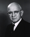 Theodore H. Bast.jpg 800 × 978; 108 KB
 Upper auricular detachment 01.jpg 412 × 600; 43 KB
 Upper auricular detachment 02.jpg 500 × 375; 27 KB
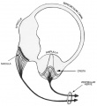 Vestibular labyrinth cartoon.jpg 600 × 657; 44 KB
 Vestibular labyrinth toadfish.jpg 600 × 480; 48 KB
 Waterston15.jpg 500 × 674; 65 KB


