Category:Bone: Difference between revisions
From Embryology
mNo edit summary |
mNo edit summary |
||
| Line 2: | Line 2: | ||
Main pages - '''{{bone}}''' | '''{{bone histology}}''' | Main pages - '''{{bone}}''' | '''{{bone histology}}''' | [[:Category:Bone|Category:Bone]] | ||
Latest revision as of 10:18, 3 May 2018
This Embryology category shows pages and media related to bone development. Note this topic is closely linked to other topics: mesoderm, cartilage, joint, blood, skull
Main pages - bone | bone histology | Category:Bone
Subcategories
This category has the following 4 subcategories, out of 4 total.
Pages in category 'Bone'
The following 162 pages are in this category, out of 162 total.
A
- AACP Meeting 2013 - Face Embryology
- ANAT2241 Bone, Bone Formation and Joints
- ANAT2241 Lymphatic Tissue and Immune System
- Template:Appearance and Fusion of Epiphyses timeline collapsetable1
- Template:Appearance and Fusion of Epiphyses timeline table1
- Template:Appendicular skeleton
- Template:Axial skeleton
B
- Template:Bone
- Bone Development
- Bone Histology
- Template:Bone Histology
- Template:Bone histology
- Template:Bone marrow
- Template:Bone Marrow Histology
- Template:Bone mineral density
- Template:Bone terms
- Template:Bone timeline
- Book - Comparative Embryology of the Vertebrates 4-15
- Book - Contributions to Embryology Carnegie Institution No.67
- Book - Manual of Human Embryology 11
- Book - Manual of Human Embryology 11D
- Book - Manual of Human Embryology 11E
C
F
- Template:Fawcett1913 figures
- Fetal Development - 12 Weeks
- Fetal Head Movie 1
- Fetal Palate Movie 1
- Template:Fetal week 10 palate links
- Template:Flecker1932 table1
- Template:Flecker1932 table2
- Template:Flecker1932 table3
- Template:Flecker1932 table4
- Template:Flecker1932 table5
- Template:Flecker1932 table57
- Template:Flecker1932 table58
- Template:Flecker1932 table6
- Template:Flecker1932 table7
- Template:Flecker1932 table8
- Template:Flecker1932 table9
- Template:Fontanel
- Template:Fontanelle
L
M
- Template:Mall1906 figures
- Template:Malleus Anatomy table
- Template:Mandible
- Mandible Growth Movie
- Template:Mandible images
- Movie - Postnatal human mandible growth
- Template:Musculoskeletal
- Musculoskeletal System - Appendicular Skeleton Development
- Musculoskeletal System - Axial Skeleton Development
- Musculoskeletal System - Bone Development
- Musculoskeletal System - Shoulder Development
- Musculoskeletal System - Skull Development
P
- Paper - A model of the left half of the human mandible at the 17 mm CRL stage
- Paper - Developmental horizons in human embryos- A review of the histogenesis of cartilage and bone
- Paper - Further observations on the ossification of the human lower jaw
- Paper - Observations on the structure and development of bone
- Paper - On Ossification Centers in Human Embryos
- Paper - On the development and morphology of the human sphenoid bone
- Paper - On the mechanism controlling the growth in length of the long bones (1934)
- Paper - Principles of growth and repair in membrane bones (1946)
- Paper - Roentgenographic observations of the times of appearance of epiphyses and their fusion with the diaphyses (1932)
- Paper - Some Gross Structural and Quantitative Aspects of the Developmental Anatomy of the Human Embryonic, Fetal and Circumnatal Skeleton
- Paper - Studies of the development of the human skeleton (1905)
- Paper - The development and ossification of the human clavicle
- Paper - The development of the human maxilla, vomer, and paraseptal cartilages (1911)
- Paper - The development of the patella
- Paper - The developmental anatomy of the human osseous skeleton during the embryonic, fetal and circumnatal periods
- Paper - The growth of the long bones in foetal life, as exemplified by a case of foetal syphilis (1929)
- Paper - The history of the earliest stages in the human clavicle
- Paper - The measurement of diaphysial growth in proximal and distal directions (1916)
- Paper - The origin and developmental mechanics of the avian sternum
- Paper - The ossification of the human frontal bone with special reference to its presumed pre- and post-frontal elements
- Template:Paraxial mesoderm
- Template:Peak Bone Mineral Density Age table1
- Template:Pelvis timeline
R
- Template:Ref-Adams1934
- Template:Ref-Arey1920
- Template:Ref-Ashley1955
- Template:Ref-Bardeen1904
- Template:Ref-Bardeen1905
- Template:Ref-Bardeen1905a
- Template:Ref-Bardeen1908
- Template:Ref-Bardeen1908a
- Template:Ref-Bardeen1910
- Template:Ref-Bast1929
- Template:Ref-Brailsford1943
- Template:Ref-Digby1916
- Template:Ref-Doan1922
- Template:Ref-Fawcett1913
- Template:Ref-Fawcett1930
- Template:Ref-Feli1939
- Template:Ref-Fell1939
- Template:Ref-Flecker1932
- Template:Ref-Geddes1912a
- Template:Ref-Geddes1913
- Template:Ref-Grüneberg1937
- Template:Ref-Hanson1919
- Template:Ref-Hanson1920
- Template:Ref-Hanson1920b
- Template:Ref-Harris1929
- Template:Ref-Hill1939
- Template:Ref-Howell1890b
- Template:Ref-Inman1937
- Template:Ref-InmanSaunders1937
- Template:Ref-Low1909
- Template:Ref-Mall1906bone
- Template:Ref-Miller1921
- Template:Ref-Noback1943 figures
- Template:Ref-Noback1944
- Template:Ref-Paterson1900
- Template:Ref-Paterson1902
- Template:Ref-PosenerWalkerWeddell1939
- Template:Ref-Prichard1946
- Template:Ref-Pryor1928
- Template:Ref-Ruth1932
- Template:Ref-Selye1934
- Template:Ref-Streeter1949
- Template:Ref-Stump1925
- Template:Ref-Sutton1885
- Template:Ref-Tomes1853
- Template:Ref-Walmsley1915
- Template:Ref-Walmsley1940
- Template:Rib
- Template:Ribs
S
U
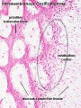
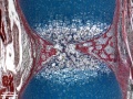
Media in category 'Bone'
The following 76 files are in this category, out of 276 total.
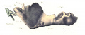
(previous page) (next page)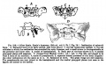 Keibel Mall 318.jpg 687 × 423; 79 KB
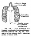 Keibel Mall 319.jpg 300 × 352; 27 KB
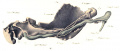
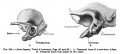 Keibel Mall 320.jpg 693 × 313; 33 KB
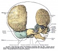 Keibel Mall 321.jpg 746 × 651; 122 KB
 Keibel Mall 322.jpg 735 × 717; 133 KB
 Keibel Mall 323.jpg 700 × 211; 27 KB
 Keibel Mall 324.jpg 722 × 377; 47 KB
 Keith1902 fig005.jpg 650 × 557; 36 KB
 Keith1902 fig013.jpg 662 × 800; 67 KB
 Keith1902 fig014.jpg 707 × 1,000; 98 KB
 Keith1902 fig243.jpg 1,000 × 631; 98 KB
 Limb patterning factors 11.jpg 1,200 × 655; 78 KB
 Low1909 fig01.jpg 900 × 371; 120 KB
 Low1909 fig02.jpg 1,059 × 889; 184 KB
 Low1909 fig03.jpg 904 × 896; 216 KB
 Low1909 fig04.jpg 1,043 × 927; 141 KB
 Low1909 fig05.jpg 893 × 891; 229 KB
 Low1909 fig06.jpg 1,371 × 1,213; 310 KB
 Low1909 fig07.jpg 897 × 893; 202 KB
 Low1909 plate01.jpg 3,574 × 2,331; 749 KB
 Low1909 plate01fig01.jpg 1,280 × 696; 115 KB
 Low1909 plate01fig02.jpg 1,280 × 518; 81 KB
 Low1909 plate01fig03.jpg 1,280 × 572; 95 KB
 Low1909 plate01fig04.jpg 1,280 × 600; 92 KB
 Low1909 plate01fig05.jpg 1,280 × 535; 73 KB
 Low1909 plate01fig06.jpg 1,280 × 546; 90 KB
 Mall1906 fig01.jpg 1,158 × 1,569; 133 KB
 Mall1906 fig02.jpg 1,317 × 1,532; 205 KB
 Mall1906 fig03.jpg 797 × 800; 47 KB
 Mall1906 fig04.jpg 1,114 × 1,044; 137 KB
 Mall1906 fig05.jpg 561 × 1,055; 45 KB
 Mall1906 fig06.jpg 522 × 966; 41 KB
 Mall1906 table01.jpg 1,319 × 1,154; 391 KB
 Mall1906 table02.jpg 2,000 × 858; 404 KB
 Mall1906 table03.jpg 2,118 × 878; 295 KB
 Mall1906 table04.jpg 2,113 × 1,125; 463 KB
 Mall1906 table05.jpg 1,325 × 1,048; 314 KB
 Mall1906 table06.jpg 2,117 × 1,074; 469 KB
 Mall1906 table07.jpg 2,118 × 1,034; 432 KB
 Meckel.jpg 800 × 667; 181 KB
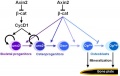
 Model coupling hematopoiesis with osteopoiesis.jpg 486 × 600; 44 KB
 Mouse E12.5 Sox9 Expression.jpg 397 × 397; 26 KB
 Mouse forelimb cartilage and bone E14.5 E18.5.jpg 1,000 × 1,300; 156 KB
 Mouse forelimb cartilage and bone E18.5.jpg 774 × 600; 70 KB
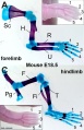
 Mouse forelimb.jpg 600 × 812; 71 KB
 Mouse hematopoietic stem cell.gif 600 × 595; 40 KB
 Mouse hindlimb cartilage and bone E18.5.jpg 774 × 600; 75 KB
 Mouse limb cartilage and bone E14.5.jpg 800 × 526; 42 KB
 Mouse limb cartilage and bone E14.5L.jpg 1,000 × 658; 73 KB
 Mouse limb cartilage and bone E18.5.jpg 774 × 1,200; 152 KB
 Mouse osteoblast 01.jpg 599 × 449; 118 KB
 Mouse osteoclast 01.jpg 600 × 453; 153 KB
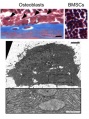 Mouse- postnatal osteoblasts.jpg 400 × 537; 59 KB
 MRI Human Embryo - upper limb 01.jpg 1,418 × 940; 106 KB
 MRI Human Embryo - upper limb 02.jpg 1,000 × 497; 74 KB
 Nail patella syndrome 02.jpg 809 × 525; 51 KB
 Noback1943-plate01.jpg 1,200 × 1,721; 274 KB
 Noback1943-plate02.jpg 1,000 × 1,436; 198 KB
 Ossification centre.jpg 450 × 600; 101 KB
 Ossification endochondral 01.jpg 817 × 613; 198 KB
 Ossification endochondral 1.jpg 750 × 1,000; 147 KB
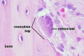 Osteoclast.jpg 500 × 333; 41 KB
 Postnatal human mandible growth 1.gif 450 × 368; 834 KB
 Postnatal human mandible growth.gif 600 × 491; 1.36 MB
 Postnatal human mandible growth.mov ; 1.2 MB
 Reticulocyte.jpg 500 × 313; 14 KB
 Rugh 146.jpg 1,000 × 373; 46 KB
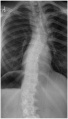 Scoliosis GH.jpg 600 × 1,053; 87 KB
 Shoulder cartoon.jpg 592 × 597; 36 KB
 Skull - osteoblast lineage model.jpg 600 × 381; 20 KB
 Skull vault defect and midface hypoplasia.jpg 800 × 798; 81 KB
 Spina bifida.jpg 800 × 633; 77 KB
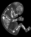
 Stage 22 image 173.jpg 1,000 × 659; 125 KB
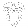
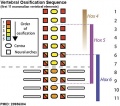 Vertebra ossification placental sequence.jpg 500 × 440; 48 KB
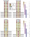 Vertebra ossification sequence.jpg 651 × 800; 97 KB
 Zebrafish- bone growth 01.jpg 634 × 1,000; 110 KB


