Brain Awareness Week 2012: Difference between revisions
mNo edit summary |
|||
| (157 intermediate revisions by the same user not shown) | |||
| Line 1: | Line 1: | ||
==Welcome to Brain Development== | ==Welcome to Brain Development== | ||
{| | {| | ||
| | | [[File:Adult_brain_animation_01.gif]] | ||
| '''In today's demonstration we will be looking at how the brain develops from a simple tube into the complex folded structure you will be seeing (and using) today.''' | | '''In today's demonstration we will be looking at how the brain develops from a simple tube into the complex folded structure that you will be seeing (and using) today.''' | ||
This animation shows a real human adult brain being "sliced", the '''cortex''' (grey matter) is on the outside. | This animation shows a real human adult brain being "sliced", the '''cortex''' (grey matter) is on the outside. | ||
''This page has been prepared as a simplified introduction to human neural development.'' | |||
The second part of the demonstration will cover comparative anatomy of the brain. | |||
[[Brain_Awareness_Week_2012#About_Brain_Awareness_Week|What is '''BAW'''?]] | |||
|} | |} | ||
--[[User:Z8600021|Mark Hill]] 11:36, 15 February 2013 (EST) Current [[K12 Brain Awareness Week]] Page. | |||
==Here is Human Development== | ==Here is Human Development== | ||
[[File:Human_development_timeline_graph_02.jpg]] | [[File:Human_development_timeline_graph_02.jpg]] | ||
This graph shows how we divide human development into different times. Key events occur in the first trimester (embryonic). The neural system continues to develop through the second and third trimester (fetal) and even after birth (postnatal). This long complex development makes it more easy to damage. | |||
==Week 3 - It begins as a '''Plate'''== | |||
{| border='0px' | {| border='0px' | ||
|- | |- | ||
| | |||
| <mediaplayer width='316' height='500' image="http://embryology.med.unsw.edu.au/embryology/images/a/a6/Neuralplate_001_icon.jpg">File:Neuralplate_001.mp4</mediaplayer> | |||
| | |||
[[Media:Neuralplate_001.mov|Quicktime]] | |||
| [[ | | valign=top| | ||
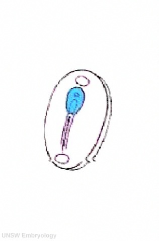
This embryo is the whole human embryo at just 3 weeks after fertilisation. | |||
* the entire nervous system will form from the flat region shown in blue. | |||
** this is called the <font color=deepskyblue>'''NEURAL PLATE'''</font>. | |||
* the <font color=deepskyblue>'''broad blue region'''</font> at the top will form the '''brain'''. | |||
* the <font color=deepskyblue>'''narrow blue region'''</font> at the bottom will form the '''spinal cord'''. | |||
| [[Image:Stage8 SEM1.jpg|240px]] | |||
This human embryo in week 3 is about 1-1.5 mm long and is viewed from the back, head end to the top. Almost all you see is the '''neural plate'''. | |||
|- | |- | ||
| | |} | ||
==Week 4 - That folds to a '''Tube'''== | |||
{| border='0px' | |||
| | |||
|- | |- | ||
| [[ | |||
| [[ | | <mediaplayer width='480' height='480' image="http://embryology.med.unsw.edu.au/embryology/images/6/6a/Neuraltube_001_icon.jpg">File:Neuraltube_001.mp4</mediaplayer> | ||
| [[ | | valign="top" | | ||
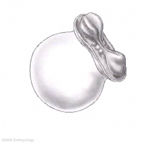
| [[ | The human embryo is now 4 weeks old and sits on top of a big yolk sac. | ||
| [[ | |||
| [[ | * the neural plate is shown on the embryo back. | ||
* the plate now folds to form a hollow '''NEURAL TUBE''' | |||
| [[File:Stage10_sem6.jpg|200px]] | |||
The same view at week 4, the embryo is now 2 - 3.5 mm long. The neural plate can be seen folding down the middle of the back, beginning to form the '''neural tube'''. | |||
|} | |||
==The tube then '''Closes at each End'''== | |||
These images show the neural tube closing leaving an opening ('''neuropore''') at each end. | |||
{| | |||
| [[File:Stage10_sem10.jpg|400px]] | |||
| [[File:Folatefruit.jpg]] | |||
<center>'''Why are these important?'''</center> | |||
|} | |||
[[File:Stage11_sem13c.jpg|250px]] [[File:Stage12_sem1.jpg|250px]] | |||
{| | |||
| [[File:Stage11_sem100.jpg|400px]] | |||
| [[File:Stage15_embryo_and_brain_01.jpg|400px]] | |||
|- | |- | ||
| '''Week 4''' - what the neural tube looks like when cut across. | |||
| '''Week 5''' - what the neural tube looks like within the embryo. | |||
|} | |} | ||
== | ==Week 6 to 8 - The brain end of the tube forms '''3 Vesicles'''== | ||
===Brain=== | |||
{| | |||
| At the brain end - the tube expands to form '''three vesicles''' (expansions, sacs or bubbles) these are described as fore-, mid- and hind-brain. Each vesicle will form different parts of the brain. Many of these parts you will not have heard of before, except the outer brain surface the Cerebrum or Cortex. | |||
# '''Forebrain''' or Prosencephalon - Telencephalon ('''Cerebrum or Cortex''', Amygdala, Hippocampus, Basal Ganglia) and Diencephalon (Epithalamus, Thalamus, Hypothalamus, Subthalamus, Pituitary, Pineal) | |||
# '''Midbrain''' or Mesencephalon - Tectum | |||
# '''Hindbrain''' or Rhombencephalon - Cerebellum, Brainstem (Pons, Medulla) | |||
| [[File:CNS_primary_vesicles.jpg|500px]] | |||
|} | |||
{| border='0px' | {| border='0px' | ||
|- | |- | ||
| < | | <mediaplayer width='520' height='540' image="http://embryology.med.unsw.edu.au/embryology/images/1/11/Human_embryo_tomography_Carnegie_stage_17.jpg">File:Human embryo tomography Carnegie stage 17.mp4</mediaplayer> | ||
| valign="top" | | |||
[[File:Human_embryo_tomography_Carnegie_stage_17.jpg|300px]] | |||
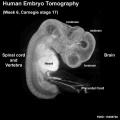
'''Week 6''' - the brain and spinal cord of the human embryo. | |||
Also visible are the heart (bright white) and placental cord containing placental blood vessels. | |||
[[Week 6]] [[Carnegie stage 17]] | |||
|- | |||
|} | |||
[[File:Stage16_embryo_and_brain_01.jpg|400px]] [[File:Stage22_embryo_and_brain_01.jpg|400px]] | |||
{| | |||
| [[File:Stage 22 image 217.jpg|400px]] | |||
|'''Week 8''' - the wall of the neural tube at the brain end. | |||
The "white matter" (thin outer layer, cortical plate) will eventually form the adult brain '''cortex'''. | |||
The other labeled layers are part of the development process and will eventually be mainly lost. | |||
The '''ventricle''' is the fluid-filled space within the neural tube and also later the brain. The smaller images (top right) show the level from the embryo. | |||
|} | |||
===Spinal Cord=== | |||
At the spinal cord end - the tube stays narrow. This region begins to put out '''motor nerves''' to innervate muscle and '''sensory nerves''' grow towards the developing spinal cord. | |||
{| | |||
| [[File:Stage22 vertebra and spinal cord 1.jpg|400px]] | |||
| '''Week 8''' wall of the neural tube at the spinal cord end. | |||
Spinal cord lies behind the vertebral body. | |||
The "grey matter" is on the inside of the spinal cord and the outside of the brain. | |||
The "grey matter" (dark central region) is where the neurons (cell bodies) are located, the "white matter" (pale outer region) is where nerve pathways run (axons). | |||
The sensory neurons lie outside the spinal cord in the '''dorsal root ganglia'''. | |||
|} | |||
==Second Trimester - Fetal brain '''Grows''' in Size== | |||
[[File:Brain ventricles and ganglia development 03.jpg|800px]] | |||
{| | |||
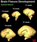
| This Scan of the living brain, shows the growth that occurs during the second trimester (red bar top right is 1 cm). | |||
* The left and right halves of the brain (hemispheres) can be clearly seen. | |||
* As the brain grows a large groove (fissure) also appears in the side surface (Sylvian fissure). | |||
* The fluid space (purple) is filled with '''cerebrospinal fluid''' or '''CSF'''. | |||
|} | |||
==Third Trimester - Fetal brain '''Grows''' in Surface Area== | |||
{| | |||
| [[File:Dev_anat_01.jpg|400px]] | |||
The brain goes from a smooth surface to begin to fold or "wrinkle". | |||
* These folds are due to the millions of cells being pushed into the cortex, increasing the surface area. | |||
* A '''groove''' is called a fissure (plural, fissures). | |||
* A '''fold''' is called a gyrus (plural, gyri). | |||
| | |||
[[File:Salt_shaker.jpg]] | |||
<center>'''Why do we need this?'''</center> | |||
|} | |||
==Week 40 on - Newborn brain '''Grows'''== | |||
[[File:WHO_motor_development_milestones.jpg|thumb|300px|Motor Development Milestones]] | |||
The brain has not finished growing at birth. | |||
'''Growth you can see!''' | |||
Much of the growth in size after birth is due to "white matter" development, the support cells of the brain, spinal cord and nerves. | |||
The skeleton containers of the nervous system, the skull (brain) and vertebral arch (spinal cord), are still flexible and can expand as the nervous system grows in size. | |||
'''There is also growth you cannot see!''' | |||
At the level of cells (neurons), the brain and spinal cord are continuing to make and break connections as the nervous system is remodelled with learning. | |||
==Here is how the human nervous system grows== | |||
{| | |||
| valign="bottom"|{{Neural plate movie}} | |||
| valign="bottom"|{{Neural Tube Closure 1 movie}} | |||
| valign="bottom"|{{Neural tube movie}} | |||
| valign="bottom"|{{Neural stage 13 movie}} | |||
| valign="bottom"|{{Stage_17_Embryo_movie}} | |||
|- | |||
| '''Week 3''' | |||
| '''Week 4''' | |||
| '''Week 4 to 5''' | |||
| '''Week 5''' | |||
| '''Week 6''' | |||
|- | |||
| valign="bottom"|{{Neural stage 22 movie}} | |||
| valign="bottom"|{{Sylvian fissure movie}} | |||
|- | |||
| '''Week 8''' | |||
| '''Week 13 to 21''' | |||
|} | |||
===Here is a developing mouse nervous system=== | |||
{| border='0px' | |||
|- | |||
| width=360px|<mediaplayer width='340' height='400' image="http://embryology.med.unsw.edu.au/embryology/images/3/34/Mouse_CT_E11.5_movie-icon.jpg">File:Mouse CT E11.5.mp4</mediaplayer> | |||
| valign="top"| | | valign="top"| | ||
'''This movie shows a | '''This movie shows a 11.5 days old mouse brain.''' | ||
(Mouse development takes 21 days) | (Mouse development takes 21 days and is a model used in research) | ||
| Line 71: | Line 246: | ||
[[File:Mouse_CT_E11.5_movie-icon.jpg|200px]] | [[File:Mouse_CT_E11.5_movie-icon.jpg|200px]] | ||
|- | |- | ||
|} | |} | ||
==Comparative Brain Anatomy== | |||
[[File:Comparative brain anatomy frog-dog.jpg|thumb|300px| Frog and Dog Brain]] | |||
In today's demonstration you will also see some models of brains from different species. Each coloured part on the brain models shows a '''different brain region''' each with a '''different function'''. | |||
Each brain region is the same colour (code) in all models. | |||
== | # Do not worry about the names of all the different structures. | ||
# Can you see the same coloured structures in all the brains? | |||
# Are the same coloured structures the same shape and size in all brains? | |||
(Link to [[Talk:Brain_Awareness_Week_2012#Cerebrum_Comparative_Anatomy|Detailed Information]], not part of demonstration) | |||
==About Brain Awareness Week== | |||
{| | |||
| [[File:BAW_icon_2012.jpg|150px]] | |||
| '''BAW''' - '''B'''rain '''A'''wareness '''W'''eek is an inspirational global campaign that unites those who share an interest in elevating public awareness about the progress and benefits of brain and nervous system research. | |||
:'''More?''' [http://www.sfn.org/index.aspx?pagename=baw_home Society for Neuroscience] | |||
|} | |} | ||
== | ===More K12 Development Topics=== | ||
{{Template:K12}} | |||
===More Detailed Neural Development=== | |||
{{Neural Links}} | |||
---- | |||
{{Glossary}} | |||
{{Footer}} | |||
[[Category:Neural]] [[Category:K12]] | |||
Latest revision as of 07:03, 12 March 2013
Welcome to Brain Development

|
In today's demonstration we will be looking at how the brain develops from a simple tube into the complex folded structure that you will be seeing (and using) today.
This page has been prepared as a simplified introduction to human neural development. The second part of the demonstration will cover comparative anatomy of the brain. |
--Mark Hill 11:36, 15 February 2013 (EST) Current K12 Brain Awareness Week Page.
Here is Human Development

This graph shows how we divide human development into different times. Key events occur in the first trimester (embryonic). The neural system continues to develop through the second and third trimester (fetal) and even after birth (postnatal). This long complex development makes it more easy to damage.
Week 3 - It begins as a Plate
| <mediaplayer width='316' height='500' image="http://embryology.med.unsw.edu.au/embryology/images/a/a6/Neuralplate_001_icon.jpg">File:Neuralplate_001.mp4</mediaplayer> |
|

This human embryo in week 3 is about 1-1.5 mm long and is viewed from the back, head end to the top. Almost all you see is the neural plate. |
Week 4 - That folds to a Tube
| <mediaplayer width='480' height='480' image="http://embryology.med.unsw.edu.au/embryology/images/6/6a/Neuraltube_001_icon.jpg">File:Neuraltube_001.mp4</mediaplayer> |
The human embryo is now 4 weeks old and sits on top of a big yolk sac.
|

The same view at week 4, the embryo is now 2 - 3.5 mm long. The neural plate can be seen folding down the middle of the back, beginning to form the neural tube. |
The tube then Closes at each End
These images show the neural tube closing leaving an opening (neuropore) at each end.

|

|


|

|
| Week 4 - what the neural tube looks like when cut across. | Week 5 - what the neural tube looks like within the embryo. |
Week 6 to 8 - The brain end of the tube forms 3 Vesicles
Brain
At the brain end - the tube expands to form three vesicles (expansions, sacs or bubbles) these are described as fore-, mid- and hind-brain. Each vesicle will form different parts of the brain. Many of these parts you will not have heard of before, except the outer brain surface the Cerebrum or Cortex.
|

|
| <mediaplayer width='520' height='540' image="http://embryology.med.unsw.edu.au/embryology/images/1/11/Human_embryo_tomography_Carnegie_stage_17.jpg">File:Human embryo tomography Carnegie stage 17.mp4</mediaplayer> |
Week 6 - the brain and spinal cord of the human embryo. Also visible are the heart (bright white) and placental cord containing placental blood vessels. |


|
Week 8 - the wall of the neural tube at the brain end.
The "white matter" (thin outer layer, cortical plate) will eventually form the adult brain cortex. The other labeled layers are part of the development process and will eventually be mainly lost. The ventricle is the fluid-filled space within the neural tube and also later the brain. The smaller images (top right) show the level from the embryo. |
Spinal Cord
At the spinal cord end - the tube stays narrow. This region begins to put out motor nerves to innervate muscle and sensory nerves grow towards the developing spinal cord.

|
Week 8 wall of the neural tube at the spinal cord end.
Spinal cord lies behind the vertebral body. The "grey matter" is on the inside of the spinal cord and the outside of the brain. The "grey matter" (dark central region) is where the neurons (cell bodies) are located, the "white matter" (pale outer region) is where nerve pathways run (axons). The sensory neurons lie outside the spinal cord in the dorsal root ganglia. |
Second Trimester - Fetal brain Grows in Size

This Scan of the living brain, shows the growth that occurs during the second trimester (red bar top right is 1 cm).
|
Third Trimester - Fetal brain Grows in Surface Area

The brain goes from a smooth surface to begin to fold or "wrinkle".
|
|

Week 40 on - Newborn brain Grows

The brain has not finished growing at birth.
Growth you can see!
Much of the growth in size after birth is due to "white matter" development, the support cells of the brain, spinal cord and nerves.
The skeleton containers of the nervous system, the skull (brain) and vertebral arch (spinal cord), are still flexible and can expand as the nervous system grows in size.
There is also growth you cannot see!
At the level of cells (neurons), the brain and spinal cord are continuing to make and break connections as the nervous system is remodelled with learning.
Here is how the human nervous system grows
|
|
|
|
| |||||||||||||||
| Week 3 | Week 4 | Week 4 to 5 | Week 5 | Week 6 | |||||||||||||||
|
| ||||||||||||||||||
| Week 8 | Week 13 to 21 |
Here is a developing mouse nervous system
| <mediaplayer width='340' height='400' image="http://embryology.med.unsw.edu.au/embryology/images/3/34/Mouse_CT_E11.5_movie-icon.jpg">File:Mouse CT E11.5.mp4</mediaplayer> |
This movie shows a 11.5 days old mouse brain. (Mouse development takes 21 days and is a model used in research)
Red - brain
|
|
Comparative Brain Anatomy

In today's demonstration you will also see some models of brains from different species. Each coloured part on the brain models shows a different brain region each with a different function. Each brain region is the same colour (code) in all models.
- Do not worry about the names of all the different structures.
- Can you see the same coloured structures in all the brains?
- Are the same coloured structures the same shape and size in all brains?
(Link to Detailed Information, not part of demonstration)
About Brain Awareness Week
| BAW - Brain Awareness Week is an inspirational global campaign that unites those who share an interest in elevating public awareness about the progress and benefits of brain and nervous system research.
|
More K12 Development Topics
More Detailed Neural Development
Glossary Links
- Glossary: A | B | C | D | E | F | G | H | I | J | K | L | M | N | O | P | Q | R | S | T | U | V | W | X | Y | Z | Numbers | Symbols | Term Link
Cite this page: Hill, M.A. (2024, June 27) Embryology Brain Awareness Week 2012. Retrieved from https://embryology.med.unsw.edu.au/embryology/index.php/Brain_Awareness_Week_2012
- © Dr Mark Hill 2024, UNSW Embryology ISBN: 978 0 7334 2609 4 - UNSW CRICOS Provider Code No. 00098G