Template:Week 4 Embryo Stages and Events table: Difference between revisions
mNo edit summary |
mNo edit summary |
||
| (7 intermediate revisions by the same user not shown) | |||
| Line 1: | Line 1: | ||
{| class="wikitable mw-collapsible mw-collapsed" | {| class="wikitable mw-collapsible mw-collapsed" | ||
! colspan=3|Week 4 - Embryo Stages and Events ({{GA}} week 6) | ! colspan=3|[[Week 4]] - Human Embryo Stages and Events ({{GA}} week 6) | ||
|- | |||
| colspan=3|{{Week}} | |||
|- | |||
|-bgcolor="FAF5FF" | |-bgcolor="FAF5FF" | ||
| <center>'''Day'''</center> | | <center>'''Day'''</center> | ||
| Line 11: | Line 14: | ||
| [[File:Stage10_dorsal1.jpg|200px]] [[File:Stage10-dorsal2.jpg|200px]] | | [[File:Stage10_dorsal1.jpg|200px]] [[File:Stage10-dorsal2.jpg|200px]] | ||
{{Neural crest}} - differentiation at spinal cord level from day 22 until day 26 | |||
{{Neural}} - neural folds begin to fuse near the junction between brain and spinal cord, when {{Neural crest}} - cells are arising mainly from the neural ectoderm | |||
{{Neural crest}} - trigeminal, facial, and postotic ganglia components visible{{#pmid:17848161|PMID17848161}} | |||
{{Neural crest}} - migration of vagal level neural crest cells begins (7-10 somite stage) | |||
{{Neural}} - Brain rostral neural tube forms 3 primary brain vesicles (week 4) | |||
{{Respiratory}} - Week 4 laryngotracheal groove forms on floor foregut. | |||
|- | |- | ||
| <center>23</center> | | <center>23</center> | ||
| | | | ||
| | | {{Heart}} - begins to beat in Humans by day 22-23, first functioning embryonic organ formed. | ||
|- | |- | ||
| Line 33: | Line 36: | ||
| [[File:Stage11_bf1c.jpg|200px|link=Carnegie stage 11]] | | [[File:Stage11_bf1c.jpg|200px|link=Carnegie stage 11]] | ||
{{Thyroid}} - thyroid median endodermal thickening in the floor of pharynx | |||
{{Neural}} - rostral (or cephalic) neuropore closes within a few hours; closure is bidirectional, it takes place from the dorsal and terminal lips and may occur in two areas simultaneously. The two lips, however, behave differently. | |||
Optic ventricle appears | Optic ventricle appears | ||
| Line 44: | Line 47: | ||
| [[File:Stage12_bf1c.jpg|200px|link=Carnegie stage 12]] | | [[File:Stage12_bf1c.jpg|200px|link=Carnegie stage 12]] | ||
{{Pituitary}} - Week 4 hypophysial pouch, Rathke's pouch, diverticulum from roof | |||
{{Liver}} septum transversum forming liver stroma and hepatic diverticulum forming hepatic trabeculae{{#pmid:9407542|PMID9407542}} | |||
{{Neural}} - caudal neuropore takes a day to close (closure is approximately at future somitic pair 31/sacral vertebra 2) | |||
{{Neural}} - secondary neurulation begins | |||
{{Neural crest}} - cardiac crest, neural crest from rhombomeres 6 and 7 that migrates to pharyngeal arch 3 and from there the truncus arteriosus{{#pmid:17848161|PMID17848161}} | |||
{{Neural crest}} - vagal neural crest enter the foregut (20-25 somite stage) | |||
|- | |- | ||
| Line 69: | Line 72: | ||
| <center>28</center> | | <center>28</center> | ||
| [[Carnegie_stage_13|Stage 13]] | | [[Carnegie_stage_13|Stage 13]] | ||
| [[File:Stage13_bf1c.jpg|200px|link=Carnegie stage 13]] [[Neural_System_Development|Neural]] - the neural tube is normally completely closed, ventricular system now separated from amniotic fluid. Neural crest at spinal level is segregating, and spinal ganglia are in series with the somites. Spinal cord ventral roots beginning to develop. | | [[File:Stage13_bf1c.jpg|200px|link=Carnegie stage 13]] [[Neural_System_Development|Neural]] - the neural tube is normally completely closed, ventricular system now separated from amniotic fluid. Neural crest at spinal level is segregating, and spinal ganglia are in series with the somites. Spinal cord ventral roots beginning to develop.{{#pmid:3354839|PMID3354839}} | ||
telencephalon cavity appears | telencephalon cavity appears | ||
{{Liver}} epithelial cord proliferation enmeshing stromal capillaries{{#pmid:9407542|PMID9407542}} | |||
{{Smell}} Crest comes from the nasal placodes{{#pmid:15604533|PMID15604533}} | |||
{{Integumentary}} - 4 weeks simple ectoderm epithelium over mesenchyme | |||
{{Integumentary}} - 1 to 3 months ectoderm - germinative (basal) cell repeated division of generates stratified epithelium; mesoderm - differentiates into connective tissue and blood vessels | |||
|- | |||
| colspan=3|Note - the day timing of stages is only approximate, system names link to first page of that specific system, and events are based upon the literature cited below. | |||
|- | |- | ||
| colspan=3|'''References''' | | colspan=3|'''References''' | ||
Latest revision as of 09:56, 26 July 2019
| Week 4 - Human Embryo Stages and Events (GA week 6) | ||
|---|---|---|
| Embryo Week: Week 1 | Week 2 | Week 3 | Week 4 | Week 5 | Week 6 | Week 7 | Week 8 | Week 9 | ||
| Event | ||
| Stage 10 |  
neural crest - differentiation at spinal cord level from day 22 until day 26 neural - neural folds begin to fuse near the junction between brain and spinal cord, when neural crest - cells are arising mainly from the neural ectoderm neural crest - trigeminal, facial, and postotic ganglia components visible[1] neural crest - migration of vagal level neural crest cells begins (7-10 somite stage) neural - Brain rostral neural tube forms 3 primary brain vesicles (week 4) respiratory - Week 4 laryngotracheal groove forms on floor foregut. | |
| heart - begins to beat in Humans by day 22-23, first functioning embryonic organ formed. | ||
| Stage 11 | 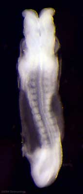
thyroid - thyroid median endodermal thickening in the floor of pharynx neural - rostral (or cephalic) neuropore closes within a few hours; closure is bidirectional, it takes place from the dorsal and terminal lips and may occur in two areas simultaneously. The two lips, however, behave differently. Optic ventricle appears | |
| Stage 12 | 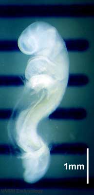
pituitary - Week 4 hypophysial pouch, Rathke's pouch, diverticulum from roof liver septum transversum forming liver stroma and hepatic diverticulum forming hepatic trabeculae[2] neural - caudal neuropore takes a day to close (closure is approximately at future somitic pair 31/sacral vertebra 2) neural - secondary neurulation begins neural crest - cardiac crest, neural crest from rhombomeres 6 and 7 that migrates to pharyngeal arch 3 and from there the truncus arteriosus[1] neural crest - vagal neural crest enter the foregut (20-25 somite stage) | |
| Stage 13 | 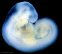 Neural - the neural tube is normally completely closed, ventricular system now separated from amniotic fluid. Neural crest at spinal level is segregating, and spinal ganglia are in series with the somites. Spinal cord ventral roots beginning to develop.[3] Neural - the neural tube is normally completely closed, ventricular system now separated from amniotic fluid. Neural crest at spinal level is segregating, and spinal ganglia are in series with the somites. Spinal cord ventral roots beginning to develop.[3]
telencephalon cavity appears liver epithelial cord proliferation enmeshing stromal capillaries[2] smell Crest comes from the nasal placodes[4] integumentary - 4 weeks simple ectoderm epithelium over mesenchyme integumentary - 1 to 3 months ectoderm - germinative (basal) cell repeated division of generates stratified epithelium; mesoderm - differentiates into connective tissue and blood vessels | |
| Note - the day timing of stages is only approximate, system names link to first page of that specific system, and events are based upon the literature cited below. | ||
References
| ||