Third Trimester: Difference between revisions
No edit summary |
mNo edit summary |
||
| (46 intermediate revisions by 2 users not shown) | |||
| Line 1: | Line 1: | ||
[[File:Frazer006_bw600.jpg| | {{Header}} | ||
==Introduction== | |||
[[File:Frazer006_bw600.jpg|thumb|The fetus in the 8th month]] | |||
The {{third trimester}} is a period of development when the fetus slows in growth in length and begins to increase substantially in weight, this is also the time where Individual variations increase. Some fetal systems, such as the respiratory system, only now begin to mature just prior to birth. | |||
Immune system, IgG to specific maternal antigens transferred passively across the placenta during the last trimester of pregnancy. | |||
{{Fetal Links}} | |||
[[:Category:Third Trimester|Category:Third Trimester]] | |||
|- | ==Some Recent Findings== | ||
{| | |||
|-bgcolor="F5FAFF" | |||
| | | | ||
* '''Synaptic development of layer V pyramidal neurons in the prenatal human prefrontal neocortex: a Neurolucida-aided Golgi study'''{{#pmid:31997813|PMID31997813}} "The prefrontal neocortex is involved in many high cognitive functions in humans. Deficits in neuronal and neurocircuitry development in this part of the cerebrum have been associated with various neuropsychiatric disorders in adolescents and adults. There are currently little available data regarding prenatal dendrite and spine formation on projecting neurons in the human prefrontal neocortex. Previous studies have demonstrated that Golgi silver staining can identify neurons in the frontal lobe and visual cortex in human embryos. In the present study, five fetal brains, at 19, 20, 26, 35, and 38 gestational weeks, were obtained via the body donation program at Xiangya School of Medicine, Central South University, China. Golgi-stained pyramidal neurons in layer V of Brodmann area 46 in fetuses were quantitatively analyzed using the Neurolucida morphometry system. Results revealed that somal size, total dendritic length, and branching points of these neurons increased from 26 to 38 gestational weeks. There was also a large increase in dendritic spines from 35 to 38 gestational weeks. These findings indicate that, in the human prefrontal neocortex, dendritic growth in layer V pyramidal neurons occurs rapidly during the third trimester of gestation." | |||
* '''Review - Second- and third-trimester biochemical and ultrasound markers predictive of ischemic placental disease.'''{{#pmid:24836829|PMID24836829}} "Ischemic placental disease is a recently coined term that describes the vascular insufficiency now believed to be an important etiologic factor in preeclampsia, intrauterine fetal growth restriction, and placental abruption. Given the increased risk for poor maternal and fetal outcomes, early prediction and prevention of this disorder is of significant clinical interest for many. In this article, we review the second- and third-trimester serum and ultrasound markers predictive of ischemic placental disease. Limited first-trimester data is also presented. While current studies report a statistical association between marker levels and various adverse perinatal outcomes, the observed diagnostic accuracy is below the threshold required for clinical utility. An exception to this generalization is uterine artery Doppler for the prediction of early-onset preeclampsia. Metabolomics is a relatively new analytic platform that holds promise as a first-trimester marker for the prediction of both early- and late-onset preeclampsia." {{ultrasound}} | |||
|} | |||
{| class="wikitable mw-collapsible mw-collapsed" | |||
! More recent papers | |||
|- | |- | ||
| | | [[File:Mark_Hill.jpg|90px|left]] {{Most_Recent_Refs}} | ||
Search term: [http://www.ncbi.nlm.nih.gov/pubmed/?term=Third+Trimester ''Third Trimester''] | |||
|} | |||
{| class="wikitable mw-collapsible mw-collapsed" | |||
! Older papers | |||
|- | |- | ||
| | | {{Older papers}} | ||
| | * '''Local tissue growth patterns underlying normal fetal human brain gyrification quantified in utero'''{{#pmid:21414909|PMID21414909}} "we applied recent advances in fetal MRI motion correction and computational image analysis techniques to 40 normal fetal human brains covering a period of primary sulcal formation (20-28 gestational weeks). Growth patterns were mapped by quantifying tissue locations that were expanding more or less quickly than the overall cerebral growth rate, which reveal increasing structural complexity. We detected increased local relative growth rates in the formation of the precentral and postcentral gyri, right superior temporal gyrus, and opercula, which differentiated between the constant growth rate in underlying cerebral mantle and the accelerating rate in the cortical plate undergoing folding. Analysis focused on the cortical plate revealed greater volume increases in parietal and occipital regions compared to the frontal lobe. Cortical plate growth patterns constrained to narrower age ranges showed that gyrification, reflected by greater growth rates, was more pronounced after 24 gestational weeks. Local hemispheric volume asymmetry was located in the posterior peri-Sylvian area associated with structural lateralization in the mature brain." {{neural}} | ||
| [ | |} | ||
==Neural== | |||
[[File:Human Fetus CRL240mm brain.jpg|thumb|Human Fetus (CRL 240 mm) Brain (scale bar 1 cm)]] | |||
{| | |||
| [[File:Dev_anat_01.jpg]] | |||
| | | '''Comparison of brain growth through the third trimester'''. | ||
| | |||
Note the increasing cortex surface area forming visible gyri (folds) and fissures (grooves) on the surface. | |||
:'''Links:''' [[Neural System Development]] | |||
|} | |||
==Respiratory== | |||
| | {| | ||
| | | [[File:Alveolar-sac-01.jpg|400px]] | ||
| | |||
Alveolar sac structure | |||
| | | | ||
'''Saccular stage''' | |||
* week 24 to near term. | |||
* most peripheral airways form widened airspaces, termed saccules. | |||
* saccules widen and lengthen the airspace (by the addition of new generations). | |||
* future gas exchange region expands significantly. | |||
* Fibroblastic cells also undergo differentiation, they produce extracellular matrix, collagen, and elastin. May have a role in epithelial differentiation and control of surfactant secretion | |||
* The vascular tree also grows in length and diameter during this time. | |||
* in late fetal development respiratory motions and amniotic fluid are thought to have a role in lung maturation. | |||
* Development of this system is not completed until late fetal just before birth. | |||
** Therefore premature babies have difficulties associated with insufficient surfactant (end month 6 alveolar cells type 2 appear and begin to secrete surfactant). | |||
:'''Links:''' {{respiratory}} | |||
|} | |||
| | |||
==Genital== | |||
| | {| | ||
| | | [[File:Testis-descent start.jpg|300px]] | ||
| | |||
| | [[File:Testis-descent end.jpg|300px]] | ||
| | | valign=top|'''Testis Descent''' | ||
| | Data from a study of male human fetal (between 10 and 35 weeks) gonad position{{#pmid:9649288|PMID9649288}} | ||
| | |||
* 10 to 23 weeks - (9.45%) had migrated from the abdomen and were situated in the inguinal canal. | |||
* 24 to 26 weeks - (57.9%) had migrated from the abdomen. | |||
* 27 to 29 weeks - (16.7%) had not descended to the scrotum. | |||
:'''Links:''' [[Testis Development]] | |||
|} | |} | ||
==Third Trimester Timeline== | |||
(Clinical Week 28-29) | |||
{{Third Trimester Timeline}} | |||
==References== | |||
<references/> | |||
===Search Pubmed=== | |||
'''Search Pubmed:''' [http://www.ncbi.nlm.nih.gov/sites/entrez?db=pubmed&cmd=search&term=Third%20Trimester Third Trimester] | |||
'''Next''': {{birth}} | |||
==External Links== | |||
{{External Links}} | |||
{{Glossary}} | |||
[[Category:Human Embryo]] [[Category:Human Fetus]] | {{Footer}} | ||
[[Category:Human Embryo]] [[Category:Human Fetus]] [[Category:Third Trimester]] | |||
Latest revision as of 00:09, 21 February 2020
| Embryology - 26 Jun 2024 |
|---|
| Google Translate - select your language from the list shown below (this will open a new external page) |
|
العربية | català | 中文 | 中國傳統的 | français | Deutsche | עִברִית | हिंदी | bahasa Indonesia | italiano | 日本語 | 한국어 | မြန်မာ | Pilipino | Polskie | português | ਪੰਜਾਬੀ ਦੇ | Română | русский | Español | Swahili | Svensk | ไทย | Türkçe | اردو | ייִדיש | Tiếng Việt These external translations are automated and may not be accurate. (More? About Translations) |
Introduction

The third trimester is a period of development when the fetus slows in growth in length and begins to increase substantially in weight, this is also the time where Individual variations increase. Some fetal systems, such as the respiratory system, only now begin to mature just prior to birth.
Immune system, IgG to specific maternal antigens transferred passively across the placenta during the last trimester of pregnancy.
| Fetal Links: fetal | Week 10 | Week 12 | second trimester | third trimester | fetal neural | Fetal Blood Sampling | fetal growth restriction | birth | birth weight | preterm birth | Developmental Origins of Health and Disease | macrosomia | BGD Practical | Medicine Lecture | Science Lecture | Lecture Movie | Category:Human Fetus | Category:Fetal | |||
|
Some Recent Findings
|
| More recent papers |
|---|
 This table allows an automated computer search of the external PubMed database using the listed "Search term" text link.
More? References | Discussion Page | Journal Searches | 2019 References | 2020 References Search term: Third Trimester |
| Older papers |
|---|
| These papers originally appeared in the Some Recent Findings table, but as that list grew in length have now been shuffled down to this collapsible table.
See also the Discussion Page for other references listed by year and References on this current page.
|
Neural


|
Comparison of brain growth through the third trimester.
|
Respiratory

Alveolar sac structure |
Saccular stage
|
Genital

|
Testis Descent
Data from a study of male human fetal (between 10 and 35 weeks) gonad position[4]
|

Third Trimester Timeline
(Clinical Week 28-29)
| Links: human timeline | first trimester timeline | second trimester timeline | third trimester timeline | ||
| Event | ||
| Clinical third trimester | 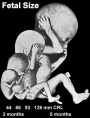 hearing 3rd Trimester - vibration acoustically of maternal abdominal wall induces startle respone in fetus. hearing 3rd Trimester - vibration acoustically of maternal abdominal wall induces startle respone in fetus.
| |
| respiratory Month 7 - respiratory bronchioles proliferate and end in alveolar ducts and sacs | ||
|
tooth Week 29 - Permanent premolars (correspond to the milk molars) appear. | ||
|
Genital male gonad (testes) descending | ||
| nail fingernails reach digit tip | ||
| neural brain cortical sulcation - primary sulci present[5] | ||
| neural brain cortical sulcation - insular, cingular, and occipital secondary sulci present[5] | ||
 Nail Development toenails reach digit tip Nail Development toenails reach digit tip
Lens Development - lens growth and interocular distance plateaus after 36 weeks of gestation[6] | ||
| Birth |  Clinical Week 40 Clinical Week 40
Heart pressure difference closes foramen ovale leaving a fossa ovalis thyroid TSH levels increase, thyroxine (T3) and T4 levels increase to 24 h, then 5-7 days postnatal decline to normal levels adrenal - zona glomerulosa, zona fasiculata present | |
References
- ↑ He LX, Wan L, Xiang W, Li JM, Pan AH & Lu DH. (2020). Synaptic development of layer V pyramidal neurons in the prenatal human prefrontal neocortex: a Neurolucida-aided Golgi study. Neural Regen Res , 15, 1490-1495. PMID: 31997813 DOI.
- ↑ Savasan ZA, Goncalves LF & Bahado-Singh RO. (2014). Second- and third-trimester biochemical and ultrasound markers predictive of ischemic placental disease. Semin. Perinatol. , 38, 167-76. PMID: 24836829 DOI.
- ↑ Rajagopalan V, Scott J, Habas PA, Kim K, Corbett-Detig J, Rousseau F, Barkovich AJ, Glenn OA & Studholme C. (2011). Local tissue growth patterns underlying normal fetal human brain gyrification quantified in utero. J. Neurosci. , 31, 2878-87. PMID: 21414909 DOI.
- ↑ Sampaio FJ & Favorito LA. (1998). Analysis of testicular migration during the fetal period in humans. J. Urol. , 159, 540-2. PMID: 9649288
- ↑ 5.0 5.1 Garel C, Chantrel E, Brisse H, Elmaleh M, Luton D, Oury JF, Sebag G & Hassan M. (2001). Fetal cerebral cortex: normal gestational landmarks identified using prenatal MR imaging. AJNR Am J Neuroradiol , 22, 184-9. PMID: 11158907
- ↑ Paquette LB, Jackson HA, Tavaré CJ, Miller DA & Panigrahy A. (2009). In utero eye development documented by fetal MR imaging. AJNR Am J Neuroradiol , 30, 1787-91. PMID: 19541779 DOI.
Search Pubmed
Search Pubmed: Third Trimester
Next: birth
External Links
External Links Notice - The dynamic nature of the internet may mean that some of these listed links may no longer function. If the link no longer works search the web with the link text or name. Links to any external commercial sites are provided for information purposes only and should never be considered an endorsement. UNSW Embryology is provided as an educational resource with no clinical information or commercial affiliation.
Glossary Links
- Glossary: A | B | C | D | E | F | G | H | I | J | K | L | M | N | O | P | Q | R | S | T | U | V | W | X | Y | Z | Numbers | Symbols | Term Link
Cite this page: Hill, M.A. (2024, June 26) Embryology Third Trimester. Retrieved from https://embryology.med.unsw.edu.au/embryology/index.php/Third_Trimester
- © Dr Mark Hill 2024, UNSW Embryology ISBN: 978 0 7334 2609 4 - UNSW CRICOS Provider Code No. 00098G