Category:Bone: Difference between revisions
From Embryology
mNo edit summary |
mNo edit summary |
||
| Line 2: | Line 2: | ||
Main pages - '''{{bone}}''' | '''{{bone histology}}''' | Main pages - '''{{bone}}''' | '''{{bone histology}}''' | [[:Category:Bone|Category:Bone]] | ||
Latest revision as of 10:18, 3 May 2018
This Embryology category shows pages and media related to bone development. Note this topic is closely linked to other topics: mesoderm, cartilage, joint, blood, skull
Main pages - bone | bone histology | Category:Bone
Subcategories
This category has the following 4 subcategories, out of 4 total.
Pages in category 'Bone'
The following 162 pages are in this category, out of 162 total.
A
- AACP Meeting 2013 - Face Embryology
- ANAT2241 Bone, Bone Formation and Joints
- ANAT2241 Lymphatic Tissue and Immune System
- Template:Appearance and Fusion of Epiphyses timeline collapsetable1
- Template:Appearance and Fusion of Epiphyses timeline table1
- Template:Appendicular skeleton
- Template:Axial skeleton
B
- Template:Bone
- Bone Development
- Bone Histology
- Template:Bone Histology
- Template:Bone histology
- Template:Bone marrow
- Template:Bone Marrow Histology
- Template:Bone mineral density
- Template:Bone terms
- Template:Bone timeline
- Book - Comparative Embryology of the Vertebrates 4-15
- Book - Contributions to Embryology Carnegie Institution No.67
- Book - Manual of Human Embryology 11
- Book - Manual of Human Embryology 11D
- Book - Manual of Human Embryology 11E
C
F
- Template:Fawcett1913 figures
- Fetal Development - 12 Weeks
- Fetal Head Movie 1
- Fetal Palate Movie 1
- Template:Fetal week 10 palate links
- Template:Flecker1932 table1
- Template:Flecker1932 table2
- Template:Flecker1932 table3
- Template:Flecker1932 table4
- Template:Flecker1932 table5
- Template:Flecker1932 table57
- Template:Flecker1932 table58
- Template:Flecker1932 table6
- Template:Flecker1932 table7
- Template:Flecker1932 table8
- Template:Flecker1932 table9
- Template:Fontanel
- Template:Fontanelle
L
M
- Template:Mall1906 figures
- Template:Malleus Anatomy table
- Template:Mandible
- Mandible Growth Movie
- Template:Mandible images
- Movie - Postnatal human mandible growth
- Template:Musculoskeletal
- Musculoskeletal System - Appendicular Skeleton Development
- Musculoskeletal System - Axial Skeleton Development
- Musculoskeletal System - Bone Development
- Musculoskeletal System - Shoulder Development
- Musculoskeletal System - Skull Development
P
- Paper - A model of the left half of the human mandible at the 17 mm CRL stage
- Paper - Developmental horizons in human embryos- A review of the histogenesis of cartilage and bone
- Paper - Further observations on the ossification of the human lower jaw
- Paper - Observations on the structure and development of bone
- Paper - On Ossification Centers in Human Embryos
- Paper - On the development and morphology of the human sphenoid bone
- Paper - On the mechanism controlling the growth in length of the long bones (1934)
- Paper - Principles of growth and repair in membrane bones (1946)
- Paper - Roentgenographic observations of the times of appearance of epiphyses and their fusion with the diaphyses (1932)
- Paper - Some Gross Structural and Quantitative Aspects of the Developmental Anatomy of the Human Embryonic, Fetal and Circumnatal Skeleton
- Paper - Studies of the development of the human skeleton (1905)
- Paper - The development and ossification of the human clavicle
- Paper - The development of the human maxilla, vomer, and paraseptal cartilages (1911)
- Paper - The development of the patella
- Paper - The developmental anatomy of the human osseous skeleton during the embryonic, fetal and circumnatal periods
- Paper - The growth of the long bones in foetal life, as exemplified by a case of foetal syphilis (1929)
- Paper - The history of the earliest stages in the human clavicle
- Paper - The measurement of diaphysial growth in proximal and distal directions (1916)
- Paper - The origin and developmental mechanics of the avian sternum
- Paper - The ossification of the human frontal bone with special reference to its presumed pre- and post-frontal elements
- Template:Paraxial mesoderm
- Template:Peak Bone Mineral Density Age table1
- Template:Pelvis timeline
R
- Template:Ref-Adams1934
- Template:Ref-Arey1920
- Template:Ref-Ashley1955
- Template:Ref-Bardeen1904
- Template:Ref-Bardeen1905
- Template:Ref-Bardeen1905a
- Template:Ref-Bardeen1908
- Template:Ref-Bardeen1908a
- Template:Ref-Bardeen1910
- Template:Ref-Bast1929
- Template:Ref-Brailsford1943
- Template:Ref-Digby1916
- Template:Ref-Doan1922
- Template:Ref-Fawcett1913
- Template:Ref-Fawcett1930
- Template:Ref-Feli1939
- Template:Ref-Fell1939
- Template:Ref-Flecker1932
- Template:Ref-Geddes1912a
- Template:Ref-Geddes1913
- Template:Ref-Grüneberg1937
- Template:Ref-Hanson1919
- Template:Ref-Hanson1920
- Template:Ref-Hanson1920b
- Template:Ref-Harris1929
- Template:Ref-Hill1939
- Template:Ref-Howell1890b
- Template:Ref-Inman1937
- Template:Ref-InmanSaunders1937
- Template:Ref-Low1909
- Template:Ref-Mall1906bone
- Template:Ref-Miller1921
- Template:Ref-Noback1943 figures
- Template:Ref-Noback1944
- Template:Ref-Paterson1900
- Template:Ref-Paterson1902
- Template:Ref-PosenerWalkerWeddell1939
- Template:Ref-Prichard1946
- Template:Ref-Pryor1928
- Template:Ref-Ruth1932
- Template:Ref-Selye1934
- Template:Ref-Streeter1949
- Template:Ref-Stump1925
- Template:Ref-Sutton1885
- Template:Ref-Tomes1853
- Template:Ref-Walmsley1915
- Template:Ref-Walmsley1940
- Template:Rib
- Template:Ribs
S
U
Media in category 'Bone'
The following 200 files are in this category, out of 276 total.
(previous page) (next page)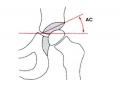 Acetabular angle.jpg 600 × 433; 22 KB
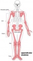 Appendicular skeleton small.jpg 220 × 407; 12 KB
 Appendicular skeleton.jpg 1,000 × 1,849; 125 KB
 Axial skeleton.jpg 1,000 × 1,434; 111 KB
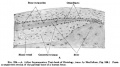
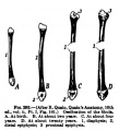
 Bailey114.jpg 690 × 433; 63 KB
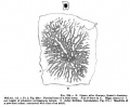
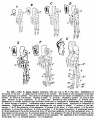
 Bailey115.jpg 630 × 767; 80 KB
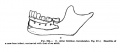
 Bailey116.jpg 835 × 434; 90 KB
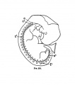
 Bailey117.jpg 623 × 527; 63 KB
 Bailey118.jpg 898 × 442; 72 KB
 Bailey119.jpg 792 × 297; 33 KB
 Bailey120.jpg 891 × 599; 214 KB
 Bailey126.jpg 903 × 693; 168 KB
 Bailey127.jpg 597 × 270; 43 KB
 Bailey128.jpg 805 × 356; 46 KB
 Bailey129.jpg 560 × 464; 44 KB
 Bailey130.jpg 714 × 439; 59 KB
 Bailey131.jpg 235 × 560; 39 KB
 Bailey132+133.jpg 940 × 570; 101 KB
 Bailey132.jpg 466 × 413; 43 KB
 Bailey133.jpg 806 × 655; 85 KB
 Bailey135.jpg 940 × 965; 216 KB
 Bailey136.jpg 835 × 566; 114 KB
 Bailey137.jpg 672 × 539; 73 KB
 Bailey138.jpg 831 × 400; 62 KB
 Bailey139.jpg 961 × 671; 96 KB
 Bailey140.jpg 793 × 505; 58 KB
 Bailey141.jpg 761 × 323; 66 KB
 Bailey142.jpg 778 × 479; 72 KB
 Bailey144.jpg 491 × 398; 39 KB
 Bailey145.jpg 777 × 654; 80 KB
 Bailey146.jpg 609 × 476; 42 KB
 Bailey147.jpg 660 × 632; 54 KB
 Bailey148.jpg 574 × 459; 38 KB
 Bailey149.jpg 576 × 520; 38 KB
 Bailey150.jpg 406 × 596; 42 KB
 Bailey151.jpg 585 × 631; 73 KB
 Bailey152.jpg 900 × 803; 300 KB
 Bailey153.jpg 799 × 585; 175 KB
 Bailey154.jpg 898 × 563; 191 KB
 Bailey155.jpg 894 × 833; 209 KB
 Baileytable02.jpg 884 × 1,109; 182 KB
 Bardeen1905 plate13.jpg 1,000 × 1,337; 85 KB
 Bone histology 001.jpg 1,280 × 1,024; 276 KB
 Bone histology 002.jpg 1,280 × 1,024; 309 KB
 Bone histology 003.jpg 1,280 × 1,024; 663 KB
 Bone histology 004.jpg 1,280 × 1,024; 605 KB
 Bone histology 005.jpg 1,280 × 1,024; 529 KB
 Bone histology 006.jpg 1,280 × 1,024; 360 KB
 Bone histology 007.jpg 1,280 × 1,024; 299 KB
 Bone histology 008.jpg 1,280 × 1,024; 550 KB
 Bone histology 009.jpg 1,280 × 1,024; 444 KB
 Bone histology 010.jpg 1,280 × 1,024; 256 KB
 Bone histology 011.jpg 1,280 × 1,024; 348 KB
 Bone histology 012.jpg 1,280 × 1,024; 165 KB
 Bone histology 013.jpg 1,280 × 1,024; 210 KB
 Bone histology 014.jpg 1,280 × 1,024; 541 KB
 Bone histology 015.jpg 1,280 × 1,024; 519 KB
 Bone histology 016.jpg 1,280 × 1,024; 379 KB
 Bone histology 017.jpg 1,280 × 1,024; 442 KB
 Bone histology 018.jpg 1,280 × 1,024; 336 KB
 Bone histology 019.jpg 1,280 × 1,024; 275 KB
 Bone histology 020.jpg 1,280 × 1,024; 272 KB
 Bone histology 021.jpg 1,280 × 1,024; 254 KB
 Bone histology 022.jpg 2,500 × 2,000; 328 KB
 Bone histology 066.jpg 2,500 × 2,000; 361 KB
 Bone histology 101.jpg 400 × 533; 59 KB
 Bone histology 111.jpg 400 × 533; 70 KB
 Bone histology 112.jpg 400 × 533; 46 KB
 Bone histology 201.jpg 400 × 533; 62 KB
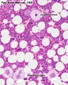
 Bone marrow histology 01.jpg 480 × 600; 114 KB
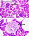
 Bone marrow histology 02.jpg 480 × 600; 109 KB
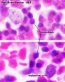
 Bone marrow histology 03.jpg 480 × 600; 81 KB
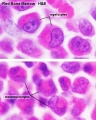
 Bone marrow histology 04.jpg 480 × 600; 61 KB
 Bone marrow histology 05.jpg 480 × 600; 62 KB
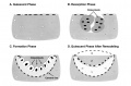 Bone remodeling cycle.jpg 958 × 628; 68 KB
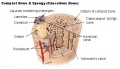 Bone structure cartoon.jpg 520 × 300; 46 KB
 Bone-bon02he.jpg 1,280 × 1,024; 348 KB
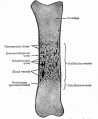
 Bone-femur.jpg 798 × 1,000; 150 KB
 Bone-structure.jpg 450 × 600; 27 KB
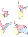
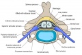 Cervical vertebra.jpg 767 × 514; 71 KB
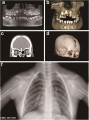 Cleidocranial dysplasia 01.jpg 518 × 700; 65 KB
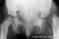 Congenital dislocation hip.jpg 400 × 265; 8 KB
 Doan-plate01.jpg 924 × 1,170; 207 KB
 Doan01.jpg 800 × 369; 79 KB
 Doan02.jpg 800 × 637; 82 KB
 Doan03.jpg 800 × 312; 31 KB
 Endochondral bone cartoon.jpg 946 × 513; 127 KB
 Endochondral ossification 1.jpg 400 × 534; 91 KB
 Endochondral ossification 2.jpg 400 × 533; 99 KB
 Endochondral ossification.jpg 400 × 533; 91 KB
 Fawcett1913 fig01.jpg 793 × 573; 67 KB
 Fawcett1913 fig02.jpg 679 × 579; 42 KB
 Fawcett1913 fig03.jpg 687 × 581; 33 KB
 Fawcett1913 fig04.jpg 928 × 664; 122 KB
 Fawcett1913 fig05.jpg 979 × 671; 98 KB
 Fawcett1913 fig06.jpg 899 × 675; 80 KB
 Fawcett1913 fig07.jpg 1,113 × 763; 97 KB
 Fawcett1913 fig08.jpg 867 × 397; 28 KB
 Fetal head lateral.jpg 632 × 447; 34 KB
 Fetal head medial.jpg 632 × 447; 34 KB
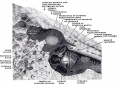
 Fetal head section 01.jpg 1,200 × 821; 186 KB
 Fetal head section.jpg 1,200 × 821; 167 KB
 Fetal week 10 hard palate 01.jpg 800 × 532; 77 KB
 Fetal week 10 hard palate 02.jpg 398 × 633; 66 KB
 Fetal week 10 hard palate 03.jpg 600 × 450; 122 KB
 Fetal week 10 hard palate 04.jpg 1,198 × 795; 196 KB
 Fetal week 10 hard palate 06.jpg 534 × 778; 88 KB
 Fetal week 10 hard palate 07.jpg 534 × 778; 97 KB
 Fetal week 10 palate 01.gif 534 × 778; 1.14 MB
 Fetal week 10 palate 01.mp4 ; 427 KB
 Fetal week 10 palate icon.jpg 534 × 778; 100 KB
 Fetal week 10 soft palate 01.jpg 571 × 784; 95 KB
 Fetal week 10 soft palate 02.jpg 534 × 778; 87 KB
 Fetal week 10 soft palate 03.jpg 534 × 778; 95 KB
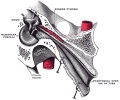
 Fetal week 14 head bone lateral 01.jpg 1,000 × 773; 107 KB
 Fetal week 9 hard palate fusion 01.jpg 661 × 400; 51 KB
 Fetal week 9 head lateral 01.jpg 700 × 600; 78 KB
 Flecker1932 fig01.jpg 700 × 498; 42 KB
 Flecker1932 fig02.jpg 702 × 500; 41 KB
 Flecker1932 fig03.jpg 691 × 500; 44 KB
 Flecker1932 fig04.jpg 508 × 700; 41 KB
 Flecker1932 fig05.jpg 508 × 700; 47 KB
 Flecker1932 fig06.jpg 700 × 513; 106 KB
 Flecker1932 fig07.jpg 700 × 506; 58 KB
 Flecker1932 fig08.jpg 300 × 363; 18 KB
 Flecker1932 fig09.jpg 300 × 374; 23 KB
 Flecker1932 fig10.jpg 750 × 541; 53 KB
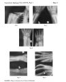 Flecker1932 plate1.jpg 2,080 × 2,569; 657 KB
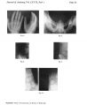 Flecker1932 plate2.jpg 2,080 × 2,535; 525 KB
 Gray0067.png 350 × 463; 46 KB
 Gray0070.jpg 800 × 796; 182 KB
 Gray0071.jpg 700 × 440; 104 KB
 Gray0130.jpg 535 × 600; 95 KB
 Gray0176.jpg 600 × 402; 58 KB
 Gray0178.jpg 617 × 368; 44 KB
 Gray0179.jpg 617 × 368; 47 KB
 Gray0180.jpg 617 × 368; 48 KB
 Gray0181.jpg 617 × 368; 52 KB
 Gray0182.jpg 600 × 329; 15 KB
 Gray0183.jpg 600 × 491; 29 KB
 Gray0184.jpg 600 × 491; 45 KB
 Gray0185.jpg 600 × 491; 36 KB
 Gray0193.jpg 719 × 1,057; 293 KB
 Gray0907.jpg 679 × 600; 110 KB
 Gray0911.jpg 651 × 400; 73 KB
 Gray0912.jpg 600 × 540; 87 KB
 Gray0913.jpg 671 × 600; 98 KB
 Gray0914.jpg 708 × 500; 102 KB
 Gray0915.jpg 720 × 600; 94 KB
 Gray1235.jpg 800 × 389; 41 KB
 Gray1236.jpg 800 × 281; 29 KB
 Gray1237.jpg 371 × 600; 57 KB
 Hematopoietic and stromal cell differentiation.jpg 1,000 × 617; 107 KB
 Human axial skeleton- axis development 01.jpg 763 × 1,095; 95 KB
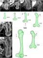 Human embryo femur CS18 to CS23.png 1,200 × 1,624; 1.42 MB
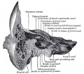
 Human fetal temporal bone and mandible 01.jpg 1,200 × 805; 170 KB
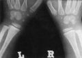
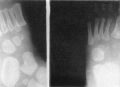
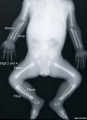 Human fetus skeleton x-ray 01.jpg 662 × 910; 54 KB
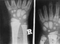
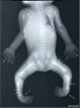 Human fetus skeleton x-ray 02.jpg 686 × 928; 70 KB
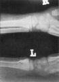
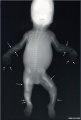 Human fetus skeleton x-ray 03.jpg 628 × 934; 81 KB
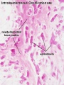 Intramembranous ossification centre.jpg 450 × 600; 69 KB
 Keibel Mall 223.jpg 727 × 1,100; 89 KB
 Keibel Mall 224.jpg 706 × 1,112; 105 KB
 Keibel Mall 224A.jpg 671 × 377; 38 KB
 Keibel Mall 224B.jpg 685 × 563; 56 KB
 Keibel Mall 224C.jpg 706 × 306; 20 KB
 Keibel Mall 231.jpg 450 × 511; 21 KB
 Keibel Mall 232.jpg 450 × 511; 35 KB
 Keibel Mall 260-263.jpg 726 × 800; 90 KB
 Keibel Mall 260.jpg 506 × 508; 25 KB
 Keibel Mall 261.jpg 506 × 508; 32 KB
 Keibel Mall 262.jpg 506 × 508; 24 KB
 Keibel Mall 263.jpg 506 × 508; 37 KB
 Keibel Mall 264-265.jpg 831 × 800; 88 KB
 Keibel Mall 264.jpg 475 × 800; 38 KB
 Keibel Mall 265.jpg 526 × 800; 51 KB
 Keibel Mall 266.jpg 740 × 608; 77 KB
 Keibel Mall 267.jpg 500 × 410; 28 KB
 Keibel Mall 271.jpg 800 × 419; 60 KB
 Keibel Mall 273-no-legend.jpg 791 × 744; 164 KB
 Keibel Mall 273.jpg 900 × 861; 200 KB
 Keibel Mall 274-278.jpg 717 × 1,072; 123 KB
 Keibel Mall 279-284.jpg 709 × 707; 84 KB
 Keibel Mall 285-288.jpg 703 × 674; 72 KB
 Keibel Mall 289.jpg 682 × 645; 85 KB
 Keibel Mall 290.jpg 800 × 452; 59 KB
 Keibel Mall 291.jpg 800 × 421; 54 KB
 Keibel Mall 292.jpg 400 × 439; 30 KB
 Keibel Mall 293.jpg 687 × 857; 137 KB
 Keibel Mall 302.jpg 300 × 233; 18 KB
 Keibel Mall 307.jpg 800 × 803; 112 KB
 Keibel Mall 308.jpg 677 × 578; 43 KB
 Keibel Mall 309.jpg 687 × 479; 52 KB
 Keibel Mall 310.jpg 686 × 760; 161 KB
 Keibel Mall 311.jpg 700 × 906; 181 KB
 Keibel Mall 312.jpg 704 × 677; 79 KB
 Keibel Mall 313.jpg 702 × 652; 84 KB
 Keibel Mall 314.jpg 701 × 625; 89 KB
 Keibel Mall 315.jpg 736 × 433; 84 KB
 Keibel Mall 316.jpg 716 × 327; 57 KB
 Keibel Mall 317.jpg 688 × 376; 53 KB


