Musculoskeletal System - Muscle Development: Difference between revisions
| Line 142: | Line 142: | ||
File:Skeletal_muscle_histology_055.jpg|Human HE x100 | File:Skeletal_muscle_histology_055.jpg|Human HE x100 | ||
File:Skeletal_muscle_histology_008.jpg|Fetal human muscle | File:Skeletal_muscle_histology_008.jpg|Fetal human muscle | ||
File:Skeletal_muscle_histology_004.jpg|Myotendinous junction HE x40 | |||
File:Skeletal_muscle_histology_006.jpg| | File:Skeletal_muscle_histology_006.jpg| | ||
File:Skeletal_muscle_histology_007.jpg| | File:Skeletal_muscle_histology_007.jpg| | ||
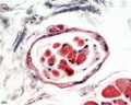
File:Skeletal_muscle_histology_077.jpg| | File:Skeletal_muscle_histology_077.jpg| | ||
File:Skeletal_muscle_histology_009.jpg|Muscle spindle HE x20 | File:Skeletal_muscle_histology_009.jpg|Muscle spindle HE x20 | ||
File:Skeletal_muscle_histology_010.jpg|Muscle spindle HE x40 | File:Skeletal_muscle_histology_010.jpg|Muscle spindle HE x40 | ||
Revision as of 17:19, 2 October 2011

Introduction
There are 3 different types of muscle: skeletal, cardiac and smooth. This page describes skeletal muscle development, descriptions of cardiac muscle and smooth muscle development can be found in other notes. Skeletal muscle forms by fusion of mononucleated myoblasts to form mutinucleated myotubes.
Differentiation/determination of mesoderm into muscle cells is thought to involve a family of basic Helix-Loop-Helix transcription factors, the first of which discovered was MyoD1. MyoD1 needs to form a dimer to be active and is maintained in an inactive state by binding of an inhibitor, Id.
Some Recent Findings
|
Myogenesis

Three different types of muscle form in the body.
- Skeletal muscle - cells originate from the paraxial mesoderm, forming somites, then dermamyotome and finally the myotome. Myoblasts undergo frequent divisions and coalesce with the formation of a multinucleated, syncytial muscle fibre or myotube. The nuclei of the myotube are still located centrally in the muscle fibre. In the course of the synthesis of the myofilaments/myofibrils, the nuclei are gradually displaced to the periphery of the cell.
- Cardiac muscle - cells originate from the prechordal splanchnic mesoderm.
- Smooth muscle - cells originate from undifferentiated mesenchymal cells. These cells differentiate first into mitotically active cells, myoblasts, which contain a few myofilaments. Myoblasts give rise to the cells which will differentiate into mature smooth muscle cells.


Muscle Groups
Epaxial Muscle
Anatomical term describing skeletal muscles which lie dorsal (posterior) to the vertebral column developing from the somite myotome. In humans, this is only a small muscle group formed by the transversospinalis, longissimus, and iliocostalis muscles. Also at the ribcage level the levatores costarum muscles involved with rib elevation during respiration. The body muscles lying ventral (anterior) to the vertebral column are the hypaxial muscles.
Hypaxial Muscle
(hypomere) Anatomical term describing skeletal muscles which lie ventral (anterior) to the vertebral column developing from the somite myotome. These muscles contribute both body (trunk) and limb skeletal muscle.
- In the trunk, these form the three anterior body muscle layers.
- In the limb, these form the extensor and flexor muscle groups.
Skeletal Muscle Stages


Myoblast - individual progenitor cells
Myotube - multinucleated, but undifferentiated contractile apparatus (sarcomere)
Myofibre (myofiber, muscle cell) - multinucleated and differentiated sarcomeres
- primary myofibres - first-formed myofibres, act as a structural framework upon which myoblasts proliferate, fuse in linear sequence
- secondary myofibers - second later population of myofibres that form surrounding the primary fibres.
Muscle Fibre Types

Muscle fiber types
- type IIB, IIA, IIX, and I fibres - based only on the myosin ATPase activity.
- Type I fibres appear red, due to the presence of myoglobin.
- Type II fibres appear white, due to the absence of myoglobin and their glycolytic nature.
- A group of individual myofibres within a muscle will be innervated by a single motor neuron (motor unit).
- The electrical properties of the motor neuron will regulate the contractile properties of all associated myofibres.
| Fibre Type | Type I fibres | Type II a fibres | Type II x fibres | Type II b fibres |
|---|---|---|---|---|
| Contraction time | Slow | Moderately Fast | Fast | Very fast |
| Size of motor neuron | Small | Medium | Large | Very large |
| Resistance to fatigue | High | Fairly high | Intermediate | Low |
| Activity Used for | Aerobic | Long-term anaerobic | Short-term anaerobic | Short-term anaerobic |
| Maximum duration of use | Hours | <30 minutes | <5 minutes | <1 minute |
| Power produced | Low | Medium | High | Very high |
| Mitochondrial density | High | High | Medium | Low |
| Capillary density | High | Intermediate | Low | Low |
| Oxidative capacity | High | High | Intermediate | Low |
| Glycolytic capacity | Low | High | High | High |
| Major storage fuel | Triglycerides | Creatine phosphate, glycogen | Creatine phosphate, glycogen | Creatine phosphate, glycogen |
| Myosin heavy chain, human genes |
MYH7 | MYH2 | MYH1 | MYH4 |
Muscle Contraction
Individual myoblasts in the developing muscle bed initial fuse together to form multi-nucleated myotubes. These myotubes then express the contractile proteins, that are organized into sarcomeres in series along the length of the myotube.
This animation shows the molecular interactions that occur within the skeletal muscle sarcomere between actin and myosin during skeletal muscle contraction.
|
Legend
|

|
Myotome
In both development and the adult, the group of skeletal muscles supplied by a specific segmental spinal nerve is referred to as a myotome. The muscle arises from a specific somite and the spinal nerve arises from a specific level of the spinal cord (identified by veretebral column).
In humans this corresponds to the following spinal nerves (from top to bottom) and muscular functions:
- C3,4 and 5 supply the diaphragm for breathing.
- C5 supply shoulder muscles and muscles to bend our elbow.
- C6 for bending the wrist back.
- C7 for straightening the elbow.
- C8 bends the fingers.
- T1 spreads the fingers.
- T1 –T12 supplies the chest wall and abdominal muscles.
- L2 bends the hip.
- L3 straightens the knee.
- L4 pulls the foot up.
- L5 wiggles the toes.
- S1 pulls the foot down.
- S3,4 and 5 supply the bladder, bowel, sex organs, anal and other pelvic muscles.
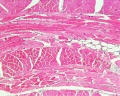
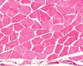
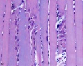
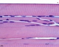
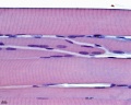
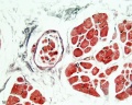
Histology Images
Human HE x4 longitudinal and transverse
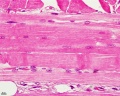
Human HE x40
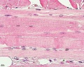
Human HE x40
Human HE x40
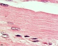
Human HE x100
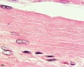
Human HE x100
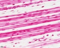
Fetal human muscle
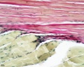
Myotendinous junction HE x40
Muscle spindle HE x20
Muscle spindle HE x40


Puberty
- Musculoskeletal mass doubles by the end of puberty
- regulated growth by - sex steroid hormones, growth hormone, insulin-like growth factors
- accumulation of (peak) bone mass during puberty relates to future osteoporosis in old age
Abnormalities
Facioscapulohumeral muscular dystrophy (FSHD)
- characterized by the progressive weakness and atrophy of a specific subset of skeletal muscles
- mostly affects the muscles of the face, scapula, and upper arms
- involvement of specific muscles that it is often used clinically to distinguish FSHD from other forms of muscular dystrophy
References
- ↑ <pubmed>21621065</pubmed>
- ↑ <pubmed>21859860</pubmed>
- ↑ <pubmed>20888930</pubmed>
- ↑ 4.0 4.1 <pubmed>18945372</pubmed>| PMC2596796 | BMC Syst Biol.
Reviews
<pubmed>21621065</pubmed> <pubmed>21183656</pubmed> <pubmed>20553711</pubmed> <pubmed>19762225</pubmed> <pubmed>16118057</pubmed>
Articles
<pubmed>21859860</pubmed> <pubmed>20195544</pubmed> <pubmed>20037161</pubmed>
Search PubMed
June 2010 " Skeletal Muscle Development" All (19316) Review (2515) Free Full Text (5587) Manage Filters Search Pubmed: Skeletal Muscle Development
Additional Images

Endochondral bone
Terms
External Links
Glossary Links
- Glossary: A | B | C | D | E | F | G | H | I | J | K | L | M | N | O | P | Q | R | S | T | U | V | W | X | Y | Z | Numbers | Symbols | Term Link
Cite this page: Hill, M.A. (2024, June 18) Embryology Musculoskeletal System - Muscle Development. Retrieved from https://embryology.med.unsw.edu.au/embryology/index.php/Musculoskeletal_System_-_Muscle_Development
- © Dr Mark Hill 2024, UNSW Embryology ISBN: 978 0 7334 2609 4 - UNSW CRICOS Provider Code No. 00098G