Gastrointestinal Tract - Liver Histology: Difference between revisions
From Embryology
| Line 35: | Line 35: | ||
|} | |} | ||
==Histology== | ==Histology Images== | ||
| Line 42: | Line 42: | ||
File:Histology-fetal_liver_HEx100.jpg|Histology-fetal liver x100 | File:Histology-fetal_liver_HEx100.jpg|Histology-fetal liver x100 | ||
</gallery> | </gallery> | ||
{{Template:Glossary}} | {{Template:Glossary}} | ||
Revision as of 12:48, 7 July 2011
Introduction
This section of notes gives an overview of liver histology
Liver Structure
Liver Lobule
| This looped animation shows the different ways of interpreting the cellular structure of the liver lobule. | 
|
Liver Blood Flow
Dual blood supply of the liver merges upon entry into the liver lobule at the portal field.
|

|
Hepatocytes
| These are the adult functional cells forming the majority of the liver (80% of the cells).
Many different functions including:
|

|
Histology Images
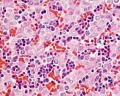
Histology-fetal liver HEx40
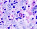
Histology-fetal liver x100


Glossary Links
- Glossary: A | B | C | D | E | F | G | H | I | J | K | L | M | N | O | P | Q | R | S | T | U | V | W | X | Y | Z | Numbers | Symbols | Term Link
Cite this page: Hill, M.A. (2024, June 10) Embryology Gastrointestinal Tract - Liver Histology. Retrieved from https://embryology.med.unsw.edu.au/embryology/index.php/Gastrointestinal_Tract_-_Liver_Histology
- © Dr Mark Hill 2024, UNSW Embryology ISBN: 978 0 7334 2609 4 - UNSW CRICOS Provider Code No. 00098G