Abnormal Development - Cleft Palate: Difference between revisions
mNo edit summary |
mNo edit summary |
||
| (42 intermediate revisions by the same user not shown) | |||
| Line 1: | Line 1: | ||
{{Header}} | {{Header}} | ||
{{ | =LA42 Cleft Palate= | ||
{| | |||
|-bgcolor="FFCC00" | |||
! {{ICD-11}} | |||
|-bgcolor="FEF9E7" | |||
| {{ICD11weblink}}1481115868 Clefts of lip, alveolus or palate] - {{ICD11weblink}}2129534948 '''LA42''' Cleft palate] | |||
:''Cleft palate is a fissure type embryopathy that affects the soft and hard palate to varying degrees.'' | |||
* LA4Y Other specified clefts of lip, alveolus or palate | |||
* LA4Z Clefts of lip, alveolus or palate, unspecified | |||
|} | |||
== Introduction == | == Introduction == | ||
[[File:Stage18 em11.jpg|thumb|300px|Human Embryo Face ([[Week 7]], [[Carnegie stage 18]], 44 - 48 days, CRL 13 - 17 mm)]] | [[File:Stage18 em11.jpg|thumb|300px|Human Embryo Face ([[Week 7]], [[Carnegie stage 18]], 44 - 48 days, CRL 13 - 17 mm)]] | ||
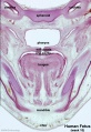
[[File:Bailey141.jpg|thumb|300px|Fetal Palate (80 mm fetus)]] | [[File:Bailey141.jpg|thumb|300px|Fetal Palate (80 mm fetus)]] | ||
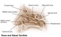
Cleft palate (palatoschisis) has many different causes and in humans occurs more frequently in females (57%) than in males (43%).{{#pmid:24179449|PMID24179449}} The palate anatomically separates the nasal cavity from the oral cavity and structurally has a bony (hard) anterior component and a muscular (soft) posterior component ending with the uvula. The oral side of the palate is covered with a squamous stratified (pluristratified) epithelium. The surface of the hard palate of most mammalian species is further thrown into a series of transversal palatal ridges or ''rugae palatinae''. Both the palatal ridge number and arrangement are also species specific. | |||
A major contribution to the palate comes from the {{neural crest}} and there are a number of molecular, mechanical and morphological steps in involving the fusion of contributing structures including a key epithelial to mesenchymal transition. In palate formation there are two main and separate times and events of development, during embryonic (primary palate) and an early fetal (secondary palate). This separation of events into embryonic and fetal period corresponds closely to the classification of associated palate abnormalities. | |||
The primary palate is formed by two parts: | The primary palate is formed by two parts: | ||
# maxillary components of the first pharyngeal arch (lateral) | # maxillary components of the first pharyngeal arch (lateral) | ||
# frontonasal prominence (midline) | # frontonasal prominence (midline) | ||
The secondary palate can also be divided in two anatomical parts: | The secondary palate can also be divided in two anatomical parts: | ||
# anterior hard palate - ossified | # anterior hard palate - ossified | ||
# posterior soft palate - muscular | ## maxilla | ||
## palatine bones | |||
# posterior soft palate - muscular | |||
## tensor veli palatini (swallowing) | |||
## palatoglossus (swallowing) | |||
## palatopharyngeus (breathing) | |||
## levator veli palatini (swallowing) | |||
## musculus uvulae (uvula movement) | |||
| Line 28: | Line 40: | ||
== Some Recent Findings == | == Some Recent Findings == | ||
[[File:Cleft_palate.jpg|thumb|300px|Cleft Palate.]] | [[File:Cleft_palate.jpg|thumb|300px|Cleft Palate.]] | ||
[[File:Cleft_lip_01.jpg|thumb|Ultrasound - Cleft Lip]] | [[File:Cleft_lip_01.jpg|thumb|Ultrasound - Cleft Lip]] | ||
{| | {| | ||
|-bgcolor="F5FAFF" | |-bgcolor="F5FAFF" | ||
| | | | ||
* ''' | * '''Identification of circular RNA-associated competing endogenous RNA network in the development of cleft palate'''{{#pmid:31074068|PMID31074068}} "Circular RNAs (circRNAs) serve as competing endogenous RNAs (ceRNAs) and indirectly regulate gene expression through shared microRNAs (miRNAs). However, the regulatory mechanisms of circRNA as ceRNA associated with the fusion of palatal shelves in palatogenesis are yet unclear. This study aimed to explore the potential mechanism underlying the role of circRNA as ceRNA in cleft palate (CP). First, we systematically analyzed RNA-seq and miRNA-seq data after high-throughput sequencing for embryonic palatal shelf tissues from a mouse CP model induced by maternal exposure to all-trans retinoic acid on embryonic gestation day 14.5 (E14.5). Thirty-nine circRNAs, 18 miRNAs, and 936 messenger RNAs (mRNAs) were significantly dysregulated (log2 [fold change {FC}] > 1; P < 0.05). Thereafter, we constructed a circRNA-associated ceRNA network. Finally, we determined the circRNA_0954-miRNA-881-3p-PRKAR1α ceRNA network as a hub involved in palatogenesis. Gene Ontology analysis revealed that ceRNA-related genes were associated with facial morphogenesis and developmental gene silencing. Kyoto Encyclopedia of Genes and Genomes pathway analysis indicated that ceRNA-related genes are involved in apoptosis (P < 0.05, fold enrichment >1). Quantitative reverse transcription polymerase chain reaction was performed to verify the results of ceRNA analysis. We found that the circRNA-miRNA-mRNA ceRNA network is involved in palatogenesis. The present results imply that circRNA_0954-miRNA-881-3p-PRKAR1α ceRNA network may cause dysfunctional palatal fusion and might facilitate the development of novel epigenetic biomarkers to treat CP in the future." | ||
* ''' | * '''Constitutive activation of hedgehog signaling adversely affects epithelial cell fate during palatal fusion'''{{#pmid:29981310|PMID29981310}} "Cleft palate is one of the most common craniofacial congenital defects in humans. It is associated with multiple genetic and environmental risk factors, including mutations in the genes encoding signaling molecules in the {{sonic hedgehog}} ({{SHH}}) pathway, which are risk factors for cleft palate in both humans and mice. However, the function of Shh signaling in the palatal epithelium during palatal fusion remains largely unknown. Although components of the Shh pathway are localized in the palatal epithelium, specific inhibition of Shh signaling in palatal epithelium does not affect palatogenesis. ...In this study, we discovered that constitutive activation of Hh signaling in the palatal epithelium results in submucous cleft palate and persistence of the medial edge epithelium (MEE). Further investigation revealed that precise downregulation of Shh signaling is required at a specific time point in the MEE during palatal fusion. Upregulation of Hh signaling in the palatal epithelium maintains the proliferation of MEE cells. This may be due to a dysfunctional p63/Irf6 regulatory loop. The resistance of MEE cells to apoptosis is likely conferred by enhancement of a cell adhesion network through the maintenance of p63 expression." | ||
* ''' | |||
* '''p63 exerts spatio-temporal control of palatal epithelial cell fate to prevent cleft palate'''{{#pmid:28604778|PMID28604778}} "Mutations in the transcription factor {{p63}} are one of the major individual causes of cleft palate; however, the gene regulatory networks in which p63 functions remain only partially characterized. Our findings demonstrate that p63 functions as an essential regulatory molecule in the spatio-temporal control of palatal epithelial cell fate to ensure appropriate fusion of the palatal shelves. Initially, p63 induces periderm formation and controls its subsequent maintenance to prevent premature adhesion between adhesion-competent, intra-oral epithelia. Subsequently, TGFβ3-induced down-regulation of p63 in the medial edge epithelia of the palatal shelves is a pre-requisite for palatal fusion by facilitating periderm migration from, and reducing the proliferative potential of, the midline epithelial seam thereby preventing cleft palate." | |||
|} | |} | ||
{| class="wikitable collapsible collapsed" | {| class="wikitable mw-collapsible mw-collapsed" | ||
! More recent papers | ! More recent papers | ||
|- | |- | ||
| [[File:Mark_Hill.jpg|90px|left]] {{Most_Recent_Refs}} | | [[File:Mark_Hill.jpg|90px|left]] {{Most_Recent_Refs}} | ||
Search term: ''Cleft Palate'' | Search term: [http://www.ncbi.nlm.nih.gov/pubmed/?term=Cleft+Palate ''Cleft Palate''] | | ||
|} | |||
{| class="wikitable mw-collapsible mw-collapsed" | |||
! Older papers | |||
|- | |||
| {{Older papers}} | |||
* '''Regulation of the Epithelial Adhesion Molecule CEACAM1 Is Important for Palate Formation'''{{#pmid:23613893|PMID23613893}} "Cleft palate results from a mixture of genetic and environmental factors and occurs when the bilateral palatal shelves fail to fuse. The objective of this study was to search for new genes involved in mouse palate formation. Gene expression of murine embryonic palatal tissue was analyzed at various developmental stages before, during, and after palate fusion using GeneChip® microarrays. Ceacam1 was one of the highly up-regulated genes during palate formation, and this was confirmed by quantitative real-time PCR. Immunohistochemical staining showed that CEACAM1 was present in prefusion palatal epithelium and was degraded during fusion. ...These results suggest that CEACAM1 has roles in the initiation of palatal fusion via epithelial cell adhesion." | |||
* '''Role of GSK-3β in the Osteogenic Differentiation of Palatal Mesenchyme'''{{#pmid:22022457|PMID22022457}} "Here, we identify a critical role for GSK-3β in palatogenesis through its direct regulation of canonical Wnt signaling. These findings shed light on critical developmental pathways involved in palatogenesis and may lead to novel molecular targets to prevent cleft palate formation." | |||
* '''Ephrin reverse signaling controls palate fusion via a PI3 kinase-dependent mechanism'''{{#pmid:21246652|PMID21246652}} "Secondary palate fusion requires adhesion and epithelial-to-mesenchymal transition (EMT) of the epithelial layers on opposing palatal shelves. This EMT requires transforming growth factor β3 (TGFβ3), and its failure results in cleft palate. Ephrins, and their receptors, the Ephs, are responsible for migration, adhesion, and midline closure events throughout development. Ephrins can also act as signal-transducing receptors in these processes, with the Ephs serving as ligands (termed "reverse" signaling). We found that activation of ephrin reverse signaling in chicken palates induced fusion in the absence of TGFβ3, and that PI3K inhibition abrogated this effect. Further, blockage of reverse signaling inhibited TGFβ3-induced fusion in the chicken and natural fusion in the mouse. Thus, ephrin reverse signaling is necessary and sufficient to induce palate fusion independent of TGFβ3." | |||
* '''A genome-wide association study of cleft lip with and without cleft palate identifies risk variants near MAFB and ABCA4'''{{#pmid:20436469|PMID20436469}} "Case-parent trios were used in a genome-wide association study of cleft lip with and without cleft palate. SNPs near two genes not previously associated with cleft lip with and without cleft palate (MAFB, most significant SNP rs13041247, with odds ratio (OR) per minor allele = 0.704, 95% CI 0.635-0.778, P = 1.44 x 10(-11); and ABCA4, most significant SNP rs560426, with OR = 1.432, 95% CI 1.292-1.587, P = 5.01 x 10(-12)) and two previously identified regions (at chromosome 8q24 and IRF6) attained genome-wide significance." | |||
* '''A dosage-dependent role for Spry2 in growth and patterning during palate development'''{{#pmid:17693063|PMID17693063}} "The formation of the palate involves the coordinated outgrowth, elevation and midline fusion of bilateral shelves leading to the separation of the oral and nasal cavities. Reciprocal signaling between adjacent fields of epithelial and mesenchymal cells directs palatal shelf growth and morphogenesis. Loss of function mutations in genes encoding FGF ligands and receptors have demonstrated a critical role for FGF signaling in mediating these epithelial-mesenchymal interactions. The Sprouty family of genes encode modulators of FGF signaling. We have established that mice carrying a deletion that removes the FGF signaling antagonist Spry2 have cleft palate." | |||
|} | |} | ||
== Textbooks == | == Textbooks == | ||
[[File: | [[File:Stage16_cleft_palate.jpg|thumb|Embryonic Human Cleft Palate Stage16 (ventral view)]] | ||
* '''The Developing Human: Clinically Oriented Embryology''' (8th Edition) by Keith L. Moore and T.V.N Persaud - Moore & Persaud Chapter Chapter 10 The Pharyngeal Apparatus pp201 - 240. | * '''The Developing Human: Clinically Oriented Embryology''' (8th Edition) by Keith L. Moore and T.V.N Persaud - Moore & Persaud Chapter Chapter 10 The Pharyngeal Apparatus pp201 - 240. | ||
* '''Larsen’s Human Embryology''' by GC. Schoenwolf, SB. Bleyl, PR. Brauer and PH. Francis-West - Chapter 12 Development of the Head, the Neck, the Eyes, and the Ears pp349 - 418. | * '''Larsen’s Human Embryology''' by GC. Schoenwolf, SB. Bleyl, PR. Brauer and PH. Francis-West - Chapter 12 Development of the Head, the Neck, the Eyes, and the Ears pp349 - 418. | ||
| Line 65: | Line 89: | ||
:'''Links:''' [[Movies]] | [[Ultrasound]] | :'''Links:''' [[Movies]] | [[Ultrasound]] | ||
==Clinical Images== | ==Clinical Images== | ||
| Line 70: | Line 95: | ||
[[File:Cleft_palate_001.jpg]] | [[File:Cleft_palate_001.jpg]] | ||
Involving only the soft palate and uvula. | Involving only the soft palate and uvula.{{#pmid:22437671|PMID22437671}} | ||
===Complete Cleft Palate=== | ===Complete Cleft Palate=== | ||
[[File:Cleft_palate_002.jpg]] | [[File:Cleft_palate_002.jpg]] | ||
Completely involving the secondary palate. | Completely involving the secondary palate.{{#pmid:22437671|PMID22437671}} | ||
[[File:Cleft palate 003.jpg|400px]] | [[File:Cleft palate 003.jpg|400px]] | ||
Surgical repair of the palate (palatoplasty). | Surgical repair of the palate (palatoplasty).{{#pmid:22437671|PMID22437671}} | ||
{ | |||
==International Classification of Diseases - Cleft Palate== | |||
Cleft lip and cleft palate (Q35-Q37) Use additional code (Q30.2), if desired, to identify associated malformations of the nose. Excludes Robin's syndrome ( Q87.0 ) | |||
{| | {| | ||
| width="100px"| '''Q37''' | | width="100px"| '''Q37''' | ||
| Line 261: | Line 149: | ||
|} | |} | ||
=== | ===Veau Classification=== | ||
The historic Veau System (1931)<ref>Veau V. Paris: Masson & Cie; '''Division Palatine'''. (1931).</ref> classifies orofacial clefting into four classes (Veau Class I - IV) according to whether the secondary and/or primary palates are affected and by laterality. | |||
# Incomplete cleft, soft palate only (no unilateral/bilateral designation) | |||
# Hard and soft palate, secondary palate only (no unilateral/bilateral designation) | |||
# Complete unilateral cleft including lip (primary and secondary palates) | |||
# Complete bilateral cleft | |||
==Cleft Palate Genetics== | |||
[[File:Cleft_palate_02.jpg|thumb|An 8 month old infant with an extensive cleft palate associated with Bamforth- Lazarus syndrome.{{#pmid:20537182|PMID20537182}}]] | |||
{| | {| | ||
! Cleft Palate Only Genes | ! Cleft Palate Only Genes{{#pmid:21331089|PMID21331089}} | ||
|- | |- | ||
| | | | ||
| Line 389: | Line 270: | ||
|} | |} | ||
'''Links:''' [http://www.ncbi.nlm.nih.gov/omim/119530 OMIM Orofacial Cleft with or without cleft palate] | |||
==Statistics== | |||
[[File:cleft_palate.jpg|thumb|Cleft palate]] | |||
* International Classification of Diseases code 749.0 | |||
* Australian national rate (1982-1992) 4.8 - 6 /10,000 births. | |||
* Of 1,530 infants 5.5% were stillborn and 11.5% liveborn died during neonatal period. | |||
* slightly more common in twin births than singleton. | |||
(Data: Congenital Malformations Australia 1981-1992 P. Lancaster and E. Pedisich ISSN 1321-8352) | |||
[[File:Australian abnormalities 81-92 git.jpg|thumb|350px|Cleft Palate - Australia (1981-1992)<ref>P. Lancaster and E. Pedisich, '''Congenital Malformations Australia''' 1981-1992, ISSN 1321-835.</ref>]] | |||
{| | |||
|- | |||
! Australian Palate Abnormalities (2002-2003)<ref>Abeywardana S & Sullivan EA 2008. [http://www.npesu.unsw.edu.au/surveillance/congenital-anomalies-australia-2002-2003 Congenital Anomalies in Australia 2002-2003]. Birth anomalies series no. 3 Cat. no. PER 41. Sydney: AIHW National Perinatal Statistics Unit.</ref> | |||
|- | |||
| '''Cleft lip with or without cleft palate''' (9.2 per 10,000 births) ICD-10 Q36.0, Q36.1, Q36.9, Q37.0–Q37.5, Q37.8, Q37.9 | |||
|- | |||
| A congenital anomaly characterised by a partial or complete clefting of the upper lip, with or without clefting of the alveolar ridge or the hard palate. Excludes a midline cleft of the upper or lower lip and an oblique facial fissure (going towards the eye). | |||
* 17% of the affected pregnancies were terminated in early pregnancy or resulted in fetal deaths. Most of the fetal deaths or terminations of pregnancy (95%) had multiple abnormalities. | |||
* more commonly seen in males than in females. | |||
* babies born before 25 weeks of gestation, 150 per 10,000 births had this anomaly. Most babies (80.0%) were born at term with a birthweight of 2,500 grams or more. | |||
* Maternal age group was not associated with the anomaly. | |||
* Rates significantly higher among Indigenous women than non Indigenous women. | |||
|- | |||
| '''Cleft palate without cleft lip''' (8.1 per 10,000 births) ICD-10 Q35.0–Q35.9 | |||
|- | |||
| A congenital anomaly characterised by a closure defect of the hard and/or soft palate behind the foramen incisivum without a cleft lip. This anomaly includes sub-mucous cleft palate, but excludes cleft palate with a cleft lip, a functional short palate and high narrow palate. | |||
* overall rate has increased to 9.1 when the rate was estimated using data from the four states that include TOP data. The reported number of fetal deaths or early terminations of pregnancy with this anomaly was small and these deaths or terminations could be due to other associated anomalies. | |||
* proportion of females with this anomaly was higher (56.9%) than males. | |||
* 52.7 per 10,000 babies born before 25 weeks of gestation. | |||
* 83.0% were born at term and most of the babies (82.7%) had a birthweight of 2,500 grams or more. | |||
* Women aged 40 years or older and women born in South Central America or the Caribbean region had the highest rates of affected births. | |||
* Multiple births had a significantly higher rate of affected babies than singleton births. | |||
* Rates did not differ significantly by Indigenous status or areas of residence. | |||
|- | |||
|} | |||
{{Australian Palate abnormalities 2002-2003}} | |||
{{10Victorian Anomalies03-04}} | |||
Cleft Risk Variants - Two genes were identified from a recent genome-wide study.<ref name="PMID20436469" /> | |||
Two genes were identified from a recent genome-wide study.<ref name="PMID20436469" /> | |||
* '''MAFB''' is expressed in the mouse palatal shelf. | * '''MAFB''' is expressed in the mouse palatal shelf. | ||
| Line 403: | Line 320: | ||
:'''Links:''' [http://www.ncbi.nlm.nih.gov/omim/608968 OMIM - MAFB] | [http://www.ncbi.nlm.nih.gov/omim/601691 OMIM - ABCA4] | |||
Folate - A recent study of periconceptional folate supplementation using the Cochrane Pregnancy and Childbirth Group's Trials Register (July 2010) identified no statistically significant evidence of any effects on prevention of cleft palate and cleft lip at birth.{{#pmid:20927767|PMID20927767}} | |||
==Cleft Palate Repair== | |||
[[File:Cleft palate 003.jpg|300px|thumb|Pre and post-operative palatoplasty.{{#pmid:22437671|PMID22437671}}]] | |||
Surgical repair of the palate is described as palatoplasty and is often carried out between 6 to 12 months of age but also during later periods (12 to 18 months). In developing countries, this may be repaired significantly later in childhood or even go unprepared. | |||
===Surgical Repair Techniques=== | |||
Some Types of Surgical Repair Techniques{{#pmid:19884664|PMID19884664}} | |||
* von Langenbeck's bipedicle flap technique | |||
* Veau-Wardill-Kilner Pushback technique | |||
* Bardach's two-flap technique | |||
* Furlow Double opposing Z-Plasty | |||
* Two-stage palatal repair | |||
* Hole in one repair | |||
* Raw area free palatoplasty | |||
* Alveolar extension palatoplasty (AEP) | |||
* Primary pharyngeal flap | |||
* Intravelar veloplasty | |||
* Vomer flap | |||
* Buccal myomucosal flap | |||
== Development Overview == | |||
:'''Links:''' [ | * '''week 4''' - pharyngeal arch formation, first pharngeal arch contributes mandible and maxilla. | ||
* '''week 6 - 7''' - primary palate formation maxillary processes and frontonasal prominence. | |||
* '''week 9''' - secondary palate shelves fuse, separating oral and nasal cavities. | |||
===Embryonic Period=== | |||
* (week 4) - pharyngeal arch formation in rostrocaudal sequence (1, 2, 3, 4 and 6) | |||
* First pharyngeal arch - upper maxillary (pair) and lower mandibular prominences | |||
* Late embryonic period - maxillary prominences fuse with frontonasal prominence forming upper jaw (maxilla and upper lip) | |||
<gallery> | |||
File:Stage16 em01.jpg|Week 5 [[:File:Stage16 em01.jpg|Stage {{CS16}}]] | |||
File:Stage17 em01.jpg|Week 6 [[:File:Stage17 em01.jpg|Stage {{CS17}}]] | |||
File:Stage18 em01.jpg|Week 6.5 [[:File:Stage18 em01.jpg|Stage {{CS18}}]] | |||
File:Stage19 em01.jpg|Week 7 [[:File:Stage19 em01.jpg|Stage {{CS19}}]] | |||
</gallery> | |||
===Fetal Period=== | |||
{| | |||
| | |||
* palatal shelves elevation | |||
* palatal shelves midline fusion | |||
| {{Fetal week 10 palate movie}} | |||
|} | |||
==Face Development== | |||
[[File:Stage16-18 face animation.gif|left]] | |||
Begins week 4 centered around stomodeum, external depression at oral membrane | |||
5 initial primordia from neural crest mesenchyme | |||
* single frontonasal prominence (FNP) - forms forehead, nose dorsum and apex | |||
* nasal placodes develop later bilateral, pushed medially | |||
* paired maxillary prominences - form upper cheek and upper lip | |||
* paired mandibular prominences - lower cheek, chin and lower lip | |||
===Neural Crest === | |||
* Mesenchyme invaded by neural crest generating connective tissue components | |||
* cartilage, bone, ligaments | |||
* arises from midbrain and hindbrain region | |||
===Frontonasal Process=== | |||
The frontonasal process (FNP) forms the majority of the superior part of the early face primordia. It later fuses with the maxillary component of the first pharyngeal arch to form the upper jaw. Failure of this fusion event during the embryonic period leads to cleft lip. Under the surface ectoderm the process mesenchyme consists of two cell populations; neural crest cells, forming the connective tissues; and the mesoderm forming the endothelium of the vascular network. | |||
A chicken developmental model study has identified a specific surface region, the Frontonasal Ectodermal Zone (FEZ), initially induced by bone morphogenetic proteins that appears to regulate the future growth and patterning of the frontonasal process. The specific frontonasal ectodermal zone was located in the frontonasal process ectoderm flanking a boundary between Sonic hedgehog (Shh) and Fibroblast growth factor 8 (Fgf8) expression domains.{{#pmid:18028903|PMID18028903}} | |||
== Embryonic Palate== | |||
{| | |||
| | |||
Human primary palate | |||
* develops between embryonic stages 15 and 18.{{#pmid:8227288|PMID8227288}} | |||
* fusion in the human embryo between stage 17 and 18, from an epithelial seam to the mesenchymal bridge. | |||
| [[File:Stage17-18 Primary palate.gif]] | |||
|} | |||
<gallery> | |||
File:Stage16 em01.jpg|Week 5 [[:File:Stage16 em01.jpg|Stage 16]] | |||
File:Stage17 em01.jpg|Week 6 [[:File:Stage17 em01.jpg|Stage 17]] | |||
File:Stage18 em01.jpg|Week 6.5 [[:File:Stage18 em01.jpg|Stage 18]] | |||
File:Stage19 em01.jpg|Week 7 [[:File:Stage19 em01.jpg|Stage 19]] | |||
</gallery> | |||
:'''EM Links:''' [[:File:Stage16 em01.jpg|Image - stage 16]] | [[:File:Stage17 em01.jpg|Image - stage 17]] | [[:File:Stage18 em01.jpg|Image - stage 18]] | [[:File:Stage19 em01.jpg|Image - stage 19]] | [[Palate Development]] | |||
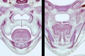
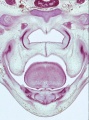
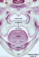
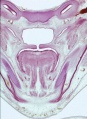
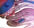
==Fetal Palate== | |||
Secondary palate, fusion in the human embryo in [[Week 9|week 9]]. This requires the early palatal shelves growth, elevation, and fusion. There are many fusion events occurring during this period between each palatal shelf, to the primary palate, and also to the nasal septum. | |||
[[:File:Palatal shelves animation.gif|palatal shelf elevation]] | [[:File:palate.gif|secondary palate]] | |||
<gallery> | |||
File:Fetal week 10 hard palate 04.jpg|Hard and soft palate | |||
File:Fetal_week_10_hard_palate_05.jpg|hard palate | |||
File:Fetal week 10 hard palate 07.jpg|hard palate labeled | |||
File:Fetal week 10 soft palate 01.jpg|soft palate | |||
File:Fetal_week_10_soft_palate_03.jpg|soft palate labeled | |||
File:Fetal week 10 hard palate 03.jpg|Detail - hard and soft palate junction | |||
File:Fetal week 10 hard palate 02.jpg|Detail - hard palate seam | |||
</gallery> | |||
==Head Growth== | |||
* continues postnatally - fontanelle allow head distortion on birth and early growth | |||
* bone plates remain unfused to allow growth, puberty growth of face | |||
==Adult== | |||
<gallery> | |||
File:Adult skull cleft palate 01.jpg | |||
File:Adult skull cleft palate 03.jpg | |||
File:Adult skull cleft palate 02.jpg | |||
</gallery> | |||
==Animal Models== | |||
[[File:Dog day0-cleft palate.jpg|thumb|Newborn dog with cleft palate.]] | |||
===Mouse Palate=== | |||
* '''E11''' - protrude from bilateral maxillary processes | |||
* '''E12.5 - secondary palatal development begins | |||
* '''E12.5-E14''' - grow vertically along the developing tongue | |||
* '''E14.5''' - they elevate, meet, and fuse at the midline, to form an intact palate shelf, reflex opening and closing movements of the mouth | |||
* '''E15.5''' - palatal fusion is complete, mesenchymal condensation followed by osteogenic differentiation occurs. | |||
[[File:Mouse_palate_gene_expression_01.jpg|600px]] | |||
Mouse (E13.5) Palatal Shelf Wnt5a, Osr2 and Pax9 Expression.{{#pmid:24433583|PMID24433583}} | |||
<gallery> | |||
File:Mouse E13.5 Bmp7 palate 1.jpg|Image - Mouse E13.5 Bmp7 palate | |||
File:Mouse E13.5 Bmp7 palate 2.jpg|Image - palate | |||
File:Mouse E13.5 Bmp7 palate 3.jpg|Image - palate detail | |||
</gallery> | |||
{| | |||
| [[File:Mouse_ruga_pattern.jpg|300px]] | |||
| [[File:Mouse_-_Spry1_cleft_palate.jpg|300px]] | |||
|- | |||
| Mouse ruga pattern (E16) | |||
| Mouse - Spry1 cleft palate | |||
|} | |||
[[File:File-Cleft palate in newborn mice.jpg]] | |||
Cleft palate in newborn mice.{{#pmid:20808828|PMID20808828}} | |||
:'''Links:''' [[Mouse Development]] | [[Developmental_Signals_-_Bone_Morphogenetic_Protein|Bone Morphogenetic Protein]] | [[Developmental Signals - Wnt|Wnt]] | [[Developmental_Signals_-_Pax|Pax]] | |||
==Molecular== | |||
<gallery> | |||
File:Mouse - palate MMP-25 expression.jpg|MMP25{{#pmid:20809987|PMID20809987}} | |||
File:Mouse E13.5 Bmp7 palate 1.jpg|Image - Mouse E13.5 Bmp7 palate{{#pmid:23516636|PMID23516636}} | |||
File:Mouse E13.5 Bmp7 palate 2.jpg|Image - palate Bmp7 palate{{#pmid:23516636|PMID23516636}} | |||
File:Mouse E13.5 Bmp7 palate 3.jpg|Image - palate detail Bmp7 palate{{#pmid:23516636|PMID23516636}} | |||
</gallery> | |||
:'''Links:''' [[Developmental_Signals_-_Bone_Morphogenetic_Protein|Bone Morphogenetic Protein]] | |||
== References == | == References == | ||
| Line 423: | Line 493: | ||
===Reviews=== | ===Reviews=== | ||
Indian J Plast Surg. 2009 October; 42(Suppl):[http://www.ncbi.nlm.nih.gov/pmc/issues/185089 Cleft Lip and Palate Issue] | Indian J Plast Surg. 2009 October; 42(Suppl):[http://www.ncbi.nlm.nih.gov/pmc/issues/185089 Cleft Lip and Palate Issue] | ||
{{#pmid:26322171}} | |||
{{#pmid:22186724}} | |||
{{#pmid:19131313}} | |||
{{#pmid:16962647}} | |||
{{#pmid:3074914}} | |||
{{#pmid:8714286}} | |||
===Articles=== | ===Articles=== | ||
{{#pmid:20149609}} | |||
{{#pmid:19341725}} | |||
===Search PubMed=== | ===Search PubMed=== | ||
| Line 458: | Line 533: | ||
==Terms== | ==Terms== | ||
{{Palate terms}} | |||
==External Links== | ==External Links== | ||
| Line 479: | Line 542: | ||
* '''Better Health Channel''' - [http://www.betterhealth.vic.gov.au/bhcv2/bhcarticles.nsf/pages/Cleft_palate_and_cleft_lip Cleft palate and cleft lip] | * '''Better Health Channel''' - [http://www.betterhealth.vic.gov.au/bhcv2/bhcarticles.nsf/pages/Cleft_palate_and_cleft_lip Cleft palate and cleft lip] | ||
* '''March of Dimes Birth Defects Foundation''' - [http://www.marchofdimes.com/baby/birthdefects_cleftpalate.html Cleft Palate] | * '''March of Dimes Birth Defects Foundation''' - [http://www.marchofdimes.com/baby/birthdefects_cleftpalate.html Cleft Palate] | ||
Latest revision as of 22:41, 12 May 2019
| Embryology - 17 Jun 2024 |
|---|
| Google Translate - select your language from the list shown below (this will open a new external page) |
|
العربية | català | 中文 | 中國傳統的 | français | Deutsche | עִברִית | हिंदी | bahasa Indonesia | italiano | 日本語 | 한국어 | မြန်မာ | Pilipino | Polskie | português | ਪੰਜਾਬੀ ਦੇ | Română | русский | Español | Swahili | Svensk | ไทย | Türkçe | اردو | ייִדיש | Tiếng Việt These external translations are automated and may not be accurate. (More? About Translations) |
LA42 Cleft Palate
| ICD-11 |
|---|
Clefts of lip, alveolus or palate - LA42 Cleft palate
|
Introduction


Cleft palate (palatoschisis) has many different causes and in humans occurs more frequently in females (57%) than in males (43%).[1] The palate anatomically separates the nasal cavity from the oral cavity and structurally has a bony (hard) anterior component and a muscular (soft) posterior component ending with the uvula. The oral side of the palate is covered with a squamous stratified (pluristratified) epithelium. The surface of the hard palate of most mammalian species is further thrown into a series of transversal palatal ridges or rugae palatinae. Both the palatal ridge number and arrangement are also species specific.
A major contribution to the palate comes from the neural crest and there are a number of molecular, mechanical and morphological steps in involving the fusion of contributing structures including a key epithelial to mesenchymal transition. In palate formation there are two main and separate times and events of development, during embryonic (primary palate) and an early fetal (secondary palate). This separation of events into embryonic and fetal period corresponds closely to the classification of associated palate abnormalities.
The primary palate is formed by two parts:
- maxillary components of the first pharyngeal arch (lateral)
- frontonasal prominence (midline)
The secondary palate can also be divided in two anatomical parts:
- anterior hard palate - ossified
- maxilla
- palatine bones
- posterior soft palate - muscular
- tensor veli palatini (swallowing)
- palatoglossus (swallowing)
- palatopharyngeus (breathing)
- levator veli palatini (swallowing)
- musculus uvulae (uvula movement)
| Palate Links: palate | cleft lip and palate | cleft palate | head | Category:Palate |
Some Recent Findings


|
| More recent papers |
|---|
 This table allows an automated computer search of the external PubMed database using the listed "Search term" text link.
More? References | Discussion Page | Journal Searches | 2019 References | 2020 References Search term: Cleft Palate | |
| Older papers |
|---|
| These papers originally appeared in the Some Recent Findings table, but as that list grew in length have now been shuffled down to this collapsible table.
See also the Discussion Page for other references listed by year and References on this current page.
|
Textbooks

- The Developing Human: Clinically Oriented Embryology (8th Edition) by Keith L. Moore and T.V.N Persaud - Moore & Persaud Chapter Chapter 10 The Pharyngeal Apparatus pp201 - 240.
- Larsen’s Human Embryology by GC. Schoenwolf, SB. Bleyl, PR. Brauer and PH. Francis-West - Chapter 12 Development of the Head, the Neck, the Eyes, and the Ears pp349 - 418.
Movies
|
|
|
|
|
- Links: Movies | Ultrasound
Clinical Images
Incomplete Cleft Palate

Involving only the soft palate and uvula.[10]
Complete Cleft Palate

Completely involving the secondary palate.[10]

Surgical repair of the palate (palatoplasty).[10]
International Classification of Diseases - Cleft Palate
Cleft lip and cleft palate (Q35-Q37) Use additional code (Q30.2), if desired, to identify associated malformations of the nose. Excludes Robin's syndrome ( Q87.0 )
| Q37 | Cleft palate with cleft lip |
| Q37.0 | Cleft hard palate with bilateral cleft lip |
| Q37.1 | Cleft hard palate with unilateral cleft lip |
| Cleft hard palate with cleft lip NOS | |
| Q37.2 | Cleft soft palate with bilateral cleft lip |
| Q37.3 | Cleft soft palate with unilateral cleft lip |
| Cleft soft palate with cleft lip NOS | |
| Q37.4 | Cleft hard and soft palate with bilateral cleft lip |
| Q37.5 | Cleft hard and soft palate with unilateral cleft lip |
| Cleft hard and soft palate with cleft lip NOS | |
| Q37.8 | Unspecified cleft palate with bilateral cleft lip |
| Q37.9 | Unspecified cleft palate with unilateral cleft lip |
| Cleft palate with cleft lip NOS |
Veau Classification
The historic Veau System (1931)[11] classifies orofacial clefting into four classes (Veau Class I - IV) according to whether the secondary and/or primary palates are affected and by laterality.
- Incomplete cleft, soft palate only (no unilateral/bilateral designation)
- Hard and soft palate, secondary palate only (no unilateral/bilateral designation)
- Complete unilateral cleft including lip (primary and secondary palates)
- Complete bilateral cleft
Cleft Palate Genetics

| Cleft Palate Only Genes[13] | ||||||||||||||||||||||||||||||||||||||||||||||||||||||||||||||||||||
|---|---|---|---|---|---|---|---|---|---|---|---|---|---|---|---|---|---|---|---|---|---|---|---|---|---|---|---|---|---|---|---|---|---|---|---|---|---|---|---|---|---|---|---|---|---|---|---|---|---|---|---|---|---|---|---|---|---|---|---|---|---|---|---|---|---|---|---|---|
|
Links: OMIM Orofacial Cleft with or without cleft palate
Statistics
- International Classification of Diseases code 749.0
- Australian national rate (1982-1992) 4.8 - 6 /10,000 births.
- Of 1,530 infants 5.5% were stillborn and 11.5% liveborn died during neonatal period.
- slightly more common in twin births than singleton.
(Data: Congenital Malformations Australia 1981-1992 P. Lancaster and E. Pedisich ISSN 1321-8352)

| Australian Palate Abnormalities (2002-2003)[15] |
|---|
| Cleft lip with or without cleft palate (9.2 per 10,000 births) ICD-10 Q36.0, Q36.1, Q36.9, Q37.0–Q37.5, Q37.8, Q37.9 |
A congenital anomaly characterised by a partial or complete clefting of the upper lip, with or without clefting of the alveolar ridge or the hard palate. Excludes a midline cleft of the upper or lower lip and an oblique facial fissure (going towards the eye).
|
| Cleft palate without cleft lip (8.1 per 10,000 births) ICD-10 Q35.0–Q35.9 |
A congenital anomaly characterised by a closure defect of the hard and/or soft palate behind the foramen incisivum without a cleft lip. This anomaly includes sub-mucous cleft palate, but excludes cleft palate with a cleft lip, a functional short palate and high narrow palate.
|
| Australian Palate Abnormalities (2002-2003) |
|---|
| Cleft lip with or without cleft palate (9.2 per 10,000 births) ICD-10 Q36.0, Q36.1, Q36.9, Q37.0–Q37.5, Q37.8, Q37.9 |
A congenital anomaly characterised by a partial or complete clefting of the upper lip, with or without clefting of the alveolar ridge or the hard palate. Excludes a midline cleft of the upper or lower lip and an oblique facial fissure (going towards the eye).
|
| Cleft palate without cleft lip (8.1 per 10,000 births) ICD-10 Q35.0–Q35.9 |
A congenital anomaly characterised by a closure defect of the hard and/or soft palate behind the foramen incisivum without a cleft lip. This anomaly includes sub-mucous cleft palate, but excludes cleft palate with a cleft lip, a functional short palate and high narrow palate.
|
|
Ten most frequently reported Birth Anomalies
- Hypospadias (More? Male movie | Genital Abnormalities - Hypospadia)
- Obstructive Defects of the Renal Pelvis (More? Renal System - Abnormalities)
- Ventricular Septal Defect (More? Cardiovascular Abnormalities - Ventricular Septal Defect)
- Congenital Dislocated Hip (More? Musculoskelal Abnormalities - Congenital Dislocation of the Hip (CDH))
- Trisomy 21 or Down syndrome - (More? Trisomy 21)
- Hydrocephalus (More? Hydrocephalus)
- Cleft Palate (More? Palate_Development)
- Trisomy 18 or Edward Syndrome - multiple abnormalities of the heart, diaphragm, lungs, kidneys, ureters and palate 86% discontinued (More? (More? Trisomy 18)
- Renal Agenesis/Dysgenesis - reduction in neonatal death and stillbirth since 1993 may be due to the more severe cases being identified in utero and being represented amongst the increased proportion of terminations (approximately 31%). (More? Renal System - Abnormalities)
- Cleft Lip and Palate - occur with another defect in 33.7% of cases.(More? Palate Development | Head Development)
(From the Victorian Perinatal Data Collection Unit in the Australian state of Victoria between 2003-2004)
Cleft Risk Variants - Two genes were identified from a recent genome-wide study.[8]
- MAFB is expressed in the mouse palatal shelf.
- ABCA4 is a member of a superfamily of transmembrane proteins, and mutations in ABCA4 play a major role in the etiology of Stargardt disease and related retinopathies. Gene produces an ATP-binding cassette (ABC) superfamily trans-membrane protein
- Links: OMIM - MAFB | OMIM - ABCA4
Folate - A recent study of periconceptional folate supplementation using the Cochrane Pregnancy and Childbirth Group's Trials Register (July 2010) identified no statistically significant evidence of any effects on prevention of cleft palate and cleft lip at birth.[16]
Cleft Palate Repair
Surgical repair of the palate is described as palatoplasty and is often carried out between 6 to 12 months of age but also during later periods (12 to 18 months). In developing countries, this may be repaired significantly later in childhood or even go unprepared.
Surgical Repair Techniques
Some Types of Surgical Repair Techniques[17]
- von Langenbeck's bipedicle flap technique
- Veau-Wardill-Kilner Pushback technique
- Bardach's two-flap technique
- Furlow Double opposing Z-Plasty
- Two-stage palatal repair
- Hole in one repair
- Raw area free palatoplasty
- Alveolar extension palatoplasty (AEP)
- Primary pharyngeal flap
- Intravelar veloplasty
- Vomer flap
- Buccal myomucosal flap
Development Overview
- week 4 - pharyngeal arch formation, first pharngeal arch contributes mandible and maxilla.
- week 6 - 7 - primary palate formation maxillary processes and frontonasal prominence.
- week 9 - secondary palate shelves fuse, separating oral and nasal cavities.
Embryonic Period
- (week 4) - pharyngeal arch formation in rostrocaudal sequence (1, 2, 3, 4 and 6)
- First pharyngeal arch - upper maxillary (pair) and lower mandibular prominences
- Late embryonic period - maxillary prominences fuse with frontonasal prominence forming upper jaw (maxilla and upper lip)
![Stage 16]]](/embryology/index.php?title=File:Stage16_em01.jpg)
![Stage 17]]](/embryology/index.php?title=File:Stage17_em01.jpg)
![Stage 18]]](/embryology/index.php?title=File:Stage18_em01.jpg)
Fetal Period
|
|
Face Development

Begins week 4 centered around stomodeum, external depression at oral membrane
5 initial primordia from neural crest mesenchyme
- single frontonasal prominence (FNP) - forms forehead, nose dorsum and apex
- nasal placodes develop later bilateral, pushed medially
- paired maxillary prominences - form upper cheek and upper lip
- paired mandibular prominences - lower cheek, chin and lower lip
Neural Crest
- Mesenchyme invaded by neural crest generating connective tissue components
- cartilage, bone, ligaments
- arises from midbrain and hindbrain region
Frontonasal Process
The frontonasal process (FNP) forms the majority of the superior part of the early face primordia. It later fuses with the maxillary component of the first pharyngeal arch to form the upper jaw. Failure of this fusion event during the embryonic period leads to cleft lip. Under the surface ectoderm the process mesenchyme consists of two cell populations; neural crest cells, forming the connective tissues; and the mesoderm forming the endothelium of the vascular network.
A chicken developmental model study has identified a specific surface region, the Frontonasal Ectodermal Zone (FEZ), initially induced by bone morphogenetic proteins that appears to regulate the future growth and patterning of the frontonasal process. The specific frontonasal ectodermal zone was located in the frontonasal process ectoderm flanking a boundary between Sonic hedgehog (Shh) and Fibroblast growth factor 8 (Fgf8) expression domains.[18]
Embryonic Palate
|
Human primary palate
|

|
- EM Links: Image - stage 16 | Image - stage 17 | Image - stage 18 | Image - stage 19 | Palate Development
Fetal Palate
Secondary palate, fusion in the human embryo in week 9. This requires the early palatal shelves growth, elevation, and fusion. There are many fusion events occurring during this period between each palatal shelf, to the primary palate, and also to the nasal septum.
palatal shelf elevation | secondary palate
Hard and soft palate
hard palate
hard palate labeled
soft palate
soft palate labeled
Detail - hard and soft palate junction
Detail - hard palate seam


Head Growth
- continues postnatally - fontanelle allow head distortion on birth and early growth
- bone plates remain unfused to allow growth, puberty growth of face
Adult


Animal Models

Mouse Palate
- E11 - protrude from bilateral maxillary processes
- E12.5 - secondary palatal development begins
- E12.5-E14 - grow vertically along the developing tongue
- E14.5 - they elevate, meet, and fuse at the midline, to form an intact palate shelf, reflex opening and closing movements of the mouth
- E15.5 - palatal fusion is complete, mesenchymal condensation followed by osteogenic differentiation occurs.

Mouse (E13.5) Palatal Shelf Wnt5a, Osr2 and Pax9 Expression.[20]
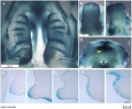
Image - Mouse E13.5 Bmp7 palate
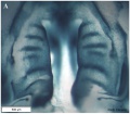
Image - palate
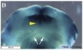
Image - palate detail


|

|
| Mouse ruga pattern (E16) | Mouse - Spry1 cleft palate |

Cleft palate in newborn mice.[21]
- Links: Mouse Development | Bone Morphogenetic Protein | Wnt | Pax
Molecular
![MMP25[22]](/embryology/images/thumb/a/af/Mouse_-_palate_MMP-25_expression.jpg/120px-Mouse_-_palate_MMP-25_expression.jpg)
MMP25[22]
Image - Mouse E13.5 Bmp7 palate[23]
Image - palate Bmp7 palate[23]
Image - palate detail Bmp7 palate[23]
![MMP25[22]](/embryology/index.php?title=File:Mouse_-_palate_MMP-25_expression.jpg)
- Links: Bone Morphogenetic Protein
References
- ↑ Kosowski TR, Weathers WM, Wolfswinkel EM & Ridgway EB. (2012). Cleft palate. Semin Plast Surg , 26, 164-9. PMID: 24179449 DOI.
- ↑ Shu X, Cheng L, Dong Z & Shu S. (2019). Identification of circular RNA-associated competing endogenous RNA network in the development of cleft palate. J. Cell. Biochem. , , . PMID: 31074068 DOI.
- ↑ Li J, Yuan Y, He J, Feng J, Han X, Jing J, Ho TV, Xu J & Chai Y. (2018). Constitutive activation of hedgehog signaling adversely affects epithelial cell fate during palatal fusion. Dev. Biol. , 441, 191-203. PMID: 29981310 DOI.
- ↑ Richardson R, Mitchell K, Hammond NL, Mollo MR, Kouwenhoven EN, Wyatt ND, Donaldson IJ, Zeef L, Burgis T, Blance R, van Heeringen SJ, Stunnenberg HG, Zhou H, Missero C, Romano RA, Sinha S, Dixon MJ & Dixon J. (2017). p63 exerts spatio-temporal control of palatal epithelial cell fate to prevent cleft palate. PLoS Genet. , 13, e1006828. PMID: 28604778 DOI.
- ↑ Mima J, Koshino A, Oka K, Uchida H, Hieda Y, Nohara K, Kogo M, Chai Y & Sakai T. (2013). Regulation of the epithelial adhesion molecule CEACAM1 is important for palate formation. PLoS ONE , 8, e61653. PMID: 23613893 DOI.
- ↑ Nelson ER, Levi B, Sorkin M, James AW, Liu KJ, Quarto N & Longaker MT. (2011). Role of GSK-3β in the osteogenic differentiation of palatal mesenchyme. PLoS ONE , 6, e25847. PMID: 22022457 DOI.
- ↑ San Miguel S, Serrano MJ, Sachar A, Henkemeyer M, Svoboda KK & Benson MD. (2011). Ephrin reverse signaling controls palate fusion via a PI3 kinase-dependent mechanism. Dev. Dyn. , 240, 357-64. PMID: 21246652 DOI.
- ↑ 8.0 8.1 Beaty TH, Murray JC, Marazita ML, Munger RG, Ruczinski I, Hetmanski JB, Liang KY, Wu T, Murray T, Fallin MD, Redett RA, Raymond G, Schwender H, Jin SC, Cooper ME, Dunnwald M, Mansilla MA, Leslie E, Bullard S, Lidral AC, Moreno LM, Menezes R, Vieira AR, Petrin A, Wilcox AJ, Lie RT, Jabs EW, Wu-Chou YH, Chen PK, Wang H, Ye X, Huang S, Yeow V, Chong SS, Jee SH, Shi B, Christensen K, Melbye M, Doheny KF, Pugh EW, Ling H, Castilla EE, Czeizel AE, Ma L, Field LL, Brody L, Pangilinan F, Mills JL, Molloy AM, Kirke PN, Scott JM, Scott JM, Arcos-Burgos M & Scott AF. (2010). A genome-wide association study of cleft lip with and without cleft palate identifies risk variants near MAFB and ABCA4. Nat. Genet. , 42, 525-9. PMID: 20436469 DOI.
- ↑ Welsh IC, Hagge-Greenberg A & O'Brien TP. (2007). A dosage-dependent role for Spry2 in growth and patterning during palate development. Mech. Dev. , 124, 746-61. PMID: 17693063 DOI.
- ↑ 10.0 10.1 10.2 10.3 Freitas JA, das Neves LT, de Almeida AL, Garib DG, Trindade-Suedam IK, Yaedú RY, Lauris Rde C, Soares S, Oliveira TM & Pinto JH. (2012). Rehabilitative treatment of cleft lip and palate: experience of the Hospital for Rehabilitation of Craniofacial Anomalies/USP (HRAC/USP)--Part 1: overall aspects. J Appl Oral Sci , 20, 9-15. PMID: 22437671
- ↑ Veau V. Paris: Masson & Cie; Division Palatine. (1931).
- ↑ Rastogi MV & LaFranchi SH. (2010). Congenital hypothyroidism. Orphanet J Rare Dis , 5, 17. PMID: 20537182 DOI.
- ↑ Dixon MJ, Marazita ML, Beaty TH & Murray JC. (2011). Cleft lip and palate: understanding genetic and environmental influences. Nat. Rev. Genet. , 12, 167-78. PMID: 21331089 DOI.
- ↑ P. Lancaster and E. Pedisich, Congenital Malformations Australia 1981-1992, ISSN 1321-835.
- ↑ Abeywardana S & Sullivan EA 2008. Congenital Anomalies in Australia 2002-2003. Birth anomalies series no. 3 Cat. no. PER 41. Sydney: AIHW National Perinatal Statistics Unit.
- ↑ De-Regil LM, Fernández-Gaxiola AC, Dowswell T & Peña-Rosas JP. (2010). Effects and safety of periconceptional folate supplementation for preventing birth defects. Cochrane Database Syst Rev , , CD007950. PMID: 20927767 DOI.
- ↑ Agrawal K. (2009). Cleft palate repair and variations. Indian J Plast Surg , 42 Suppl, S102-9. PMID: 19884664 DOI.
- ↑ Foppiano S, Hu D & Marcucio RS. (2007). Signaling by bone morphogenetic proteins directs formation of an ectodermal signaling center that regulates craniofacial development. Dev. Biol. , 312, 103-14. PMID: 18028903 DOI.
- ↑ Diewert VM & Lozanoff S. (1993). A morphometric analysis of human embryonic craniofacial growth in the median plane during primary palate formation. J. Craniofac. Genet. Dev. Biol. , 13, 147-61. PMID: 8227288
- ↑ Almaidhan A, Cesario J, Landin Malt A, Zhao Y, Sharma N, Choi V & Jeong J. (2014). Neural crest-specific deletion of Ldb1 leads to cleft secondary palate with impaired palatal shelf elevation. BMC Dev. Biol. , 14, 3. PMID: 24433583 DOI.
- ↑ Jiang YH, Pan Y, Zhu L, Landa L, Yoo J, Spencer C, Lorenzo I, Brilliant M, Noebels J & Beaudet AL. (2010). Altered ultrasonic vocalization and impaired learning and memory in Angelman syndrome mouse model with a large maternal deletion from Ube3a to Gabrb3. PLoS ONE , 5, e12278. PMID: 20808828 DOI.
- ↑ Brown GD & Nazarali AJ. (2010). Matrix metalloproteinase-25 has a functional role in mouse secondary palate development and is a downstream target of TGF-β3. BMC Dev. Biol. , 10, 93. PMID: 20809987 DOI.
- ↑ 23.0 23.1 23.2 Kouskoura T, Kozlova A, Alexiou M, Blumer S, Zouvelou V, Katsaros C, Chiquet M, Mitsiadis TA & Graf D. (2013). The etiology of cleft palate formation in BMP7-deficient mice. PLoS ONE , 8, e59463. PMID: 23516636 DOI.
Journals
- The Cleft Palate-Craniofacial Journal Homepage | Available issues
Reviews
Indian J Plast Surg. 2009 October; 42(Suppl):Cleft Lip and Palate Issue
Funato N, Nakamura M & Yanagisawa H. (2015). Molecular basis of cleft palates in mice. World J Biol Chem , 6, 121-38. PMID: 26322171 DOI.
Bush JO & Jiang R. (2012). Palatogenesis: morphogenetic and molecular mechanisms of secondary palate development. Development , 139, 231-43. PMID: 22186724 DOI.
Meng L, Bian Z, Torensma R & Von den Hoff JW. (2009). Biological mechanisms in palatogenesis and cleft palate. J. Dent. Res. , 88, 22-33. PMID: 19131313 DOI.
Dudas M, Li WY, Kim J, Yang A & Kaartinen V. (2007). Palatal fusion - where do the midline cells go? A review on cleft palate, a major human birth defect. Acta Histochem. , 109, 1-14. PMID: 16962647 DOI.
Ferguson MW. (1988). Palate development. Development , 103 Suppl, 41-60. PMID: 3074914
Hay ED. (1995). An overview of epithelio-mesenchymal transformation. Acta Anat (Basel) , 154, 8-20. PMID: 8714286
Articles
Steding G & Jian Y. (2010). The origin and early development of the nasal septum in human embryos. Ann. Anat. , 192, 82-5. PMID: 20149609 DOI.
Xiong W, He F, Morikawa Y, Yu X, Zhang Z, Lan Y, Jiang R, Cserjesi P & Chen Y. (2009). Hand2 is required in the epithelium for palatogenesis in mice. Dev. Biol. , 330, 131-41. PMID: 19341725 DOI.
Search PubMed
Search Pubmed: palate development | cleft palate development |
Additional Images
Nasal cavities and palate
Palate, tongue and Meckel's cartilage
Historic Pharynx cartoon
Unilateral cleft lip and palate
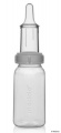
Cleft palate feeder


Historic
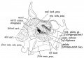
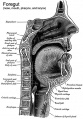
Fig. 6. Showing the structures formed in the Lateral Nasal Processes.
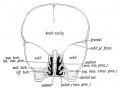
Fig. 7. Coronal section of the skull of a 7th month human foetus to show the cartilages of the Lateral and Mesial Nasal Processes and the bones formed round them.
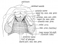
Fig. 8. Showing the ingrowth of the palatal plates of the two maxillary processes early in the 2nd month. (After Kollmann.) .
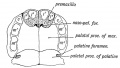
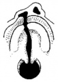
Fig. 9. Showing the Hard Palate at birth. The premaxillary part is formed from the Mesial Nasal Processes ; the remainder by the Palatal Plates of the Maxillary Processes.
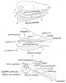
Fig. 10, a, b, c. Showing what become of the skeletons of the Mandibular Arch (Meckel's Cartilage) and Maxillary Process (Palato-quadrate Cartilage).
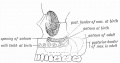
Fig. 11. Showing the manner in which the development of the Maxillary Antrum affects the size of the palate and position of the molar teeth.


Terms
| Palate Development (expand to see terms) |
|---|
|
| Other Terms Lists |
|---|
| Terms Lists: ART | Birth | Bone | Cardiovascular | Cell Division | Endocrine | Gastrointestinal | Genital | Genetic | Head | Hearing | Heart | Immune | Integumentary | Neonatal | Neural | Oocyte | Palate | Placenta | Radiation | Renal | Respiratory | Spermatozoa | Statistics | Tooth | Ultrasound | Vision | Historic | Drugs | Glossary |
External Links
External Links Notice - The dynamic nature of the internet may mean that some of these listed links may no longer function. If the link no longer works search the web with the link text or name. Links to any external commercial sites are provided for information purposes only and should never be considered an endorsement. UNSW Embryology is provided as an educational resource with no clinical information or commercial affiliation.
- Prof Virginia Diewert - Professor of Orthodontics, University of British Columbia, who recently visited the Lab and helped with content, organisation and development of the Palate Development section.
- Medline Plus - Cleft Lip and Palate
- Better Health Channel - Cleft palate and cleft lip
- March of Dimes Birth Defects Foundation - Cleft Palate
Glossary Links
- Glossary: A | B | C | D | E | F | G | H | I | J | K | L | M | N | O | P | Q | R | S | T | U | V | W | X | Y | Z | Numbers | Symbols | Term Link
Cite this page: Hill, M.A. (2024, June 17) Embryology Abnormal Development - Cleft Palate. Retrieved from https://embryology.med.unsw.edu.au/embryology/index.php/Abnormal_Development_-_Cleft_Palate
- © Dr Mark Hill 2024, UNSW Embryology ISBN: 978 0 7334 2609 4 - UNSW CRICOS Provider Code No. 00098G