Skeletal Muscle Histology: Difference between revisions
mNo edit summary |
|||
| (26 intermediate revisions by 2 users not shown) | |||
| Line 1: | Line 1: | ||
==Introduction== | ==Introduction== | ||
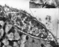
[[File:Skeletal muscle histology 016.jpg|thumb|Skeletal muscle sarcomeres]] | |||
[[File:Skeletal muscle structure cartoon.jpg|thumb|Skeletal muscle structure cartoon]] | |||
This page describes skeletal muscle histology which can identify cross-striational structure and /or properties related to contractile speed and fatigue. | |||
Development of skeletal muscle, cardiac muscle and smooth muscle can be found in other notes. | |||
Development of skeletal muscle from mesoderm occurs by mononucleated '''myoblasts''' fusing together to form mutinucleated '''myotubes''' that express contractile proteins forming sarcomeres within '''myofibers'''. | |||
The regular organisation of filament structures within the sarcomeres gives skeletal muscle, and cardiac muscle, the striated appearance. | |||
{{Muscle Histology links}} | |||
{{ | {{Musculoskeletal Links}} | ||
{{SkMhistolinks}} | |||
==Skeletal Muscle Stages== | ==Skeletal Muscle Stages== | ||
| Line 26: | Line 29: | ||
* primary myofibres - first-formed myofibres, act as a structural framework upon which myoblasts proliferate, fuse in linear sequence | * primary myofibres - first-formed myofibres, act as a structural framework upon which myoblasts proliferate, fuse in linear sequence | ||
* secondary myofibers - second later population of myofibres that form surrounding the primary fibres. | * secondary myofibers - second later population of myofibres that form surrounding the primary fibres. | ||
==Muscle Contraction== | |||
Skeletal, cardiac and smooth muscle all contract using the same mechanism: actin thin filaments being drawn together by myosin thick filaments. | |||
* In skeletal and cardiac muscle these thick and thin filaments '''are organised''' in series into '''sarcomeres'' along the length of the muscle cell. This regular organization gives the muscle cells a striated appearance. | |||
* In smooth muscle these thick and thin filaments '''are not organised''' into '''sarcomeres'' but are spread throughout the cell cytoplasm. | |||
This animation shows the molecular interactions that occur within the skeletal muscle sarcomere between actin and myosin during skeletal muscle contraction. This irregular organization gives the muscle a non-striated appearance. | |||
This animation shows the molecular interactions that occur within the skeletal muscle sarcomere between actin and myosin during skeletal muscle contraction. This irregular organization gives the muscle a non-striated appearance. | |||
{| | |||
| | |||
'''Legend''' | |||
* <font color=mediumvioletred>'''Moving blob and stick'''</font> - myosin complex. | |||
* <font color=red>'''Moving blob and stick'''</font> - myosin complex with ATPase activation. | |||
* <font color=green>'''Ball binding myosin and splitting'''</font> - ATP losing a phosphate to form ADP. | |||
* <font color=orange>'''Twisted string of beads'''</font> - actin helix. | |||
* <font color=blue>'''Blue string'''</font> - tropomyosin. | |||
* <font color=magenta>'''Beads stacked on large bead on blue string'''</font> - troponin. | |||
* <font color=gold>'''Small ball binding troponin'''</font> - Calcium ion (Ca<sup>2+</sup>). | |||
* <font color=grey>'''Grey pyramid'''</font> - Magnesiun ion (Mg<sup>2+</sup>). | |||
| [[File:Actin_myosin_crossbridge_3D_animation.gif]] | |||
|} | |||
==Muscle Fibre Types== | ==Muscle Fibre Types== | ||
| Line 87: | Line 114: | ||
* S3,4 and 5 supply the bladder, bowel, sex organs, anal and other pelvic muscles. | * S3,4 and 5 supply the bladder, bowel, sex organs, anal and other pelvic muscles. | ||
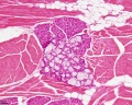
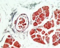
== | ==Histology Images== | ||
{{SkMhistolinks}} | |||
<gallery> | |||
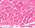
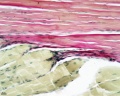
File:Skeletal_muscle_histology_014.jpg|Human HE x4 longitudinal and transverse | |||
File:Skeletal_muscle_histology_011.jpg|Human HE x40 transverse | |||
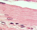
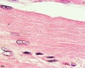
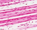
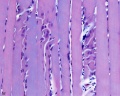
File:Skeletal_muscle_histology_012.jpg|Human HE x40 longitudinal | |||
File:Skeletal_muscle_histology_013.jpg|Human HE x40 longitudinal | |||
File:Skeletal_muscle_histology_015.jpg|Muscle Spindle HE x40 | |||
File:Skeletal_muscle_histology_001.jpg|Human HE x4 longitudinal and transverse | |||
File:Skeletal_muscle_histology_002.jpg|Human HE x40 | |||
File:Skeletal_muscle_histology_022.jpg|Human HE x40 | |||
File:Skeletal_muscle_histology_003.jpg|Human HE x40 | |||
File:Skeletal_muscle_histology_005.jpg|Human HE x100 | |||
File:Skeletal_muscle_histology_055.jpg|Human HE x100 | |||
File:Skeletal_muscle_histology_008.jpg|Fetal human muscle | |||
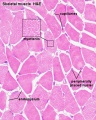
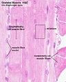
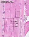
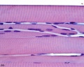
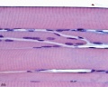
File:Skeletal_muscle_histology_004.jpg|Myotendinous junction HE x40 | |||
File:Skeletal_muscle_histology_006.jpg| | |||
File:Skeletal_muscle_histology_007.jpg| | |||
File:Skeletal_muscle_histology_077.jpg| | |||
File:Skeletal_muscle_histology_017.jpg|Tongue x10 | |||
File:Skeletal_muscle_histology_018.jpg|Tongue x100 | |||
File:Skeletal_muscle_histology_009.jpg|Muscle spindle HE x20 | |||
File:Skeletal_muscle_histology_010.jpg|Muscle spindle HE x40 | |||
</gallery> | |||
==Electron Micrographs== | |||
<gallery> | |||
File:Muscle_satellite_cell_EM01.jpg|Skeletal muscle and satellite cell | |||
File:Muscle_satellite_cell_EM02.jpg|Skeletal muscle and satellite cell | |||
</gallery> | |||
===Electron Microscopy Virtual Slides=== | |||
{| | |||
| valign=bottom|{{SlideSkeletalMuscleEM01}} | |||
| valign=bottom|{{SlideSkeletalMuscleEM02}} | |||
| valign=bottom|{{SlideSkeletalMuscleEM03}} | |||
|- | |||
| valign=bottom|{{SlideSkeletalMuscleEM04}} | |||
| valign=bottom|{{SlideSkeletalMuscleEM05}} | |||
|} | |||
== References == | == References == | ||
| Line 100: | Line 164: | ||
'''Search Pubmed:''' [http://www.ncbi.nlm.nih.gov/sites/entrez?db=pubmed&cmd=search&term=skeletal%20muscle%20development Skeletal Muscle Development] | '''Search Pubmed:''' [http://www.ncbi.nlm.nih.gov/sites/entrez?db=pubmed&cmd=search&term=skeletal%20muscle%20development Skeletal Muscle Development] | ||
== | ==Terms== | ||
[[File:Skeletal muscle histology 016.jpg|thumb|Skeletal muscle sarcomeres]] | |||
* '''A-band''' - (anisotropic band, Greek, ''anisos'' = unequal + ''tropos'' = turning) meaning having not equal properties in every direction,transverse bands in living skeletal muscle which rotate the plane of polarised light. | |||
* '''H-band''' - (Hell-band, German, ''hell'' = light + band; also Henle's band) light band within A-band of the myofibril. | |||
* '''I-band''' - (isotropic band, Greek, ''isos'' = equal + ''tropos'' = a turning, direction) Meaning having equal properties in every direction, of transverse bands in skeletal muscle which do not rotate the plane of polarised light. | |||
* '''sarcolemma''' - (Greek, ''sarkos'' = flesh + ''lemma'' = rind, husk) The plasma membrane plus basement membrane of a single muscle cell. | |||
* '''sarcomere''' - (Greek, + ''meros'' = a part) The structural subunit of striated muscle, repeating unit (segment) of myofibril from one Z-disc to the next. | |||
* '''sarcoplasm''' - (Greek, + ''plasma'' = a thing formed) the cytoplasm of a muscle cell. | |||
* '''sarcoplasmic reticulum''' - the endoplasmic reticulum of a muscle cell. | |||
* '''Z-disc''' - (Z-band, Z-line, Zwischenscheibe German, ''Zwischenscheibe'' = a between-disc) dark disc in centre of I-band; end disc of a sarcomere; Dobbie's line; Krause's line. | |||
==External Links== | |||
{{External Links}} | |||
* Blue Histology - [http://www.lab.anhb.uwa.edu.au/mb140/CorePages/Muscle/Muscle.htm Muscle] | |||
{{Template:Glossary}} | {{Template:Glossary}} | ||
Latest revision as of 00:53, 17 April 2014
Introduction


This page describes skeletal muscle histology which can identify cross-striational structure and /or properties related to contractile speed and fatigue.
Development of skeletal muscle, cardiac muscle and smooth muscle can be found in other notes.
Development of skeletal muscle from mesoderm occurs by mononucleated myoblasts fusing together to form mutinucleated myotubes that express contractile proteins forming sarcomeres within myofibers.
The regular organisation of filament structures within the sarcomeres gives skeletal muscle, and cardiac muscle, the striated appearance.
- Muscle Histology: Muscle Development | Human HE x4 longitudinal and transverse | Human HE x40 transverse | Human HE x40 longitudinal | Human HE x40 longitudinal | Human HE x4 longitudinal and transverse | Muscle Spindle HE x40 | Human HE x40 | Human HE x40 | Human HE x40 | Human HE x100 | Human HE x100 | Fetal human muscle | Myotendinous junction label | Myotendinous junction HE x40 | Whipf 1 | Whipf 2 | Whipf 3 | Tongue HE x10 transverse | Tongue x100 | Muscle spindle HE x20 | Muscle spindle HE x40
Skeletal Muscle Stages


Myoblast - individual progenitor cells
Myotube - multinucleated, but undifferentiated contractile apparatus (sarcomere)
Myofibre (myofiber, muscle cell) - multinucleated and differentiated sarcomeres
- primary myofibres - first-formed myofibres, act as a structural framework upon which myoblasts proliferate, fuse in linear sequence
- secondary myofibers - second later population of myofibres that form surrounding the primary fibres.
Muscle Contraction
Skeletal, cardiac and smooth muscle all contract using the same mechanism: actin thin filaments being drawn together by myosin thick filaments.
- In skeletal and cardiac muscle these thick and thin filaments are organised' in series into sarcomeres along the length of the muscle cell. This regular organization gives the muscle cells a striated appearance.
- In smooth muscle these thick and thin filaments are not organised' into sarcomeres but are spread throughout the cell cytoplasm.
This animation shows the molecular interactions that occur within the skeletal muscle sarcomere between actin and myosin during skeletal muscle contraction. This irregular organization gives the muscle a non-striated appearance. This animation shows the molecular interactions that occur within the skeletal muscle sarcomere between actin and myosin during skeletal muscle contraction. This irregular organization gives the muscle a non-striated appearance.
|
Legend
|

|
Muscle Fibre Types

Muscle fiber types
- type IIB, IIA, IIX, and I fibres - based only on the myosin ATPase activity.
- Type I fibres appear red, due to the presence of myoglobin.
- Type II fibres appear white, due to the absence of myoglobin and their glycolytic nature.
- A group of individual myofibres within a muscle will be innervated by a single motor neuron (motor unit).
- The electrical properties of the motor neuron will regulate the contractile properties of all associated myofibres.
| Fibre Type | Type I fibres | Type II a fibres | Type II x fibres | Type II b fibres |
|---|---|---|---|---|
| Contraction time | Slow | Moderately Fast | Fast | Very fast |
| Size of motor neuron | Small | Medium | Large | Very large |
| Resistance to fatigue | High | Fairly high | Intermediate | Low |
| Activity Used for | Aerobic | Long-term anaerobic | Short-term anaerobic | Short-term anaerobic |
| Maximum duration of use | Hours | <30 minutes | <5 minutes | <1 minute |
| Power produced | Low | Medium | High | Very high |
| Mitochondrial density | High | High | Medium | Low |
| Capillary density | High | Intermediate | Low | Low |
| Oxidative capacity | High | High | Intermediate | Low |
| Glycolytic capacity | Low | High | High | High |
| Major storage fuel | Triglycerides | Creatine phosphate, glycogen | Creatine phosphate, glycogen | Creatine phosphate, glycogen |
| Myosin heavy chain, human genes |
MYH7 | MYH2 | MYH1 | MYH4 |
Myotome
In both development and the adult, the group of skeletal muscles supplied by a specific segmental spinal nerve is referred to as a myotome. The muscle arises from a specific somite and the spinal nerve arises from a specific level of the spinal cord (identified by veretebral column).
In humans this corresponds to the following spinal nerves (from top to bottom) and muscular functions:
- C3,4 and 5 supply the diaphragm for breathing.
- C5 supply shoulder muscles and muscles to bend our elbow.
- C6 for bending the wrist back.
- C7 for straightening the elbow.
- C8 bends the fingers.
- T1 spreads the fingers.
- T1 –T12 supplies the chest wall and abdominal muscles.
- L2 bends the hip.
- L3 straightens the knee.
- L4 pulls the foot up.
- L5 wiggles the toes.
- S1 pulls the foot down.
- S3,4 and 5 supply the bladder, bowel, sex organs, anal and other pelvic muscles.
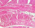
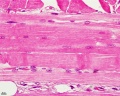
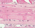
Histology Images
- Muscle Histology: Muscle Development | Human HE x4 longitudinal and transverse | Human HE x40 transverse | Human HE x40 longitudinal | Human HE x40 longitudinal | Human HE x4 longitudinal and transverse | Muscle Spindle HE x40 | Human HE x40 | Human HE x40 | Human HE x40 | Human HE x100 | Human HE x100 | Fetal human muscle | Myotendinous junction label | Myotendinous junction HE x40 | Whipf 1 | Whipf 2 | Whipf 3 | Tongue HE x10 transverse | Tongue x100 | Muscle spindle HE x20 | Muscle spindle HE x40
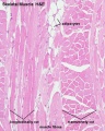
Human HE x4 longitudinal and transverse
Human HE x40 transverse
Human HE x40 longitudinal
Human HE x40 longitudinal
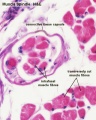
Muscle Spindle HE x40
Human HE x4 longitudinal and transverse
Human HE x40
Human HE x40
Human HE x40
Human HE x100
Human HE x100
Fetal human muscle
Myotendinous junction HE x40
Tongue x10

Tongue x100
Muscle spindle HE x20
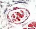
Muscle spindle HE x40


Electron Micrographs
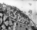
Skeletal muscle and satellite cell
Skeletal muscle and satellite cell


Electron Microscopy Virtual Slides
|
|
| |||||||||
|
|
References
- ↑ 1.0 1.1 <pubmed>18945372</pubmed>| PMC2596796 | BMC Syst Biol.
Search PubMed
June 2010 " Skeletal Muscle Development" All (19316) Review (2515) Free Full Text (5587) Manage Filters Search Pubmed: Skeletal Muscle Development
Terms
- A-band - (anisotropic band, Greek, anisos = unequal + tropos = turning) meaning having not equal properties in every direction,transverse bands in living skeletal muscle which rotate the plane of polarised light.
- H-band - (Hell-band, German, hell = light + band; also Henle's band) light band within A-band of the myofibril.
- I-band - (isotropic band, Greek, isos = equal + tropos = a turning, direction) Meaning having equal properties in every direction, of transverse bands in skeletal muscle which do not rotate the plane of polarised light.
- sarcolemma - (Greek, sarkos = flesh + lemma = rind, husk) The plasma membrane plus basement membrane of a single muscle cell.
- sarcomere - (Greek, + meros = a part) The structural subunit of striated muscle, repeating unit (segment) of myofibril from one Z-disc to the next.
- sarcoplasm - (Greek, + plasma = a thing formed) the cytoplasm of a muscle cell.
- sarcoplasmic reticulum - the endoplasmic reticulum of a muscle cell.
- Z-disc - (Z-band, Z-line, Zwischenscheibe German, Zwischenscheibe = a between-disc) dark disc in centre of I-band; end disc of a sarcomere; Dobbie's line; Krause's line.
External Links
External Links Notice - The dynamic nature of the internet may mean that some of these listed links may no longer function. If the link no longer works search the web with the link text or name. Links to any external commercial sites are provided for information purposes only and should never be considered an endorsement. UNSW Embryology is provided as an educational resource with no clinical information or commercial affiliation.
- Blue Histology - Muscle
Glossary Links
- Glossary: A | B | C | D | E | F | G | H | I | J | K | L | M | N | O | P | Q | R | S | T | U | V | W | X | Y | Z | Numbers | Symbols | Term Link
Cite this page: Hill, M.A. (2024, June 20) Embryology Skeletal Muscle Histology. Retrieved from https://embryology.med.unsw.edu.au/embryology/index.php/Skeletal_Muscle_Histology
- © Dr Mark Hill 2024, UNSW Embryology ISBN: 978 0 7334 2609 4 - UNSW CRICOS Provider Code No. 00098G