Embryology Education: Difference between revisions
mNo edit summary |
mNo edit summary |
||
| Line 123: | Line 123: | ||
==Educational Presentations== | ==Educational Presentations== | ||
# [[Anatomy and Embryology Goettingen - 2013_Seminar|2013 Goettingen Anatomy and Embryology Seminar]] | # [[Anatomy and Embryology Goettingen - 2013_Seminar|2013 Goettingen Anatomy and Embryology Seminar]] | ||
# [[Museum_of_Natural_History_Berlin - 2013_Seminar|2013 Museum of Natural History Embryology Seminar]] | # [[Museum_of_Natural_History_Berlin - 2013_Seminar|2013 Museum of Natural History Embryology Seminar]] | ||
# [[ANZACA_Meeting_2012 - Embryology|2012 Australian and New Zealand Association of Clinical Anatomists (ANZACA) Meeting]] | # [[ANZACA_Meeting_2012 - Embryology|2012 Australian and New Zealand Association of Clinical Anatomists (ANZACA) Meeting]] | ||
Revision as of 23:22, 26 November 2013
Introduction


The very size and dynamic nature of embryonic development has always made it a difficult topic to teach. The structures are always small and the changing nature of development means that a feature you identify at any one time changes and becomes something else at a later stage. Students take some time to come to grips with all the new terminology and the sequence of events. Interestingly, the discipline of embryology was quick utilise each new technology as as they came along to help with understanding development. The internet today is the latest technology which can allow a wide distribution to students to be used at any time and quickly linked to other related topics. This page provides a broad introduction to some of these educational concepts that are covered in detail elsewhere, identified by internet links. I have also included on this page links to meeting presentations that cover specific topics drawing upon the many available resources on this site.
Historically models made from many different media (papier-mâché, ceramic, wax or plastic) not the "animal models" described elsewhere on this site, were very useful in that they make tangible the microscopic structures to the students. Models have been in use for some time in the medical teaching of human development with the original plaster models now replaced by commercial modern plastic versions. Many of the early teaching models were not about development, but about the process of childbirth for doctor and midwife education.
An alternative to the use of teaching models, was the use of plates and drawings found in historic textbooks and papers from the late 19th century onwards. These often beautifully drawn images gave a semi-realistic view of embryo anatomy, often with sections removed to show anatomical relationships or systems drawn in isolation. These early papers often included drawings of histological sections from specific embryos, providing a prelude to later photomicrographs.
At the beginning of the 20th century photography also came into play and can be seen associated with embryo collections, such as the Carnegie and Kyoto collection. These photographic embryo images even today are unique tools in understanding human embryonic development. Animal embryos were far more easy to both obtain and to characterise. The realisation that most developmental processes are conserved across species, just differing in timing, led to growth in "animal models". In particular establishing a staging sequence allowed more consistent comparison of research findings in the literature.
Today there are many teaching and research movies developed to show specific developmental processes. This use of dynamic media most closely links to the dynamics seen in development.
| Education Links: Educational Presentations | Embryology Education | Embryology iBooks | Embryology Models | Textbooks | Embryology History | Category:Education |
Embryology in Medicine

There have been extensive changes in Anatomy core subject teaching in recent years, with a general trend to a decrease in overall contact hours in all disciplines and also increasing cross-discipline "integration" that makes it more difficult to easily identify actual contact hours.
Embryology dropped 50% by the 1970's and has remained stable at 20-24 contact hours, which suggests that this is the absolute minimum required to adequately cover this discipline. Interestingly, while histology and anatomy remain relatively stable in actual content, embryology now has many new topics to cover:
- Assisted Reproductive Technology (In vitro fertilization)
- Stem Cells
- Molecular Development (genetics, epigenetics, inheritance)
- Prenatal Diagnosis (genetic, ultrasound, MRI, fetal cells in maternal)
- Fetal Surgery (prenatal medicine)
USA (2009) - total of 130 allopathic and 25 osteopathic medical schools in USA.
Australia (2012) - UNSW Medicine contact hours - (lecture and practicals, including foundations and phase 2) is 24 hours.
Textbooks
There are many different textbooks that have used in preparing this Embryology resource, that can be found listed on the Textbooks page. I do not intend to endorse any specific textbook other than listing and linking too, those I have seen and/or used. Note that UNSW students also have full access to electronic versions of some textbooks subscribed to through library resources.
Embryology Models

Models made from many different media (papier-mâché, ceramic, wax or plastic) are very useful in that they make tangible the microscopic structures to the students. Models have been in use for some time in the medical teaching of human development with the original plaster models now replaced by commercial modern plastic versions. Many of the early teaching models were not about development, but about the process of childbirth for doctor and midwife education.
Note "animal models" is a term used to describe animals that "model" either human development or developmental processes.
A historic alternative to the use of teaching models, were series of plates and drawings found in textbooks and papers from the late 19th century onwards. At the beginning of the 20th century photography also came into play, seen mainly associated with embryos within the Carnegie collection.
- LInks: Embryology Models
Movies
![]()
![]()
Development is a dynamic process with structures changing (shape, name and relationships) over time and the best way to show this is with dynamic images, such as movies. Movies are generally uploaded in two main formats, MP4 and Quicktime, with some simple animations appearing as GIF format.
Here is an example of a movie showing atrial septation available in all 3 formats: MP4 Movie page | MP4 version | Quicktime version | Animated GIF version
There are excellent cartoons illustrating developmental processes correlating with concepts students will be learning from textbooks. There are also many movies from research studies in animal models and early human development. In addition, there are sets of prenatal ultrasound and MRI studies as well as postnatal neurological development.
Student Teaching Projects
Science students have been preparing online group projects as part of their ANAT2341 Embryology course since 2009. Each year a main topic is selected in consultation with the course coordinator. Each student group (4-5 students) then selects a specific topic within the main theme that they then research and work collaboratively on that topic throughout the semester. The project is then assessed by their peers before being finally edited for submission as their group assessment component for the course.
Medical students have also worked on individual long term projects as part of an Independent Learning Project (ILP) that can be carried out during their undergraduate studies.
Mobile Access

The current website will work for all online access through any browser enabled: desktop, laptop, netbook, iPad, tablet, iPhone and mobile (cell) phone. There are additional options (free applications) for viewing on mobile devices and for viewing content off-line (PDF).
We are also currently attempting to implement the Wiki mobile browsing extension that will not require additional software. This will allow detection of the operating system accessing a page and displaying appropriately.
- Links: Mobile Access | Cloud Embryology
K12 Students
Some pages have been designed specifically for K12 student and include both simplified terminology and suitable images. In addition there are currently two tutorial pages on Brain Growth and Comparative Embryology.
The Comparative Embryology tutorial page includes easy to print worksheets for students.
Please contact the editor to request specific topics or pages for K12 students.
- 2013 Brain Awareness Week - High school student presentation.
- 2012 Brain Awareness Week - Museum of Human Disease High school student presentation.
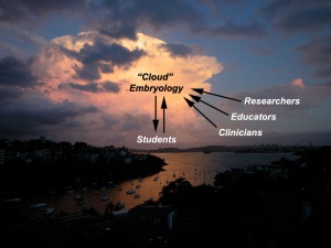
Cloud Embryology
- "Cloud embryology is a model for enabling ubiquitous, convenient, on-demand network access to a shared pool of configurable embryology resources"
This embryology resource has now been online since 1997 and transferred to the new Wiki format in 2009. The 1997 site was purely a website with no interaction available to the reader other than playing and controlling animations. The 2009 site initially was a transfer and reformatting of the original content pages. This was enhanced by addition of quizzes and followed by student online collaborative project work. The next step is now to access of educators from Universities and High Schools to prepare their own online presentations from the vast resources (9,000+ uploaded files) already available on this Embryology website.
- LInks: Cloud Embryology
Massive Open Online Course
We are currently exploring the possibility of offering a MOOC version of Introduction to Embryology. This will provide a background introduction to concepts in human development.
- Links: The Conversation
Educational Presentations
- 2013 Goettingen Anatomy and Embryology Seminar
- 2013 Museum of Natural History Embryology Seminar
- 2012 Australian and New Zealand Association of Clinical Anatomists (ANZACA) Meeting
- 2012 Medicine Learning and Teaching Forum - Online Projects
- 2012 Learning & Teaching Seminar
Meeting Presentations
- 2013 Australian Sonographers Association (ASA) Meeting
- 2013 Australian Chapter, American Academy of Craniofacial Pain (AACP) Meeting
- 2012 Fetal ECHO Meeting
- 2011 Maternal-Fetal Medicine Trainees
Online Video Tutorials
2013 I have developed a number of online video tutorials, beginning with Histology material as this is the latest course material to be added to the website. These tutorial videos are initially designed to help students with the "nuts n bolts" of everyday tools in their undergraduate courses. The tutorials are not designed to replace content or face-to-face teaching, but to provide additional resources for student self-directed learning and helping with those "How do I...." questions they may have with their work. I will also be preparing a similar "How do I...make online tutorials?" for other teachers to develop their own materials. The first link below is to the page containing the external links (listed next).
- Histology Tutorial 1 UNSWTV Introduction to online support | YouTube
- Histology Tutorial 2 UNSWTV Using the Virtual Microscope
- Links: UNSWTV
Awards
2012 UNSW Vice-Chancellor’s Awards for Initiatives that Enhance Learning
2011 ALTC Citation for Outstanding Contributions to Student Learning
References
- ↑ <pubmed>19890982</pubmed>
External Links
External Links Notice - The dynamic nature of the internet may mean that some of these listed links may no longer function. If the link no longer works search the web with the link text or name. Links to any external commercial sites are provided for information purposes only and should never be considered an endorsement. UNSW Embryology is provided as an educational resource with no clinical information or commercial affiliation.
Glossary Links
- Glossary: A | B | C | D | E | F | G | H | I | J | K | L | M | N | O | P | Q | R | S | T | U | V | W | X | Y | Z | Numbers | Symbols | Term Link
Cite this page: Hill, M.A. (2024, June 10) Embryology Embryology Education. Retrieved from https://embryology.med.unsw.edu.au/embryology/index.php/Embryology_Education
- © Dr Mark Hill 2024, UNSW Embryology ISBN: 978 0 7334 2609 4 - UNSW CRICOS Provider Code No. 00098G