U
| Embryology - 28 Apr 2024 |
|---|
| Google Translate - select your language from the list shown below (this will open a new external page) |
|
العربية | català | 中文 | 中國傳統的 | français | Deutsche | עִברִית | हिंदी | bahasa Indonesia | italiano | 日本語 | 한국어 | မြန်မာ | Pilipino | Polskie | português | ਪੰਜਾਬੀ ਦੇ | Română | русский | Español | Swahili | Svensk | ไทย | Türkçe | اردو | ייִדיש | Tiếng Việt These external translations are automated and may not be accurate. (More? About Translations) |
Glossary Links
- Glossary: A | B | C | D | E | F | G | H | I | J | K | L | M | N | O | P | Q | R | S | T | U | V | W | X | Y | Z | Numbers | Symbols | Term Link
U
UAC
- An acronym for umbilical arterial catheter.
UCB
- An acronym for umbilical cord blood
UVC
- An acronym for umbilical venous catheter.
uhrf1
- An acronym for ubiquitin-like protein containing PHD and ring finger domains-1 (Np95 in mouse, ICBP90 in human) a cell cycle regulator required for liver outgrowth in embryonic and adult zebrafish. Also a transcriptional activator of top2a expression.
- (More? Liver Development | PMID17242348)
ulipristal
- A new analog of mifepristone that acts as a selective progesterone receptor modulator. This drug has been identified as a second generation emergency contraceptive.
- (More? Mifepristone)
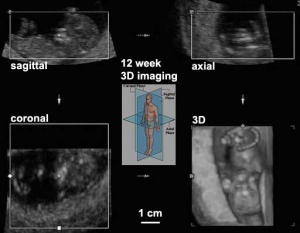
ultrasound
- A non-invasive technique for visualizing and prenatal diagnosis of several features of development including: follicles in the ovaries, the gestational sac, fetus in the uterus, fetal parameters, and the placenta. The technique uses high-frequency sound waves that are reflected off internal structures. These reflections can then be analysed and displayed by computer. Modern ultrasound machines can also carry out 3 dimensional reconstructions and measure "flow" (blood) using doppler measurements.
- (More? Ultrasound)
umami
- An historical Japanese word describing the taste in seaweed, used to describe the taste sensation of "savoury". Stimulated by the amino acid glutamate and monosodium glutamate.
- (More? Sensory - Taste Development)
Umbilical cord acid-base analysis
- A clinical perinatal test that can be used to assessing intrapartum hypoxia, measuring one or several indices: arterial umbilical cord blood pH, lactate, and base deficit. Hypoxia is indicated by a low pH, high base deficit and high lactate.
- (More? Placenta Development | Prenatal Diagnosis)
umbilical cord blood
- (UCB) The blood from cord and placenta which can be collected at birth as a source of cord stem cells.
umbilical cord
- (placental cord) The placental cord is the structure connecting the embryo/fetus to the placenta. It is initially extra-embryonic mesoderm forming the connecting stalk within which the placental blood vessels (arteries and veins) form. In human placental cords the placental blood vessels are initially paired, later in development only a single placental vein remains with a pair of placental arteries. This structure also contains the allantois, an extension from the hindgut cloaca then urogenital sinus. Blood collected from the placental cord following delivery is a source of cord blood stem cells.
umbilical arterial catheter
- (UAC) A catheter sometimes used if newborn infant has significant respiratory disease or requiring repeated early blood sampling. Catheter from umbilical artery connects to internal iliac artery then the aorta.
- (More? Placenta Development | Lecture - Placenta Development | NZ National Women's Health - Umbilical Artery Catheters)
umbilical arteries
umbilical vein
umbilical venous catheter
- (UVC) A catheter sometimes used if newborn is sick and requires central access. Catheter from umbilical vein connects to ductus venosus then to left branch of the portal vein.
umbilicus
- Term used to describe the navel region, in the embryo anatomically associated with the placental cord, midgut herniation and the allantois.
unexplained cause of infertility
- A type of infertility for which no cause has been determined despite a comprehensive evaluation.
unicornuate uterus
- An abnormality of uterine development where the paramesonephric ducts (Mullerian ducts) fail to fuse. A single paramesonephric duct can fuse with the vaginal plate and will opens into the vagina, while the other duct forms a diverticulum. There are a range of additional uterine abnormalities based upon the degree of initial duct fusion and regression. Uterus didelphys (double uterus) is a rare condition where the entire tract is separated.
uniparental disomy
- Genetic term referring to cells containing both copies of a homologous pair of chromosomes from one parent and none from the other parent. See also disomy.
universal precautions
- Safety term, used when dealing with biological materials, in particular human specimens. These are a set of precautions designed to prevent transmission of human immunodeficiency virus (HIV), hepatitis B virus (HBV), and other bloodborne pathogens when providing first aid or health care and when carrying out basic research on these tissues. Involve the use of protective barriers (PPE, personal protective equipment) such as gloves, gowns, aprons, masks, or protective eyewear, which can reduce the risk of exposure of the health care worker's skin or mucous membranes to potentially infective materials. Under universal precautions, it is recommended that all health care workers take precautions to prevent injuries caused by needles, scalpels, and other sharp instruments or devices.
unstimulated cycle
- An artifical reproductive technology (ART) cycle in which the woman does not receive drugs to stimulate her ovaries to produce more follicles. Instead, follicles develop naturally.
upper jaw
- The head and face region formed in the embryo by the paired maxillary processes of the pharyngeal arch 1 and the frontonasal prominence. The upper jaw is where developmental clefting typically occurs. The lower jaw is formed by the larger mandibular processes of the first pharyngeal arch.
- (More? Head Development)
ureter
- The ureters are the paired hollow tubes that link and carry urine from each kidney to the urinary bladder. The ureters develop from the ureteric buds and the adult tubes have a muscular wall lined with transitional epithelium.
ureteric bud
- The embryonic structure which develops into the nephron of the renal (kidney) system. An epithelial outgrowth from the caudal mesonephric duct (Wolffian duct) extending into the intermediate mesoderm metanephric mesenchyme, that reciprocally regulates mesenchymal differentiation in early kidney development. The bud begins as a local thickening of the mesonephric duct that elongates and branches into the intermediate mesoderm.The tyrosine kinase receptor Ret is locally expressed and thought to have a role in ureteric bud initiation.
- (More? Renal System Development | Lecture - Renal Development | PMID 12094231)
urethra
- The single muscular tube that links and carries urine from the bladder to the exterior. The opening in the urinary bladder forms part of the trigone. In humans, the urethral length differs between the sexes (male longer, female shorter).
urethral plate
urethro-ejaculatory duct reflux
(UER) Male clinical condition uncommonly reported condition in children, results in reflux of urine into the ejaculatory ducts and may result in recurrent orchitis and sterility. Diagnosed by using a micturating cystourethrogram (MCUG) to show the reflux of contrast into any of the ejaculatory ducts.
- (More? PMID 19830659)
urinary
- Term used to describe all components of the kidney system including the bladder, ureters and urethra.
urinary bladder
- (bladder) The muscular sac for the storage of urine. In early development, the hindgut cloaca is divided by down growth of urorectal septum into a posterior rectum and an anterior urogenital sinus. The superior end of the urogenital sinus is continuous with the allantois and initially the paired mesonephric ducts also fuse into the wall, later replaced by the ureteric buds becoming incorporated. The bladder is surrounded by smooth muscle forming the detrusor muscle required for bladder emptying, urination.
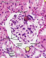
urinary pole
- Renal term for the side of the nephron Bowman's capsule where the proximal convoluted tubule starts.. The opposite "pole" is the vasular pole.
urinary tract dilation
- (UT dilation) Renal abnormality detected by ultrasound in 1–2% of fetuses and can be due to a number of possible uropathies (Congenital Abnormalities of the Kidney and Urinary Tract or CAKUT). This clinical term has been recently suggested (PMID 25435247) as replacing the following terms: hydronephrosis, pyelectasis, pelviectasis, uronephrosis, UT fullness or prominence, and pelvic fullness.
- (More? Renal Abnormalities | Renal System Development | PMID 25435247)
urine
- Term used to describe the liquid waste produced by the kidney, stored in the bladder and excreted from the body through the urethra.
urogenital fold
- (urogenital ridges, urethral folds) The ventral portion of the original cloacal folds, which contribute to the formation of the urethral groove on the ventral aspect of the genital tubercle. In females, these folds remain separate. In males, these folds will later fuse, failure of complete fusion leads to the male genital abnormality hypospadia.
- (More? Genital System Development)
urorectal septum
- (URS) The structure which develops to separate or partition the cloaca into an anterior urinary part (common urogenital sinus) and a posterior rectal part.
urorectal septum malformation
- The abnormalities associated with the urorectal septum (URS) and urogenital organs due to developmental abnormality.
URSMS
- An acronym for urorectal septum malformation sequence, clinically describing abnormalities of the urorectal septum (URS) and urogenital organs.
ureteropelvic junction
- (UPJ) Anatomical junction of the ureter and the kidney. During development this is the most common site for obstruction causing hydronephrosis.
UT-PI
- Acronym for Uterine Artery Pulsatility Index an ultrasound technique for monitoring placental and fetal function.
uteric bud
- Renal (kidney) development term for paired lateral diverticulum epithelial tubes arising from each mesonephric duct near its cloacal connection. This branch from the mesonephric duct extends into the intermediate mesoderm (metanephric mesenchyme) inducing the surrounding this mesoderm (metanephric blastema) to differentiate. The uteric bud gives rise to the renal collecting ducts, calyces, pelvis and developing ureters.
- (More? Renal Development | Lecture)
uterine activity
- (UA) The contractile activity pattern of uterine muscular wall occuring mainly during during labor for birth (parturition). This contractility can also be electrically monitored externally.
- (More? Birth | Lecture - Birth)
uterine artery resistance index
- (RI) This is a measurement made by Doppler ultrasound and is one of several clinical indices (uterine artery blood flow volume, average velocity, vessel cross-sectional area, resistance index, and spiral artery resistance index) that can be used to determine placental function and pregnancy status.
- (More? Placenta Development | Ultrasound)
uterine body
- Anatomical term describing the region of the uterus above the uterine isthmus and below the opening of the uterine tubes.
uterine duplication
- (uterus didelphys, double uterus, uterus didelphis, bicorporeal uterus) Rare uterine developmental abnormality where the paramesonephric ducts (Mullerian ducts) completely fail to fuse generating two separate uterus parts each connected to the cervix and having an ovary each. Failure of fusion of lower paramesonephric ducts, with either double or single vagina. ESHRE/ESGE classification of uterine anomalies - U3.
- (More? Uterus Development)
uterine factor
- A disorder in the uterus that reduces fertility.
uterine isthmus
- Anatomical term describing the region between the uterine body (corpus) and the cervix.
uterine dehisence
- Clinical term for the disruption of the uterine muscular wall with an intact serosa that can occur during pregnancy and birth. See also uterine rupture
- (More? Birth)
uterine evacuation
- Clinical surgical term associated with abortion treatment, often with first trimester miscarriage (early fetal loss).
uterine fibroids
- A non-cancerous tumor that can develop within the wall of the uterus composed of muscle cells or other tissues. Their location can be either submucosal, intramural or subserosal.
- (More? Menstrual Cycle | Genital System Development | Medline Plus)
uterine gland
- (endometrial gland) The simple tubular glands formed by invagination of the uterine endometrium (a columnar epithelium of ciliated cells and secretory cells). The glands extend into the underlying thick vascular stromal layer. The glands line the uterus body and change in appearance and secretion during the menstrual cycle. The glands secretions function to provide the initial nutritional support of the conceptus and may have a role in maintaining adhesion.
uterine horn
- (fallopian tube, oviduct, salpinx) see uterine tube. Alternative term used to describe the uterine tube.
uterine leiomyomata
- (UL, uterine fibroids) Pathology term for non-malignant tumours that arise from the uterine smooth muscle wall. These are the most common neoplasm in reproductive-age women, a proportion ( 40%) show recurrent cytogenetic abnormalities (deletions, inversions and translocations) including the deletion 7q (q22q32).
- (More? Uterus Development)
uterine natural killer cells
- (uNK) main lymphocytes present in the uterus during early pregnancy and in the secretory phase of the menstrual cycle. These cells have low cytotoxicity, constitutively secrete cytokines, chemokines and angiogenic molecules. and differ from blood NK cells (CD56 high) in the killer cell immunoglobulin-like receptor repertoire and hormonal gene regulated expression. Thought to have a role in decidualization, association with spiral arteries and interaction with trophoblast cells.
- (More? Menstrual Cycle | Uterus Development | Medline Plus)
uterine peristalsis
- The rhythmic muscular contraction of the uterus, which occurs during the menstrual cycle and is maximal just before ovulation, in the non-pregnant uterus.
uterine artery pulsatility index
- (UT-PI) A clinical ultrasound technique used for monitoring placental and fetal function. Can be used in second trimester screening in combination with maternal factors and fetal biometry to predict stillbirths and in particular those associated with impaired placentation.
- (More? Ultrasound | Placenta Development | Stillbirth and Perinatal Death | PMID 27601282)
uterine endometrial thickness
- A clinical measurement used in Assisted Reproductive Technology related to successful embryo implantation following in vitro fertilization. At the time of the hCG (human chorionic gonadotropin) trigger, an endometrial thickness of more than 8 to 9 mm correlates with a successful pregnancy,
uterine rupture
- The disruption of the uterus wall muscle which can all include uterine serosa and extension to the bladder or broad ligament. Can occur during pregnancy and birth in women who have had a previous caesarean section or other uterine procedures. Induction can increase the risk (increases twofold to threefold) and can lead to maternal death (haemorrhage, blood loss) and fetal death (asphyxiation).
- (More? Uterus Development | Birth | PMID 3594331 | PMID 15598960)
uterine tube
- (uterine horn, oviduct, fallopian tube, salpinx) A pair of tubular structures that transport the oocyte (egg) from the ovary to the uterus body. They are located laterally on the upper uterus and consist medial to lateral of three main parts: isthmus (medial constricted third), ampulla (intermediate dilated portion) and infundibulum (containing the abdominal opening/ostium, surrounded by finger-like fimbriae). The tube has structurally several layers: a lining mucosa (mix of ciliated and secretory epithelium), a middle muscularis layer (inner circular muscle layer and an outer longitudinal layer) and outer serous layer (peritoneal).
ureterocele
- (ureteroceles) Developmental urinary bladder abnormality where the ureter enlarges or "balloons" at the site of opening into the urinary bladder.
uterodome
- (pinopod) Cellular feature seen on the apical uterine epithelium surface, the presence of these structures in other species is thought to be a marker for endometrial receptivity. These transient micro-protrusions inter-digitate with microvilli on the apical syncytiotrophoblast surface of the blastocyst during adplantation and implantation process.
- (More? Week 2 | Implantation | Blastocyst | Reproductive Cycles)
uterus
- The female internal genital (reproductive) tract forming a hollow muscular walled organ, embryonically derived from the paramesonephric ducts. The human uterus has two uterine tubes (fallopian tubes, oviducts) where the first week of development occurs and a single hollow body where implantation of the blastocyst normally occurs. Following puberty, the non-pregnant uterus (epithelium and underlying stroma) undergoes cyclic changes under the influence of hormones, the menstrual cycle. This cycle of uterine changes ceases during pregnancy. In other species females of non-primate vertebrates (eg rats, mice, horses, pig) have a reproductive cycle called the estrous cycle (oestrous, British spelling). In pregnancy, the uterus contributes the maternal component of the placenta.
uterus didelphys
- (double uterus, uterus didelphis) A rare uterine developmental abnormality where the paramesonephric ducts (Mullerian ducts) completely fail to fuse generating two separate uterus body parts. Both parts are connected to a single cervix and each part is connected its own uterine tube and ovary.
Glossary Comments
Use this page to access brief definitions of specific embryology terms. Additional information can be accessed from links listed at the end of each definition. Glossary from the UNSW Embryology program compiled and written by Dr Mark Hill. Reference material used in preparing this glossary list includes: texts listed on page 1 "Reading" of each notes section, Department of Anatomy Publications, WWW resources from NCBI, NIH, OMIM, NHMRC (Australia), AMA (USA), Office of Rare Diseases (USA), PubMed Medline Dictionaries, MSDS, Merck Manual home edn. and WHO ART terminology (2009).
These notes are for Educational Purposes Only Please email Dr Mark Hill if you wish to make a comment about this current project.
Glossary Links
- Glossary: A | B | C | D | E | F | G | H | I | J | K | L | M | N | O | P | Q | R | S | T | U | V | W | X | Y | Z | Numbers | Symbols | Term Link
Cite this page: Hill, M.A. (2024, April 28) Embryology U. Retrieved from https://embryology.med.unsw.edu.au/embryology/index.php/U
- © Dr Mark Hill 2024, UNSW Embryology ISBN: 978 0 7334 2609 4 - UNSW CRICOS Provider Code No. 00098G