Paper - The changes in the mesonephric tubules of human embryos ten to twelve weeks old
| Embryology - 28 Apr 2024 |
|---|
| Google Translate - select your language from the list shown below (this will open a new external page) |
|
العربية | català | 中文 | 中國傳統的 | français | Deutsche | עִברִית | हिंदी | bahasa Indonesia | italiano | 日本語 | 한국어 | မြန်မာ | Pilipino | Polskie | português | ਪੰਜਾਬੀ ਦੇ | Română | русский | Español | Swahili | Svensk | ไทย | Türkçe | اردو | ייִדיש | Tiếng Việt These external translations are automated and may not be accurate. (More? About Translations) |
Altschule MD. The changes in the mesonephric tubules of human embryos ten to twelve weeks old. (1930) Anat. Rec. 46(1): 81-91.
| Online Editor |
|---|
| This historic 1930 paper by Altschule describes the early human mesonephros developing in the embryonic period between 10-12 days.
|
| Historic Disclaimer - information about historic embryology pages |
|---|
| Pages where the terms "Historic" (textbooks, papers, people, recommendations) appear on this site, and sections within pages where this disclaimer appears, indicate that the content and scientific understanding are specific to the time of publication. This means that while some scientific descriptions are still accurate, the terminology and interpretation of the developmental mechanisms reflect the understanding at the time of original publication and those of the preceding periods, these terms, interpretations and recommendations may not reflect our current scientific understanding. (More? Embryology History | Historic Embryology Papers) |
The Changes in the Mesonephric Tubules of Human Embryos Ten to Twelve Weeks Old
Mark David Altschule
Department Of Anatomy, Harvard Medical School, Boston
Three figures
In 1912, Felix, in his treatise “The Development of the Urogenital Organs,” stated that the mesonephric tubules in man begin degenerating at 7 mm. and that by the time the 22—mm. stage is reached none is intact. This was generally accepted until 1916, when Bremer expressed the opinion that sound mesonephric tubules may persist as late as the 37 or 40—mm. stage. His conclusions were based on the observation that about a dozen apparently healthy glomeruli are present in embryos of that size. He was able, moreover, to trace intact tubules from some of these glomeruli to the wolffian duct. Other observers, including Lewis and Shikinami, have since called attention to the presence of numerous healthy tubules in the mesonephroi of embryos of about 23 mm., and both Von Winiwarter and Wilson have noted what looked like healthy mesonephric tissue as late as the 35- or 40—mm. stage, but the testimony of the two latter, based merely on histological evidence, must be considered inconclusive, as will be seen later.
In an attempt to throw additional light on the subject, it was decided to study carefully the number, disposition, and condition of the tubules in the mesonephroi of human embryos at various stages from about 30 to 40 mm.
The material used in this investigation consisted of sections of the following embryos:
| No. | Sex | Crown-rump Length | Sections | Stain |
|---|---|---|---|---|
| H.E.C. 2043 | ♀ | 31 mm | 10 μ transverse | Alum cochineal and orange G |
| H.E.C. 2050 | ♂ | 36 mm | 10 μ transverse | Alum eocliineal and orange G |
| H.E.C. 1917 | ♀ | 40 mm | 12 μ frontal | Alum cochineal and orange G |
| H.E.C. 838 | ♂ | 42 mm | 14 μ transverse | Borax earmine and Lyons blue |
Camera-lucida. drawings were made from these serial sections at a magnification of 150 diameters and were used in making graphic reconstructions of the mesonephroi studied.
Observations
Microscopic examination shows the Wolffian body to be rather sharply divided into two regions, an anterior, which later gives rise to the Vasa efferentia. and ductus epididymidis or to the epoophoron, and a posterior, in which the tubules ultimately either disappear or contribute to the paradidymis or paroophoron.
The changes that occur in the tubules of the anterior portions of the mesonephroi of embryos 31 to 42 mm. long have been adequately described by von Winiwarter and by Wilson and need not be gone into at any great length here. They consist of a gradual dedifferentiation, beginning anteriorly and proceeding caudally to about the middle of the Wolffian body. The picture is essentially the same in all of the embryos studied, except that the older embryos have proportionately more tubules in the last stages of the dedifferentiation.
Lewis has described the mature human mesonephric tubule as consisting of a. capsule, a thick secretory tubule, and a thinner collecting tubule. On microscopic examination, the glomerulus is seen to ‘be lined with a flat epithelium and to contain a capillary tuft full of blood corpuscles. Afferent and efierent arterioles can be seen. The secretory tubule is lined with cells, each of which contains a round or oval nucleus surrounded by a fairly large amount of granular cytoplasm. The nucleus may be situated either centrally or basally. Cell boundaries are indistinct, except, of course, where they impinge upon the lumen. Here, though well defined, they may be quite irregular. This, coupled with the fact that the cells differ considerably in height, tends to make the Width of the lumen vary appreciably from point to point.
The junction of the secretory and collecting tubules is fairly well marked, as the appearance of the former is quite different from that of the latter. The epithelium of the collecting tubule consists of small cuboid cells containing round centrally placed nuclei in cytoplasm that is denser, though less granular, than that of the cells of the secretory tubule. Cell boundaries are well defined and the lumen is quite regular. The entire mesonephric tubule, from capsule to Wolffian duct, is separated from the surrounding connective tissue by a fine continuous basement membrane.
The first stage in the process of dedifferentiation observed in the anterior halves of the wolffian bodies studied is a thickening of the epithelium of the capsule. A little later, the number of capillaries in the glomerular tuft becomes smaller. Simultaneously the cells in the secretory tubule begin to decrease in size and stain like the epithelium of the connecting tubule. The lumen of the tubule is seen to contain much debris. Reconstruction of the tubules undergoing these changes shows them to be diminishing in length, although the continuity from duct to capsule is still maintained (figs. 1 and 2). The presence of debris in the lumen must be due to the loss of cells from the tubule, since the marked decrease in the length of the tubule can be accounted for only to a slight extent by the shrinkage of the individual cells. A considerable number of them must drop out before the tubule can become as short as it does.
This process continues. The tubule becomes thinner and much shorter and loses some of its curves; the cells of the tubular epithelium become more uniform in size and staining reaction; the capsular epithelium becomes more definitely cuboid and columnar; and the tuft becomes increasingly dense as its cells thicken and its capillaries withdraw. The remains of the glomerulus are finally extruded, the capsule peeling itself off the tuft to become a spherical vesicle enclosed by a single layer of cells now indistinguishable from those of the rest of the tubule.
The gonadic tissue has in the meantime been encroaching on the wolffian body so that now a number of the dedifferentiated capsules are surrounded by cells derived from the testis.
At about this time a few of the changing tubules lose their continuity, becoming broken either at the junction of the tubule and the mesonephric duct or of the tubule and the capsule. This usually occurs in two or three of the most anteriorly situated tubules and less frequently in the other portions of what is to become the epididymis or epoophoron. These broken tubules will, of course, contribute to the paradidymis or paroophoron, while some or all of the dozen or so tubules that still maintain their continuity will give rise to the vasa efferentia of the epididymis.
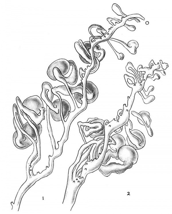
Fig. 1 Reconstruction of part of the right mesonephros of a human embryo of 36 mm.
Fig. 2 Reconstruction of part of the right mesonephros of a human embryo of 31 mm.
The extreme anterior tip of the wolffian duct may degenerate, thus cutting off a tubule or two. A tubule isolated in this way, or by degeneration of its own tissue at its junction with the wolffian duct, may swell up and become vesicular. This has been observed only at the anterior end of the organ and is interpreted as being an early stage in the development of a hydatid of Morgagni.
This account of the metamorphosis of the cephalic portion of the wolfiian body in human embryos is entirely in harmony with that of Von VViniwarter and differs only slightly from that of Wilson.
The process going on in the posterior halves of the mesonephroi of the embryos studied differs entirely from that just described. On microscopic examination the caudal portions of embryos of 31 to 42 mm. seem to consist largely of healthy tubules and glomeruli embedded in a stroma of young connective tissue. There are numerous capsules containing blood—laden capillary tufts, most of which may be seen to join normal afferent or efferent arterioles. Scattered about in the sections are segments of secretory and collecting tubules, each lined with its characteristic epithelium.
If reconstructions of this region are made, it is found that a. number of the apparently sound tubules are not continuous from wolffian duct to glomerulus, but are broken in one or more places. Occasionally there is no actual discontinuity, the tubule being reduced to a thin lumenless cord of cells.
It is not possible to distinguish between the intact and most of the broken tubules histologically. This and the fact that the fragments of the broken tubules often maintain their relative positions make it appear that there are more sound ones than there really are. Thus embryos of 40 and 42 mm. were at first thought to contain about a dozen intact tubules, but careful reconstruction showed that there was none.
Undoubtedly the discrepancies in the published statements as to the final time of degeneration of all tubules are due to the persistence of the normal appearance of glomeruli and tubules after actual continuity has been lost.
At 36 mm. it is possible to demonstrate ten continuous tubules. figure 1 is a reconstruction of the right mesonephros of a male embryo of that size and shows five of these.
It is seen that at this stage the wolffian body has a. wavy duct running from a dorsolateral position in the cephalic portion of the organ to a ventrolateral position more posteriorly. The thirty—one tubules spring from it at irregular intervals, generally from its dorsal or medial aspect. The most posteriorly situated tubules run forward from their points of origin and those of the anterior portion run caudally, thus tending to make the mesonephros more compact. The tubules are not arranged linearly, the various parts of one being overlapped by portions of several others when viewed from the side. The healthy tubules, which are located posteriorly, are seen to be of exactly the same shape a11d arrangement as those described by Lewis in embryos of 23 mm.
Only ten of the eighteen tubules in the posterior portion of this mesonephros are found to be intact, but a larger number can be demonstrated in younger embryos. The organ pictured in figure 2 is that of a female embryo of 31 mm. Among its twenty caudal tubules are fifteen sound ones, of which four are shown. Its duct is less wavy than that of the 36—mm. embryo, it has six more tubules, and the entire organ is shorter and hence more compact, but its general arrangement is the same. In the stages studied there is no difference between the sexes in regard to the wolffian body, although the gonad already shows considerable differentiation.
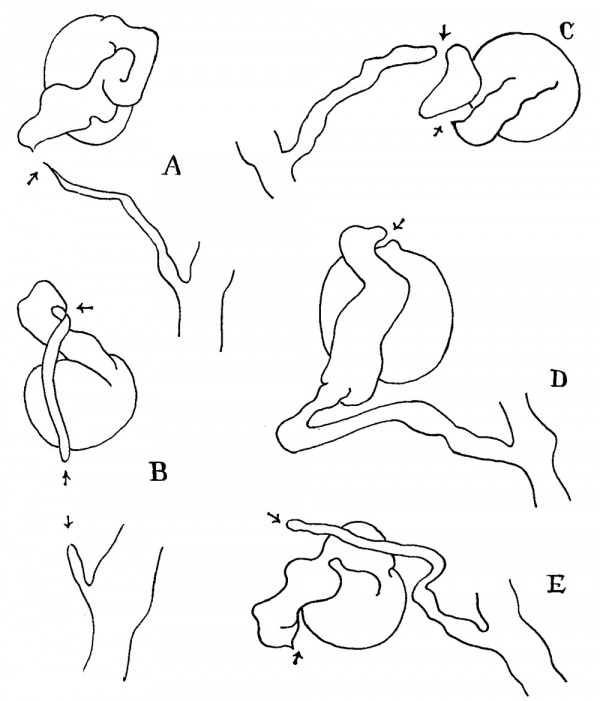
The point at which the tubules become discontinuous varies. Breaks have been noted at the junction of the glomerulus and secretory tubule, in the secretory tubule, at or immediately below the junction of the secretory and collecting tubules (the most frequent site), and in the collecting tubule itself. Some tubules are broken at more than o11e point. A number of these discontinuous tubules are shown in figure 3.
Fig. 3 Reconstructions of broken mesonephric tubules. A and E are broken immediately below the junction of secreting and collecting tubules. B is reduced to :2. lumenless cord at the junction of the secreting and collecting tubules. In addition, the collecting tubule itself is broken somewhat lower down. C is broken at the junction of the secreting and collecting tubules as well as in the secreting tubule itself. D is broken at the junction of the glomerulus and secreting tubule.
Generally the fragments maintain their relative positions, so that there is no doubt as to which tubule each is derived from, but occasionally the broken portions are widely separated because of either an extensive loss of substance or an actual movement of the parts, probably as a result of pressure exerted by adjacent glorneruli. In such cases it is always possible to determine with certainty the relationship of separated parts by the process of elimination.
There can occasionally be observed a somewhat distended capsule in which the glomerulus is disintegrating in situ, but the last stages of this process are not found in the embryos studied.
Discussion
It has been definitely shown that continuous and apparently healthy wolffian tubules persist up to the 36-min. stage in man. Numerous observers, including Bremer, Lewis, Shikinami, von Winiwrarter, and Wilsoii, have expressed the opinion, based on histological study, that the human mesonephros is an organ capable of functioning. Felix, on the other hand, believed that it is not, reasoning that since (as he believed) the mesonephric tubules have degenerated completely by the time the 22—mm. stage is reached and the metanephric glomeruli cannot function until about 30 mm., to assume activity on the part of the former would be to assume that the human embryo is left without an excretory organ between the 22- and 30—mm. stages. This line of reasoning is completely invalidated by the evidence presented in this study.
On the other hand, this work does not prove that human rnesonephrie tubules actually do function as urinary organs. It merely shows that, as far as can be determined morphologically, some of them are capable of doing so. The question is compliea.ted by the fact that there are, as first noted by Bremer, epithelial plates in the chorion of embryos as early as the 29—mm. stage or perhaps even earlier. Bremer has called attention to the fact that these plates and their situation immediately adjacent to capillaries are characteristic of organs in which osmotic interchanges occur. It is therefore quite evident that these structures can perform the function usually ascribed to the renal glomerulus. Indeed, it is fairly certain that they do so, as the exceedingly small size in the human embryo of the reservoir for mesonephrictand metanephric urine, the allantois, makes it impossible to believe that the embryonic kidneys actually function.
We thus have a situation in which the mesonephros, capable, as far as can be determined, of secreting urine, apparently remains inactive. The reason for this is not known, but is possibly connected in some way with the level of the blood pressure and the constitution of the blood plasma in the human embryo.
There is some evidence that under suitable circumstances the human mesonephros may actually secrete. Spitzer and Wallin have reported a case of apparent persistence of a pair of actively secreting mesonephroi in a young adult female. Unfortunately, the gross morphological study, which seemed to indicate quite definitely that the bodies in question were mesonephroi, could not be confirmed by histological study, as permission to remove the organs was not obtained. This piece of evidence must therefore also be regarded as inconclusive. However, the sum total of information thus far gathered does seem to favor the view that the human mesonephros actually can function as an excretory organ at one time or another.
The exact nature of this function is unknown. It is probably different from that of the adult amphibian mesonephros, in view of the morphological differences existing in the two forms. Thus the tubule in the human embryo consists of a capsule containing a capillary tuft, a slightly convoluted secretory tubule, and a. collecting tubule, while that of the mature amphibian is, according to Huber, divided into capsule, proximal convoluted tubule, straight tubule, distal convoluted tubule, and collecting tubule. The work of White, White and Schmitt, ‘Wearn and Richards, and others has shown that each of these portions has a function of its own.
The human mesonephros, since it differs morphologically from the adult amphibian wolffian body, probably also differs functionally. It is possible that some light can be thrown on the nature of the physiologic activities of the human wolflian body by a careful study of the mesonephroi of such mammals as the opossum, in which the organs persist and function for a considerable period after birth.
Summary and Conclusions
- The mesonephros of the human embryo ten to twelve weeks old and the changes occurring in it are described.
- It is shown that the mesonephroi of embryos of 31 mm. crown—rump length have fifteen sound tubules, while those of 36 mm. have only ten. No intact tubules were found at 40 or 42 mm. These conclusions are based on evidence derived from reconstructions as well as histological examination of the embryos studied. It is also shown that conclusions as to the persistence of mesonephric tubules which are based merely on the study of isolated histological sections are invalid.
- The bearing of this evidence 011 the question as to whether or 1not the human wolffian body functions is discussed.
This problem was suggested to me by Prof. John Lewis Bremer. I wish to express my gratitude for his aid and encouragement, without which this study could not have been completed.
Bibliography
BREMER, J. L. 1916 The interrelations of the mesonephros, kidney, and placenta in diiferent classes of animals. Am. Jour. Ana.t.., vol. 19, p. 179.
Felix W. The development of the urinogenital organs. In Keibel F. and Mall FP. Manual of Human Embryology II. (1912) J. B. Lippincott Company, Philadelphia. pp 752-979.
FELIX, W. 1912 The development of the urogenital organs. Keibel and Mall’s Human Embryology, vol. 2, p. 752.
HUBER, G. C. 1928 Renal tubules. Cowdry’s Special Cytology, vol. 1, p. 663.
LEWIS, F. T. 1920 The course of the wolflian tubules in mammalian embryos Am. Jour. Anat., vol. 26, p. 423.
SHIKINAMI, J. 1926 Detailed form of the wolffiaii body in human embryos of the first eight weeks. Contrib. to Embryology, Carnegie Institution, vol. 18, p. 49.
SPITZER, W. M., AND WALLIN, I. E. 1928 Supernumerary ectopic ureters. Annals of Surgery, vol. 88, p. 1053.
WEARN, J. T., AND RICHARDS, A. N. 1924 Observations on the composition of glomerular urine with particular reference to the problem of reabsorp« tion in the renal tubules. Amer. Jour. Physiol., vol. 71, p. 209.
1925 The concentration of chlorides in the glomerular urine of frogs. Jour. Biol. Chem., vol. 66, p. 247.
WHITE, H. W. 1929 The question of water reabsorption by the renal tubule and its bearing on the problem of tubular secretion. Amer. Jour. Physiol., vol. 88, p. 267.
WHlTE, H. W., AND ScHM1'r'r, F. 0. 1926 Kidney function in Necturus maeulosus. Amer. Jour. Physiol., vol. 76, p. 220.
1926 The site of reabsorption in the kidney tubule of Necturus. Amer. Jour. Physiol., vol. 76, p. 483.
WILSON, K. M. 1926 Origin and development of the rete ovarii and rete testis in the human embryo. Contrib. to Embryology, Carnegie Institution, vol. 17, p. 69.
voN WINIWARTER., H. 1910 La constitution et involution du corps de Wolff et le développemcnt du canal de Miiller dons l’espece hum:Line. Arch. Biol., T. 25, p. 169.
Cite this page: Hill, M.A. (2024, April 28) Embryology Paper - The changes in the mesonephric tubules of human embryos ten to twelve weeks old. Retrieved from https://embryology.med.unsw.edu.au/embryology/index.php/Paper_-_The_changes_in_the_mesonephric_tubules_of_human_embryos_ten_to_twelve_weeks_old
- © Dr Mark Hill 2024, UNSW Embryology ISBN: 978 0 7334 2609 4 - UNSW CRICOS Provider Code No. 00098G


