Paper - Stapes, fissula ante fenestram and associated structures in man 5
| Embryology - 28 Apr 2024 |
|---|
| Google Translate - select your language from the list shown below (this will open a new external page) |
|
العربية | català | 中文 | 中國傳統的 | français | Deutsche | עִברִית | हिंदी | bahasa Indonesia | italiano | 日本語 | 한국어 | မြန်မာ | Pilipino | Polskie | português | ਪੰਜਾਬੀ ਦੇ | Română | русский | Español | Swahili | Svensk | ไทย | Türkçe | اردو | ייִדיש | Tiếng Việt These external translations are automated and may not be accurate. (More? About Translations) |
Anson BJ. and Cauldwell EW. Stapes, fissula ante fenestram and associated structures in man: V . From the fetus of 160 mm to term. (1948) 48(3): 263-300.
| Historic Disclaimer - information about historic embryology pages |
|---|
| Pages where the terms "Historic" (textbooks, papers, people, recommendations) appear on this site, and sections within pages where this disclaimer appears, indicate that the content and scientific understanding are specific to the time of publication. This means that while some scientific descriptions are still accurate, the terminology and interpretation of the developmental mechanisms reflect the understanding at the time of original publication and those of the preceding periods, these terms, interpretations and recommendations may not reflect our current scientific understanding. (More? Embryology History | Historic Embryology Papers) |
Stapes, Fissula ante fenestram and Associated Structures in man: V . From the Fetus of 160 mm to Term
Barry J. Anson, Ph.D. (Med. Sc.) and Earl W. Cauldwell, M.D. Chicago
Contribution no. 506 from the Anatomical Laboratory of Northwestern University Medical School.
Dr. T. H. Bast, of the Department of Anatomy of the University of Wisconsin, gave us permission to study his excellent series of temporal bones.
This investigation was conducted under the auspices of the Central Bureau of Research of the American Otological Society. During the course of this study, Dr. Cauldwell served on a fellowship endowed by the late Dr. George J. Dennis and, subsequently, by Mrs. Dennis.
Introduction
In continuation of an investigation into the developmental and adult anatomy of the auditory ossicles, of the otic capsule and of the extracapsular portion of the temporal bone, attention has been focused on the stapes, the vestibular (oval) window and the anteriorly situated fissular tract, which regularly opens into the fenestra. Two introductory articles in the series dealt with the general features of stapedial and fissular morphogenesis[1]; in more recent articles, through a study of more than 200 otologic series of graded age levels, details were supplied to account for the steps in development from the stage of primordial appearance in cartilage to the assumption of adult form and dimensions.[2]
From a selected set of 75 specimens thirty-two crucial stages were especially studied as the basis for the present contribution; of these twenty-one are represented in the illustrations.
Material and Methods
All of the series described and the figures presented in this paper are contained in the otologic collection at the University of Wisconsin. The order of presentation of the observations follows the graded increase in recorded fetal length. However, since crown-rump length does not provide an accurate indication of ossicular and capsular development, the statements made in the concluding division of the paper are arranged in the order of the successive steps in morphogenetic advance.
Reconstructions of the stapes, of surrounding capsular structures and of adjacent fissular anatomyiwere prepared by the wax plate method. Comparable, or additional, anatomic features in intermediate stages are demonstrated by drawings and photomicrographs of selected sections. All reconstructions were prepared at a magnification of 125 diameters by the wax plate method from tracings made with an Edinger projection apparatus. Each reconstruction originally included not only the stapes but also the capsular wall of the vestibular window and the adjacent fissula ante fenest-ram. For several of the figures, these reconstructions were dismantled or cut in order that either the form or the topographic relationships could be more advantageously recorded.
The semidiagrammatic drawings were prepared from Edinger tracings, at a magnification of 50 diameters, from sections similar to those shown in the two introductory plates of photomicrographs (original magnification, 65 diameters).
The following stages are illustrated by photomicrographs:
| Length (mm) | Wisconsin Series No. | Figure No. |
|---|---|---|
| 167 | 105 | 1a (see fig. 3a to c, from same series) |
| 175 | 104 | 1b (See fig. 4a. and b) |
| 160 | 41 | 1c (See figs. 5a to 4:, 9a and b, 10a and b) |
| 170 | 131 | 1d (See fig. 5d to f) |
| 210 | 51 | 1e (See figs. 2b, 7a, 8b, 13a and b) |
| 290 | 59 | If (See fig. 8e) |
| 202 | 70 | 2a. (See fig. 6b) |
| 210 | 51 | 2b (See figs. 1e, 70., ab, 13:; and b) |
The following stages are illustrated by semidiagrammatic line drawings of sections:
| Length (mm) | Wisconsin Series No. | Figure No. |
|---|---|---|
| 167 | 105 | 3a. to c (see fig. 1a, from same series) |
| 163 | 33 | 3d |
| 175 | 104 | 4a and 12 (See fig. lb) |
| 160 | 41 | 5a to c (See figs. 1c, 9a. and 1;, 10a and b) |
| 170 | 131 | 5d to f (See fig. 1d) |
| 190 | 29A | 6a |
| 202 | 70 | 6b (See fig. 2a) |
| 205 | 129 | 6c |
| 215 | 677 | 6d |
| 210 | 51 | 7a (See figs. 1e, 2b, 8b, 130', and b) |
| 222 | 46 | 7b |
| 275 | 4 | 7c (See flg. 150. to c) |
| 290 | 59 | 7d |
| 345 | 61 | 7a (see fig. 8d) |
| 205 | 129 | 8a |
| 210 | 51 | 8b (see figs. le, 20) |
| 290 | 59 | 8c (See fig. if) |
| 345 | 61 | 8d. (See fig. 7e) |
| Newborn | 315 | 8e |
The following specimens are represented by drawings of reconstructions:
| Length (mm) | Wisconsin Series No. | Figure No. |
|---|---|---|
| 160 | 41 | 9a, and b, 100. and b (See figs. Io, 5a to c, from same series) |
| 180 | 45B | 11a to c, 12a to d |
| 210 | 51 | 13a and b (See figs. 1e, -2b, 7a, Sb) |
| 240 | 112 | 14a to c |
| 275 | 4 | 15a to c (See flg. 7c) |
| Term | 102 | 16a: and b |
As will be apparent from a preliminary survey of the illustrations, it is our plan to depict important levels in the fissular channels (e. g., fig. 3a to 3 c, of the 167 mm. stage) and to record pictorially the morphologic features of the several portions of the stapes (e. g., fig. 4a and b, of the 175 mm fetus).
Observations and Comment
160 mm Fetus
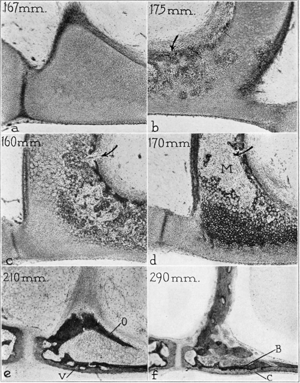
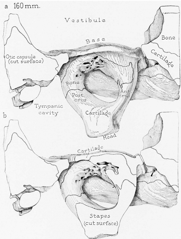
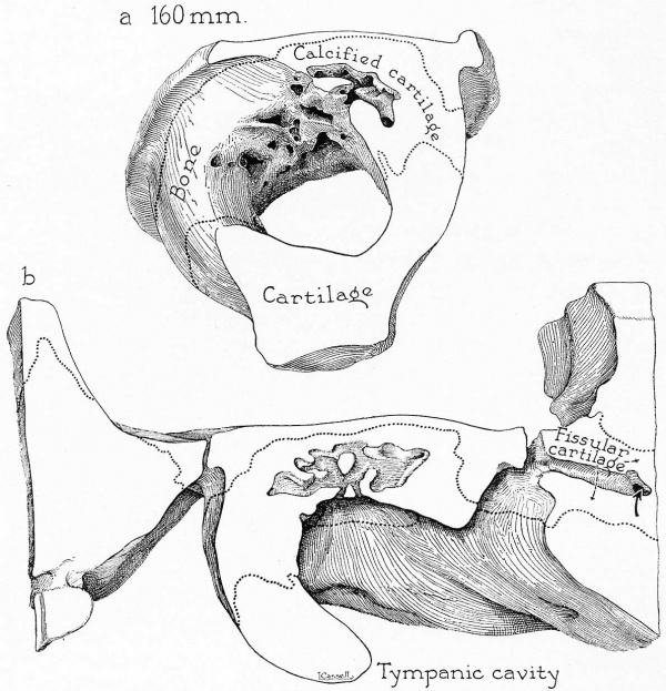
(19.5 ; weeks; series 41) — At this crucial stage the stapes exhibits the initial histologic changes whereby a solid cartilaginous structure becomes converted into a hollowed ossicle, whose structure will be that of a foraminous shell of bone, with stapedial form. New bone forniatioh is found principally on the obturator surface of the base and is of highly irregular, foraminous appearance (fig. lc). The solitary center of ossification extends along the base to encroach on the vestibular rim of stable cartilage (fig. 5 b) ; it involves principally the inferior (caudal) portion of the base along a line immediately proximal to the cartilaginous articular flange (figs. 9a and 13; 10a and b). A narrow band of perichondrial bone surrounds the basal portion of the posterior crus. Ossification of the corresponding portion of the anterior crus forms an irregular pattern; the lower, or inferiorly directed, portion of the crus is first involved, with perichondrial ossification of a narrow zone anteriorly. As a consequence, a narrow strip of cartilage persists to separate two wings of perichondrial bone encroaching on the basal extremity of the anterior crus (fig. 9 a). Hyaline cartilage of the primordial ossicle remains unaltered on the vestibular aspect of the base as a layer approximately one-eighth the total basal thickness; its free (vestibular) surface is covered by a dense perichondrium, which is continuous marginally with the stapedial ligament and with the fibrous covering of the vestibular Wall. The opposite, or tympanic, aspect of the base is the scene of active osteogenic change; already a narrow, irregular plate of endochondral bone has been formed over the area of modified cartilage. Subjacent thereto, at the core of the base, the related tissues are predominantly a vascular marrow and a calcifying cartilage (fig. lc). Here true endochondral bone is sparse, a fact which indicates that the process is one of rapid destruction preparatory to rebuilding. Toward the marginal portion of the base, and at the broad areas of continuity of the base and the crura, the process is less pronounced. That portion of the now undisturbed hyaline cartilage which forms a thick stratum on the vestibular aspect of the base is destined to be retained throughout life. In this relatively inactive part of the basal cartilaginous layer, the sparse and palestaining matrix, with small, densely packed chondrocytes, resembles closely the immature cartilage of antecedent stages.[2] The matrix is coarsely fibrillar peripherally, the fibers blending with those of the basal periosteum.
The newly formed bone, no more than a mere pellicle on the external aspect of the crus, is still uninterrupted by foramens (compare posterior crus of the 160 mm. stage, 9a). On the external aspect it is a thicker layer. However, here the osseous “collar” is foraminous, being perforated by vessels which enter the opening from the vascular mesenchyrna of the intercrural (obturator) space. The small vessels, supported by clumped mesenchymal cells, form the invasive osteogenic buds. The line of separation between the new bone and the old cartilage (now calcified) is very distinct. In the excavated portion of the base there occur occasional spicules of early intrachondrial bone (fig. 1c).
The capital extremities of the crura and the entire head of the stapes are still wholly cartilaginous (figs. 9a, 10 (1). There is an In I) the arrow points to an area of peripheral erosion on the obturator aspect of the base. In c the arrow is directed toward an invasive bud and the newly made foramen, which transmits the osteogenic tissue with its blood vessels. In d the arrow passes through such a foramen from the circumstapedial mesenchymal tissue into the primitive marrow of the stapedial crus.
Fig. 1. Photomicrographs of the base and crus of the stapes, showing progressive stages in the removal of cartilage and the formation of bone. X 40. (a) Base at posterior crus, left ear; 167 mm. fetus (Wisconsin series 105, slide 19, section 9). (b) Base and anterior crus, left ear; 175 mm. fetus (Wisconsin series 104, slide 17, section 5). (c) Posterior crus and base, left ear; 160 mm. fetus (Wisconsin series 41, slide 18, section 5). (d) Posterior crus and base, left ear; 170 mm. fetus (Wisconsin series 131, slide 21, section 10). (e) Posterior crus and base, left ear; 210 mm. fetus (Wisconsin series 51, slide 38, section 6). (f) Posterior crus and base, left ear; 290 mm. fetus (Wisconsin series 59, slide 36, section 1).
Abbreviations: B and C indicate bone and cartilage, respectively, of the bilaminar plate of the stapedial base; M represents the base of the stapes; V, vestibular layer of endochondral (endosteal) bone which forms one of the two constituent lamellas in the base of the stapes.
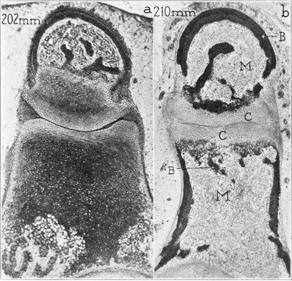
Fig. 2. Photomicrographs of the neck and head of the stapes and of the lentieular process of the incus, showing erosion of the cartilage and its ultimate replacement (except where articular) by bone. x40 (a) 202 mm fetus (Wisconsin series 70 slide 37, section 6); (b) 310 mm fetus (Wisconsin series 51, slide 38, section 6).
Abbreviations: B indicates bone (perichondrial in the incus, endochondral in the head of the stapes; C, cartilage (of the articular plates of the ineus and stapes); M, marrow.
€lC\'£1'El(')l1 at the point of attachment ml" ligament.
the (level01)i11;_>; staperlial
At this stage for the ilrst time there is ex-'ide1'1t the succession of steps by which the osteogenie process will ultimately im-'0lve the several clivisions of the stapes, namely’. basal, erural and, finally, capital portions. In the present iI1St{111C@ the head has not yet been im'0l\'ed. On the mi-clpart of the l'>a;'~“~e em.~:.i0n is deep: ])eripl1e1‘z1ll}' it is still superficial. A thin vestibular lamina of hyaline cartilage remains unaffected. The excavated area contains a primitive fibrous marrow and some irregular deposits of calcified cartilage. Mainly, the process is still one of calcification and erosion of cartilage, and not of bone formation. The greater number of vascular buds enter from the intercrural (obturator) space, that is, from the internal aspect of base and crura; only an occasional bud enters from the external aspect. Such perichondrial bone as does exist is in the form of a relatively intact plate; sites of periosteal erosion are few and small in caliber.
The fissula ante fenestrarn is a narrow, fibrous seam in a rather bulky cartilaginous capsular mass (fig. 5 a). At the vestibular extremity, the peripheral cartilage is continuous with that forming the marginal cartilage of the vestibular window (fig. 5c). The mass is being separated from the chondral shell of the cochlea through the activity of osteogenic buds. At the level of the auxiliary, or fenestral, opening of the fissula the relation of cartilage to bone is fundamentally similar (fig. 5 b). Peripherally the cartilage becomes calcified; farther away from the hyaline core the calcified tissue is being converted into intrachondrial bone. At the level of the tympanic opening the fissular cartilage is not yet segregated; it is continuous deeply (anteriorly) with the cochlear mass of cartilage. However, its vascular fibrous core communicates narrowly with that of the marrow at the cochlear end of the mass. The fibrous fissula, as it may conveniently be termed, has three orifices opening, respectively, on the tympanic cavity, on the vestibular (oval) window and on the vestibule at points not distantly separated from one another. The cartilage of the fissula (for which the fibrous tissue constitutes a core) extends without interruption from the tympanic cavity, through the otic capsule to the vestibule; at this stage its mass is being cut off from more anterior portions of the original capsule, as, at a slightly earlier stage, it was separated on those surfaces which now face marrow.
At this stage vacuolation of cartilage cells in the fissular region of the optic capsule is a prominent feature; the process is especially evident at the transverse level of the vestibular orifice of the fissula. At the level of the tympanic opening, however, there is no zone of altered cartilage to indicate where the cochlear shell will later be demarcated from the chondral shell of the fissula and from that of the vestibular window.
163 mm Fetus
(20 weeks; series 33) — At the 163 mm. stage the basal and crural portions of the stapes have been excavated by vascular buds. The process of erosion extends a considerable distance through these portions of the ossicles, beginning on the inner (intercrural) wall (fig. 3d).
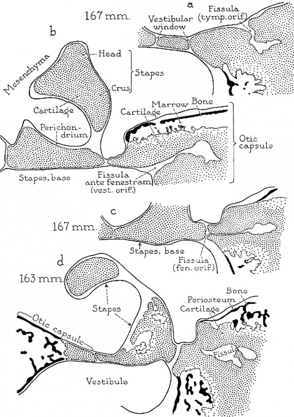
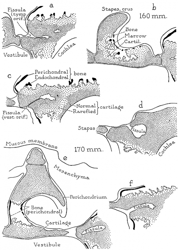
Fig. 3. Drawings (semi-diagrammatic) from Edinger tracings of the stapes and the adjacent fissular region of the otic capsule, showing developmental changes in the ossicle and in the fissular part of the capsule; X 6.6. Further developmental steps are recorded in the succeeding five plates of figures. In this, and in the five following plates, regular stippling represents unaltered cartilage; less dense stippling stands for rarefied cartilage; the areas treated in black represent bone. Parts a to c are from a 167 mm. (20 weeks) fetus (Wisconsin series 105) ; (a) slide 23, section 8; (b) slide 21, section 9; (C) slide 19, section 9). Part d is from a 163 mm. (19 week) fetus (Wisconsin series 33; slide 17, section 8). Here, a is taken at the tympanic (cranial. or superior) orifice of the fissula ante fenestram; b, at the fenestral (intermediate) orifice; c, at the vestibular (caudal, or inferior) orifice and throifigh the anterior crus of the stapes, and d, through the body, or midportion, of the fissula.
Abbreviations in this and in succeeding plates are interpreted as follows: Ant. or Ant. crus, anterior crus; Cartil, cartilage; fen. orif., fenestral orifice (of fissula ante fenestram); Post. or Post. crus, posterior crus; Tymp. cav., or Tymp. cavity, tympanic cavity (middle ear); tymp. orif., tympanic orifice (of fissula); V est, vestibule; vest. orifi, vestibular orifice (of fissula); V. w., vestibular (oval) window.
The stapes of the 167 mm. fetus is still wholly cartilaginous; in the capsule, on the contrary, bone is replacing carti1age( seen on the vestibular surface in a, on the tympanic aspect in c and on both surfaces in b). In the course of this process the cartilage of the fissula becomes separated.
In the 163 mm fetus ossification of the capsule has progressed further; the base and crus of the stapes have been excavated, but bone has not yet appeared.
167 mm Fetus
(20 weeks; series 105) — In this specimen the developmental processes are less advanced than those represented by the 160 mm specimen (series 41). Calcification of the cartilage has not yet occurred; instead, merely enlargement and vacuolation of the chondrocytes are evidenced. These steps presage those of calcification. This preparatory process occurs on the tympanic aspect of the base, where the center of ossification later appears (fig. 1 a). The relatively precocious ossification of the capsule in the fissular area stands in sharp contrast to the lag in stapedial development.[3]
Reconstruction of subsequent stages demonstrates that full dimensions of the stapes have been attained in the 167 mm. fetus. All portions of the stapes are thick, and the intercrural space, as a result, is relatively small. Subsequent changes, therefore, are in the category of differentiation rather than of growth.
The fissular cartilage is continuous with that of the cochlea. Erosion, which is narrowing the fissular mass, is most active near the cochlear extremity (fig. 3 19). Between this mass and the perichondrial shell of the capsule the initial changes in osteogenesis are in evidence. There is a continuous fissular cleft from the tympanic to the vestibular surface, traversing the fenestral margin in its course (fig. 3c). The fissula itself is a stripe of differentiated tissue in the cochlear division of the otic capsule. The cartilage in which the fissular fibrous tissue is lodged is continuous from the fenestral and vestibular walls to the cochlear wall. Bone is being formed around the fissula, segregating the mass on the lateral and medial sides. The vestibular extremity is of typically elongate form (fig. 3 b). The tympanic extremity is small (fig. 3 a); it is an almost circular orifice, a form typical of the adult fissula. The fenestral opening is narrow (fig. 3 c).
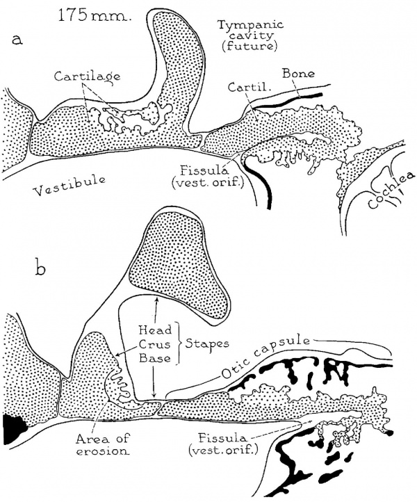
Fig. 4. Drawings (continued) of developmental stages from a 175 mm. (20 week) fetus (Wisconsin series 104: (a) slide 17, section 5; (b) slide 16, section 2); x 6.6.
The stapes is now excavated on the obturator surface of the base (a and b), at the basal end of the anterior crus (a) and in the corresponding portion of the posterior crus (b). However, bone has not yet formed over the eroded area. In the antefenestral portion of the capsule, periosteal bone, which appears in the form of thin larninas, does not extend to the vestibular window. In the latter portion of the capsule the fenestral shell of cartilage is continuous with that which encloses the fissular tract of connective tissue. The fissular shell of cartilage is becoming detached, at its opposite (or anterior) extremity, from the cartilaginous wall of the cochlea (a). In the region between the vestibule and the cochlea, spicules of intrachondrial bone now are present, having been formed as a result of rapid ossification of persistent spicules of cartilage (b).
With respect to form and structure, the fissula in this specimen is important both as a stage and as a type. A crucial phase in development is represented by the early erosion of the medial and lateral aspects of the main mass of cartilage in which the fissula (fibrous tissue) is lodged. In this specimen, the fissula constitutes a continuous stripe between the fenestral and the cochlear part of the original capsule. The fissula does not possess separate orifices. Correspondingly, an “opening” occurs in an oblique line downward and medialward, as an uninterrupted cleft from the tympanic wall of the otic capsule, across the fenestral, to the vestibular wall.
Retention of the narrow stripe of fibrous tissue, characteristic of the embryonic type of fissula, is an unexpected feature at this stage, in View of the fact that perichondrial bone will be soon added to the tympanic wall and endochondral bone will be abundantly laid down adjacent to the fissular tract.
170 mm Fetus
(20 weeks; series 131) — Stapedial development is further advanced in the 170 mm fetus than it is at the 160 mm stage {series 41). The cartilage of the crura has been almost completely removed, leaving hollow cylinders of periosteal bone to invest fragments of hyaline cartilage and calcified cartilaginous remnants (fig. l d ). The bone of the internal surface of the osseous crura is foraminous; that of the external aspect remains unbroken. The process of excavation is more advanced in the posterior than it is in the anterior crus (fig. 5 (3). Whereas perichondrial bone surrounds the crura and covers the tympanic aspect of the base, it has not yet extended to the stapedial head.
In the otic capsule, perichondrial bone approaches the tympanic and vestibular orifices of the fissula (fig. 5 d and f). Destruction of cartilage is followed chiefly by the formation either of primitive marrow spaces or of intrachondrial bone; endochondral bone is inconspicuously present in the form of small spicules. The fissular cartilage is broad; its contained connective tissue is vascular. The vestibular orifice is wide (fig. Sf) and is continuous with a well defined fenestral opening (fig. 5 e). The tympanic extremity is narrow; however, it broadens quickly as it extends into the adjacent mass of cartilage (fig. 5d). This type of fissula ante fenestram is very unlike that in which fibrous tissue appears as a mere streak within the fissular cartilage. The cartilage of the fissula is still broadly continuous with that of the cochlea.
175 mm Fetus
(20 weeks; series 104) — Although the stapes is still entirely cartilaginous, the process of osteogenic excavation is appreciably advanced (fig. 4a and b). - Osteogenesis affects the tympanic aspects of the base and the internal surface of the basal portion of each crus (fig. lb). The head of the stapes is unchanged. The surrounding mesenchyme, now highly vascular, is the source of the abundant osteogenic buds which invade the basal and crural portions of the stapes.
Fig. 5. Drawings, continued, of the stapes and the adjacent fissular region of the otic capsule, depicting developmental stages; X 6.6. Parts a to c are from a 160 mm. (19.5 week) fetus (Wisconsin series 41: (a) slide 19, section 4; (b) slide 18, section 8; (c) slide 17, section 9). d to f, from a 170 mm. (20 week) fetus (Wisconsin series 131: (d) slide 25, section 5; (e) slide 21, section 10; (f) slide 20, section 8). Here, a is taken at the level of the tympanic orifice of the fissula ante fenestram; 19, through the middle of the fissular tract and the posterior crus of the stapes; c, through the vestibular orifice of the fissula, and d to f, from sections of the 170 mm. fetus which pass through similar levels of the fissula and ossicle (in succession, the tympanic extremity, the body and the vestibular extremity of the fisstula).
In the 160 mm specimen destruction of cartilage is under way; bone is formed on the obturator aspect of the base and crus of the stapes and on the tympanic and vestibular walls of the otic capsule. Concurrently, the cartilage of the fissula has become partially separated; while almost detached at its cochlear end, the fissular cartilage is still broadly continuous with similar tissue at the vestibular window.
In the 170 mm fetus newly formed bone is present on both aspects of the crus as a complete shell externally, as a foraminous wall internally (i. e., toward the obturator foramen).
The fissular cartilage has become almost separated from the cochlear cartilage by the process of gradual erosion. Perichondrial bone has spread to the anterior aspect of the tympanic orifice, but has not yet encroached as deeply on the cartilage of the vestibular window as it has on the chondral wall of the vestibular extremity. In the latter situation bone has almost reached the vestibular orifice of the fissula. The fissular shell of cartilage appears as an elongate stripe extending from the cochlea to the vestibular window (fig. 4a). Its connective tissue approaches, but does not reach, the fenestral margin, there being, consequently, no auxiliary (fenestral) orifice.
179 mm Fetus
(20 weeks; series 135 B) — Osteogenesis has advanced beyond the stage seen in the 170 mm. fetus (series 131) ; the stapes is very similar to that of the 205 mm. specimen (series 7
The fissula, like that of the 167 mm. fetus (series 105), is a narrow seam, whose usually separate orifices are continuous. The fibrous tissue within the obliquely coursing cleft is thereby applied to the perichondrium throughout its extent (from the tympanic Wall, across the window, to the vestibule).
180 mm Fetus
(21 weeks; series 137 and 45 B) — The stapes is similar in its developmental stage to that of the 205 mm. specimen (series 7). Fenestrated periosteal bone, present on the crura, encloses marrow tissue. However, in fetus 137 the ossifying process has not yet involved the entire wall of the intercrural space; it fails to reach the capital portion. In fetus 45 B there is rapid spread of periosteal and endochondral bone, involving the base, the crura and the basal portion of the head (fig. 12a to d). The accompanying extensive excavation of the cartilage converts these portions of the ossicle into hollowed members, whose contained cavities are continuous. Irregular masses of calcified cartilage occur in the crurocapital and basal areas. The periosteal shells of bone, which form the peripheral portions of the crura, are becoming foraminous. These features would place this stage, developmentally, immediately antecedent to the 205 min. specimen (series 7).
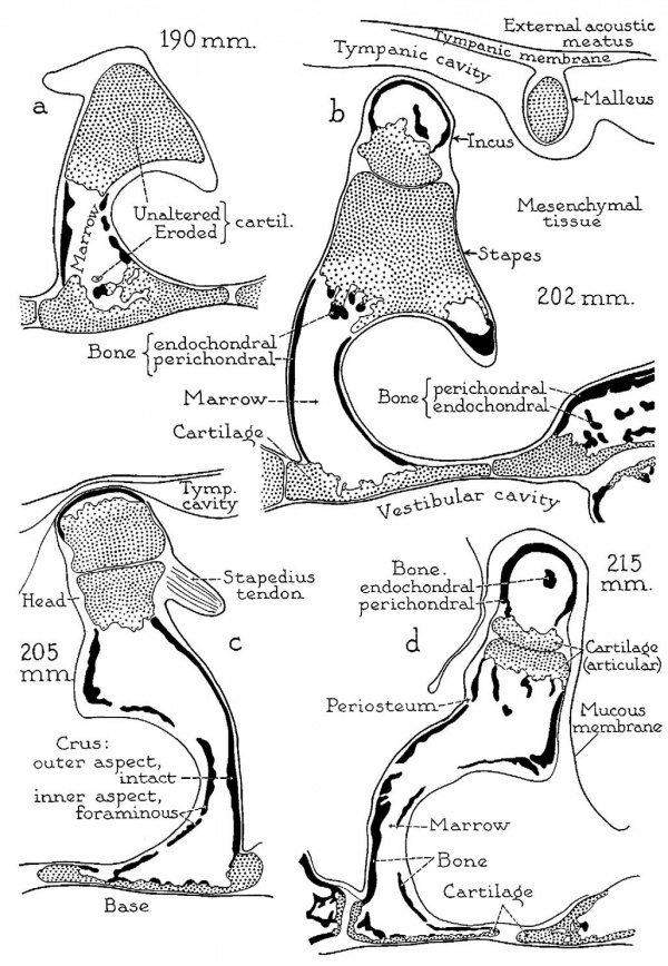
Fig. 6. Drawings of crucial developmental stages, continued: X 6.6. (a) 190 mm. (21 week) fetus (Wisconsin series 29A, slide 18, section 1); (b) 202 mm. (23 week) fetus (Wisconsin series 70, slide 27, section 6); (c) 205 mm. (23 week) fetus (Wisconsin series 129, slide 20, section 3); (d) 215 mm. (24 week) fetus (Wisconsin series 62, slide 28, section 4). Parts (1, b and d represent sections from series of the left ear; c is from the right ear; all represent the transverse level of the posterior crus of the stapes.
Four further steps in the progress of stapedial ossification are illustrated. In the 190 mm. specimen, the process of ossification has spread to the base of the stapes, but has not yet affected either the neck or the head of the ossicle (a). At ‘the 202 mm. stage excavation of the neck is in progress. (b) This developmental phase has been completed in the 205 mm. specimen; additionally, endochondral bone is being deposited on the internal surface of the excavated basal plate of cartilage. (c) In the 215 mm. fetus the articular plate of cartilage on the head of the stapes, now fully excavated, is being converted by a similar process into a bilaminar articulation. (d) Concurrently, a like series of changes is taking place in the lenticular process of the incus. Destruction of periosteal bone on the obturator surface of the stapes keeps pace with the formation of endochondral bone within the capital and basal portions of the ossicle.
The vestibular window is now set off sharply from the remainder of the differentiating otic capsule; it has assumed the form of a cartilaginous ring lodged in a framework of periosteal and endochondral bone. The periosteal bone meets, and slightly overlaps, the cartilage of the fenestral rim in exactly the same way that comparable bone of the stapedial head overlaps the cartilage which is there being gradually replaced. Endochondral bone is present in the form of discontinuous collections surrounded by primitive marrow.
The fissular tract of connective tissue is embedded in a considerable cartilaginous mass. The cartilage appears especially massive because of its association with a surrounding collection of delicate endochondral fragments. On the tympanic wall the fissula ends in two small orifices (fig. ll a to c). The tympanic extremity meets the body of the fissula at a right angle, as it does regularly in postnatal specimens. This observation indicates that the form of the fissula is fixed during the stage at which the cartilaginous otic capsule is being converted into an osseous “box.” A wide fibrocartilaginous cupola extends cranially for a considerable distance above the tympanic orifice (fig. 12 a).
In fetus 137 the fissula approaches, but does not actually reach, the vestibular window; it also fails to open on the tympanic surface of the otic capsule. This is an important fact in the interpretation of the adult condition in some specimens, since in several such specimens previously studied the tympanic opening was wanting. Formerly, we had ascribed this “aberrancy” to a late, obliterative overgrowth of periosteal bone. Now it is clear that it may be due to embryonic failure to establish, or to retain, such an opening during the period when the surrounding tissue is still cartilage.
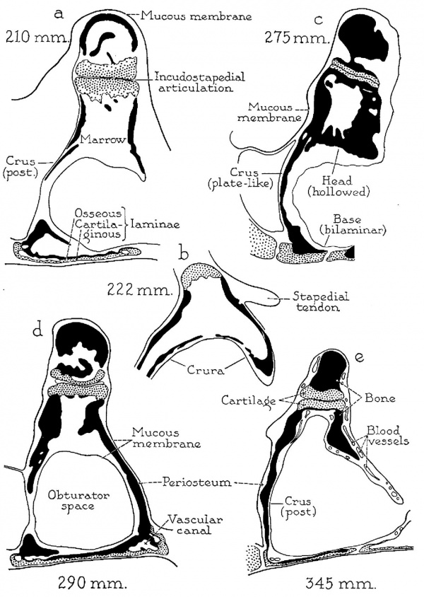
Fig. 7. Drawings of developmental stages, continued; X 6.6: (a) 210 mm. (23 week) fetus (Wisconsin series 51, slide 38, section 5); (b) 222 mm. (25 week) fetus (Wisconsin series 46, slide 19, section 10); (c) 275 mm. (30 week) fetus (Wisconsin series 4, slide 25, section 4); (d) 290 mm. (32 week) fetus (Vlfisconsin series 59, slide 36, section 5); (e) 345 mm. (38 week) fetus (Wisconsin series 61,. slide 42, section 1).
In the 275 mm specimen investment of the cartilaginous lamina of the stapedial base by endochondral bone has been completed (a) ; fusion of the peripheral remnant of perichondrial bone on the obturator aspect of the base (a) with the newly formed endochondral bone has resulted in the formation of osseous canals for the transmission of blood vessels (d). The mucous membrane and associated submucosal tissue, which in the 210 mm. specimen have already replaced the primitive marrow of the crura (a), later spread medialward to invest the endochondral and other bone of the base (290 mm., d); they ultimately invade the excavated neck and head of the stapes (345 mm., e). Thus, with regard to form, the stapes is essentially an adult ossicle in the 290 mm. fetus (d) ; with respect to mucosal relations, the ossicle attains adulthood at the 345 mm. stage (e) (compare with fig. 8e, from the newborn).
190 mm Fetus
(22 weeks; series 29 A) — The process of excavation of cartilage in the stapes has not progressed so far as in the 205 mm. fetus (series 7, to be described later) and is well behind that in the 180 mm. fetus (series 45 B, described in preceding section). The head and neck of the stapes are still composed of unaltered cartilage (fig. 6 a). The crura show the effects of extensive excavations; in each crus periosteal bone is intact on the outer aspect,‘but is foraminous on the inner (obturator) surface. The excavated crura contain marrowSmall remnants of calcified cartilage and intrachondral bone are found. Excavation of the base has progressed beyond that represented by the 160 mm. stage (series 41), but not so far as in the 180 mmstage (series 45 B).
The fissula possesses narrow orifices and a relatively wide midportion, or body. The fissular cartilage is being invaded peripherally by osteogenic buds. The cartilage does not quite reach the cochlear wall. Since the entire capsule was originally formed in cartilage (all cartilage persisting at later stages being remnants), it may be said with certainty that the reduction in length of the fissular cartilage is due to replacement of cartilage by bone, as is evidenced, at this 190 mm. stage, in the reduction of width of fissular mass.
193 mm Fetus
(22 weeks; series 85 B) — In general structure, this stage is more advanced than the 190 mm. fetus (series 29 A) and the 180 mm. specimen (series 137 or series 45B); with respect to certain features it is even beyond the 205 mm. fetus (series 7) in development. The stapedial crura are completely channeled. The old foraminous wall of the base remains in part. Internal to this disappearing wall, on the tympanic (lateral) cartilaginous lamella of the base, bone is spreading rapidly to form an osseous plate. The cartilage of the head of the stapes is also deeply excavated; like the crura, it has attained the form characteristic of the adult ossicle. The base retains but part of its fenestrated wall. In subsequent stages the foraminous walls of the crura and base will be wholly removed, through widening and coalescence of the foramens, to render the resultant space continuous around the entire obturator aspect of the ossicle. Concurrently, marrow will be replaced by loose fibrous tissue.
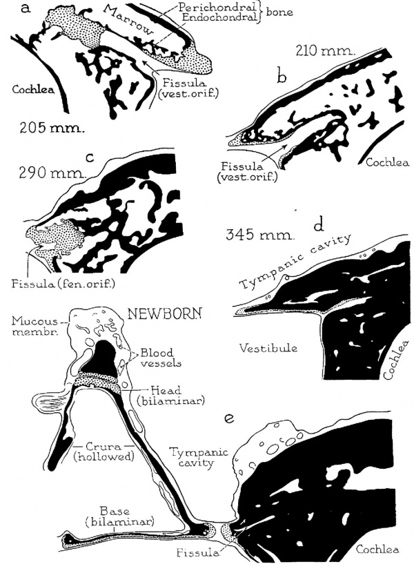
Fig. 8. Drawings of developmental stages concluded. X 6.6‘. (a) 205 mm. (23 week) fetus (Wisconsin series 129, slide 20, section 3); (b) 210 mm. (23 week) fetus (Wisconsin series 51, slide 38, section 5); (C) 290 mm. (32 week) fetus (Wisconsi11 series 59, slide 36, section 5); (d) 345 mm. (38 week), fetus (Wisconsin series 61, slide 42, section 1); (6) newborn infant (Wisconsin series 315. slide 20', section 8). Parts a, b and d are from sections through the vestibular orifice of the fissula; (C) and (e), from sections at the fenestral orifice.
The cartilage which surrounds the fissula ante fenestram in the 202 mm. specimen (fig. 6b) as a thick chondral layer, bordered by spicules of intrachondrial bone, undergoes rapid reduction in bulk to become, at the vestibular extremity, an osseous shell lined by a thin stratum of persistent cartilage or by a perichondrium (205 and 210 mm., a and b). Cartilage still persists at the cochlear extremity of the fissula as a considerable mass (205 mm., a); similarly, it still remains at the fenestral extremity, where the fissular cartilage is still continuous with similar tissue which lines the vestibular window (290 mm., c). While the cartilage is being gradually reduced in bulk, the marrow space, which everywhere surrounds the fissula, becomes occupied by endochondral bone. At first, spicules of_intrachondrial bone are sparsely distributed in the area between plates of perichondrial bone (205 and 210 mm., a and b); endochondral bone next forms around these spicules (290 mm., C), finally converting the capsule into a petrous structure (345 mm., d; newborn, 9). Thus, in the newborn (c), the capsule has acquired an adult appearance, as has the stapes.
The cartilaginous fissula is likewise in a more advanced stage of development. Bone of the capsule has spread into the fenestral and fissular areas, leaving cartilage as a relatively inconsiderable remnant. The fenestral cartilage is thinned through circumferential encroachment of bone. The surface area of the cartilage exposed to the vestibule and to the tympanic cavity has become greatly reduced in comparison with its extent in the earlier stages, having virtually assumed the form exhibited by that cartilage in the fetus at term and in the infant.
The fissular cartilage, likewise, is greatly diminished in bulk and is receding from the originally continuous cochlear cartilage. At the vestibular extremity of the fissula, periosteal bone approaches the orifice, and cartilage, continuous with that of the fissula, extends over a rather small, adjacent area of the vestibular wall. The exterior of the cartilaginous mass is invested with intrachondrial bone, a circumstance w-hich indicates rapid deposition of bone on partially destroyed cartilage. The two tissues remain thus associated in the adult ear, with, however, relative reduction in the bulk of the cartilage.
Near the fissular cartilage are situated separate spicules, consisting of a combination of intrachondrial and endochondral bone. As such spicules increase in number and in dimensions, they will close the space which at this stage exists between the periosteal shells of tympanic cavity, cochlea and vestibule. Even after coalescence of the now separate elements the constituent tissue will contain the cartilage islands, which here are encountered in their earliest stage of formation.
Fig. 9. Drawings of a reconstruction of the stapes and adjacent portions of the otic capsule in a 160 mm. (19.5 week) fetus (Wisconsin series 41); superior (cranial) views; X 7: (a) of the stapes entire; (b) with an upper segment of the stapes removed (in the plane of the constituent transverse section), showing erosion of the stapedial base and of the basal extremities of the crura. The dotted lines on the stapes indicates the approximate limits of the ossifying area; to either side of the area bounded by these lines, the cartilage of the base (on the vestibular surface) and of the head, neck and capital extremities of the crura (toward the tympanic aspect) is as yet unaltered. Similarly, osseous and cartilaginous portions of the capsule are indicated. In I) the arrow enters the tympanic orifice of the fissula; in figure 10b the arrow enters the junction of the vestibular extremity and the body of the fissula.
202 mm Fetus
(23 weeks; series 14 and 70) — In fetus 70 stapedial morphogenesis is not so far advanced as in the 205 mm. fetus (series 7). However, fissular development is further advanced in the former specimen. The stapedial base is excavated deeply. The portions of the osseous crura and base facing the obturator space are extensively foraminous. The capital cartilage is not so deeply excavated as are the crura and base.
The stapedial head is still entirely cartilaginous (fig. 2 a). Osteogenic vascular buds have encroached on the future neck of the stapes, with resultant patchy change to calcified cartilage. The crura are completely excavated internally; these tubes are filled with marrow, which now extends through the space from the neck to the base (fig. «6 b). At the latter site, the basocrural portions are made up of unaltered cartilage, covered by a thin and irregular plate of calcifying cartilage. The greater part of the base is excavated, leaving an obturator wall of foraminous periosteal bone. Small portions of endochondral bone are formed in the capital extremities of the crura (fig. 2 a).
The fissular cartilage remains as a shell for the contained connective tissue and forms a terminal bulbous mass at its cochlear end. It is lined with bone on the medial aspect of the vestibular orifice.
Bone Covers the cartilaginous rim of the vestibular window. This shell is mergent on its attached, or deep, aspect with spicules of intrachondrial bone, some of which still constitute a bridge between fissular cartilage and the now osseous cochlear wall.
In series 14 osteogenesis of the stapes is more advanced than in series 70 (figs. 2 a, 6 19). Almost no endochondral bone remains within the crura or head; the stage is preparatory to that in which the osseous obturator wall and contained marrow become, respectively, removed and replaced. The perichondrial bone of the head approaches the plaque of cartilage which, of the entire chondral mass, will alone be retained to serve as an articular plate. The base is deeply excavated from within, the cartilage now being thickest peripherally, where it forms the flangelike projection toward the fenestral rim.
Thus, there remains a basal plate of cartilage, not niuch thicker than it will be when definitive endochondral bone is laid down on its obturator surface. This means that crural development is most rapid and capital development slowest and that the base stands developmentally. in an intermediate position.
205 mm Fetus
(23 weeks; series 7) — On the capital extremity of the stapes unaltered cartilage persists as a relatively limited area, adjacent to which is a narrowing zone of calcifying cartilage (fig. 6c). Ossification is complete in the crura, these once separate osseous cylinders now being connected by bone across the lateral (capital) aspect of the obturator foramen. The obturator surface of each crus is strikingly foraminous. Excavation of the base has progressed to the point at which the obturator wall is extensively foraminous, and the vestibular portion is a cartilaginous plate on the internal surface of which numerous small zones of calcified cartilage are being replaced by endochondral bone (fig. 6 c).
This stage is a critical one in the whole process of reconstruction of the stapedial base. Endochondral bone is extending across the eroded internal surface of the cartilaginous lamina. This new bone is connected with the original, now foraminous, primary (perichondrial) layer by a few elongate spicules. Between the old cartilage and the new bone, vessels remain in widened canals. Some of these, judging from the later appearance of the base, are of transitory nature; others will remain as nutrient ‘vessels. The vessels will become surrounded by endochondral bone, thus coming to lie in channels which are comparable to Volkrnann’s canals.
Fig. 10. Drawings, continued; same specimen (160 mm.) as in figure 9 (Land 2). X 7; (a) at the same level as figure 91;, viewed from a more lateral position, in order to reveal the number and size of the foramens present in the periosteal plate on the obturator surface of the base and on the basal extremities of the crura; (b) .a more inferior segment of the stapes and of the otic capsule (viewed as are 9 a and 19), showing cavities produced in the calcifying cartilage preceding the stage of complete excavation of the base. Here, and in figure 9 a and b, the osseous shell externally and the calcifying cartilage internally merge imperceptibly in a transitional zone. In I) of figure 10 the hummocks exposed in the basal portion of the stapes are composed chiefly of calcified cartilage; their summits are capped by either endochondral or intrachondrial bone.
At this stage of development the following alterations have been completed: The crura have become hollow, osseous cylinders, cartilage and spicules of bone having been removed from their interior; the conjoined portions of the crura at the neck of the ossicle have attained similar structure; the head is composed of solid cartilage and is, therefore, the least altered portion of the stapes; the base is hollowed, its vestibular wall (the future definitive base) is thinned in the intercrural area to one-third or one-fourth the thickness of its peripheral (articular, or fenestral) portion. Within the base, on the tips of the eroded cartilage, endosteal bone is being deposited, these spicules being the fragmentary forerunners of the continuous lamina which will eventually cover the tympanic surface of the cartilage. In like manner, bone will later be deposited on the internal surface of the cartilage of the head, but only after this cylindric portion of the ossicle has become hollowed toward the obturator space. Consequently, the head will be converted into bone, except where its lateral, foveate, surface forms an articular area for the incus. Bone will be added in two ways: by growth inward from the periosteal bone which clasps the remaining. disklike piece of capital cartilage, and by deposition of islets of endochondral bone on the irregular inner surface of the articular cartilage. In the base, the process of fusion of part of the foraminous lateral wall (periosteal bone) with the bone (endosteal) deposited on the lateral surface of the basal cartilage is under way. In the current specimen, the approximation of the two osseous strata is witnessed in its initial stages. Ultimately, only the peripheral part of the foraminous periosteal bone will remain; it will sink to the level of the newly formed plate of endochondral bone which, then, has covered the internal surface of the basal layer of cartilage. Circumferential vascular spaces of the base are being formed through the inclusion of vessels between the merging periosteal and endosteal layers of bone.
The fissular channel (containing fibrous tissue) is l111.1’l‘l€(lla'{€l}' invested with a cartilaginous tube. In turn, the chondral tube is surrounded by endochondral bone and intermittent patches of intrachondrial bone, cartilage and marrow (fig. 8a).
210 mm Fetus
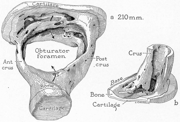
(23 weeks; series 51) — ln this specimen there is evidenced the next important step in the process of ossification (fig. 13 a) : The bone on the obturator portion of the crura and neck has been resorbed (figs. 2 b, 7 a) ; irregular ledgelike and bridgelike portions are all that remain of the corresponding portion of the base (fig. 1 e). These perforated and irregular bridgelike remnants of the obturator portion of the base pass obliquely from the basal extremity of the inferior, crural margin to a corresponding position on the superior, crurobasal margin posteriorly (fig. 13 17). From the same (anteroinferior) crural margin a wedge-shaped fragment extends perpendicularly, to become continuous with the basal bony plate. Similar remnants, of various forms, are common in other specimens of a comparable stage of development.
This specimen represents another crucial step in stapedial development. It is advanced over the 215 mm. fetus (series 62), principally in respect to configuration of the crura and base. The obturator portion of crural periosteal bone has been removed, leaving open, grooved crura. Absorption has also taken place in the corresponding bone on the basal surface; endosteal bone forms a continuous plate over the cartilage lamella of the base (fig. le).
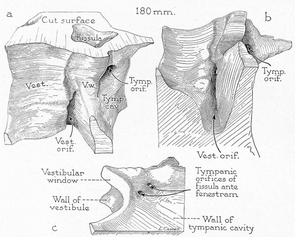
Fig. 11. Reconstruction of the fissular region of the otic capsule in a 180 mm. (21 week) fetus (Wisconsin series 45-b) with stapes removed; X 7: (a) viewed from a posterosuperior position, that is, looking toward the anterior wall of the vestibular window and into both orifices of the fissula ante fenestram; (b) viewed from a posteroinferior position, looking upward into the vestibular orifice of the fissula; (c) viewed from a lateral (tyrnpanic) position, looking toward the outer wall of the capsule, where occur, in this specimen, paired tympanic openings of the fissula. Part c is of a block less inclusive than that shown in a and b.
In this specimen the fissula is of aberrant form, being both taller and wider than usual. A cupola-like extension is prolonged cranialward beyond the horizontal level of the tympanic orifice. It contains cartilage (not represented in the reconstruction).
The fibrocartilaginous fissula is invested with endochondral bone(fig. 8 b). The fenestral articular cartilage forms a circumferential rim for the vestibular orifice. It is continuous across the adjacent part of the anterior wall of the vestibule with cartilage which extends for a short distance into the vestibular extremity of the fissula ante fenestram ; it is no longer carried through the length of the fis.sula, as. it was in earlier developmental stages.
215 mm Fetus
(24 weeks; series 62).~—The stapes is but slightly advanced beyond the stage of the 202 mm. fetus (series 14), and not so far advanced as that of the 205 mm. fetus (series 7) stage (compare fig. 66 and d).
Excavation of the stapedial head has continued, leaving a wideplate of articular cartilage, internal to which are persistent spicules of endochondral bone (fig. 6d). The neck and crura are completely hollowed, but they still contain marrow. Numerous foramens appears on the outer aspect of each crus; since the opposite (obturator) wall is the one which is regularly resorbed, it is probable that the existent foramens would have been obliterated in the course of later development. There is progressive resorption of bone on the obturator aspect. The interval between the thinned bony plate of the obturator surface and the adjacent basal plate of cartilage is gradually lessening, thereby bringing these laminas into closer apposition. Spicules of endochondral bone remain attached to the basal articular plate on the obturator surface of the latter. Comparable advances in osteogenesis are observed in the adjacent capsule and in the incus. The osseous walls of the fissula face a marrow space which is decidedly fetal in character, since it possesses virtually no bone. Its vestibular orifice is continuous with a wide fenestral opening.
222 mm Fetus
(25 weeks; series 46) —This stapes is not so far advanced as it is in the 210 mm. fetus (series 51). The base retains a residuum of fenestrated periosteal bone. This bone occurs. as a plate which no longer extends uninterrupted between crura; instead, reduced in amount, it passes abruptly downward to an attachment on the newly formed lamina of endosteal bone of the base. The latter forms an almost continuous layer, independent of any contribution from the periosteal bone of earlier genesis. The crura are channeled, the facing walls being entirely removed. The capital extremity is widely excavated, leaving a broad cartilaginous (articular) plate, o-n the inner surface of which there is early, sparse deposition of endosteal bone (fig. 7b).
230 mm Fetus
(26 weeks; series 2) —The stapes is similar to that of the 222 mm. stage (series 46). The stapedial crura are channeled, and the marrow has been removed. The head, likewise, is hollowed; cartilage remains only on the articular aspect. At the junction of crura and base, and along the basal plate, final architectural alterations are in progress; at the periphery of the crurobasal junction, bone is replacing the chondral remnant, with the formation of voluminous vascular channels; in the midportion of the base, an incomplete endosteal lamina is applied to the obturator surface of the thinned basal cartilage, especially in the crypts remaining from an earlier stage of erosion.
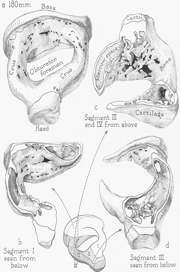
Fig. 12. Drawings of a reconstruction of the stapes in the 180 mm. (21 week) fetus (Wisconsin series 45 B); X 7: (a) the entire stapes, in superolateral view; (1)) segment 1 (see inset), viewed from below (that is, here inverted) ; (c) segments 2, 3 and 4 in superomedial view; (d) segment 3, from below (reconstruction inverted), with segment 4 removed; inset, reconstruction with segments lettered and with arrows recording the direction of View in the correspondingly lettered figures.
The drawings show the extent to which periosteal bone has been removed on the obturator surface of the base (a and b), crura and neck (c and d) and the degree to which the stapes throughout has been hollowed (I) to d). Cartilage now remains only on the articular surface of the head and on the vestibular surface and fenestral margin of the base of the ossicle (b and c). The addition of endochondral bone has converted the base into a thinned bilaminar plate ((2 and c). Such bone appears in limited amount as spicules in the area where the crura join the neck of the stapes (d).
The fissula ante fenestram has attained adult form. At the vestibular end of the fissula, bone surrounds the connective tissue as a complete shell but is, in turn, lined by an incomplete lamina of cartilage. The vestibular extremity ascends to approach the customary site of a fenestral orifice; however, a true fenestral opening is not established, cartilage, or tissue intermediate between cartilage and perichondrium, intervening between the fissular connective tissue and the annular ligament. The tympanic extremity opens by a definite orifice into the semicanal for the tensor tympani muscle.
The bone of the fissular part of the capsule is largely intrachondrial. Marrow space outbulks bone, the bone occurring as fine spicules, sparsely distributed, some of which are directly continuous with the cartilage which surrounds the fissular connective tissue. A fissula of this type undergoes relatively little subsequent change. Once formed, the fibrocartilaginous component of the fissula remains dormant until birth; then the changes which do occur affect chiefly the bone external to that of the fissula. Only in those instances in which cartilage remains as a mass will notable fetal or postnatal modifications of fissular tissuesoccur.
240 mm Fetus
(27 weeks ; series 112) In stapedial development, this series would precede the 205 mm. fetus (series 7) and follow the 210 mm. specimens (series 51 and 21); in all major otologic features the present (240 mm.) specimen resembles the 210 mm. fetus. The walls of the crura are only moderately fenestrated, but the internal (corresponding) wall of the base is rather foraminous. Although the effects of erosion are clearly evident, no osteoclasts are discoverable. The base of the stapes is bilaminar. The new bone has been applied as a thin layer to the cartilaginous lamella, but is removed by an appreciable space from the old periosteal bone. Within the space between periosteal and endosteal bone, marrow tissue still persists. A vascular mesenchyma occupies the circumstapedial “space” of the future tympanic cavity (middle car). In the areas of implantation of the crura into the base, the bone of the remaining (external) part of the crus and that of the base are continuous, a fact whch suggests an inward spread from the periphery. In the base, the newly formed osseous layer is only one fourth as thick as the older layer of cartilage, to which it is now applied. In the peripheral part of the base, intra chrondrial bone occurs in association with the endochondral type. Spaces exist marginally between the bone and the cartilage where the crura meet the base; these, as already mentioned, serve for the transmission of blood vessels in adult stapes. The head is deeply excavated, there being little more of the cartilage left than would be present for articular purposes. Bone has ‘not yet invested the internal surface.
The fenestral cartilage is separated from the capsular wall by intervening formation of periosteal bone. It is, however, continuous with a still bulky fissular cartilage. The cartilaginous portion of the fenestral rim and that of the adjacent base of the stapes are of the same hyaline structure as that of the auxiliary fenestral orifice of the fissula.
Fig. 13. Drawings of a reconstruction of the stapes in a 210 mm. (23 week) fetus (Wisconsin series 51) ; X 7: (a) reconstruction entire, seen from a superclateral position; (b) basal portion of the posterior crus and the adjacent posterior third of the base, seen as though from the obturator space.
Almost all of the obturator wall has been resoi-bed; that portion which remains is foraminous (arrow through a foramen in a). The obturator surface of the base is irregularly eroded; a portion of it persists as the crista stapedis (beneath which crest an arrow passes in a and b). The cartilage. which remains on the articular surface of the head is permanent (a); on the base not only does it cover the vestibular surface, but also is carried over the periphery of the base along the surface related to the vestibular window (I), at cut edge). A remnant portion of the original periosteal bone (encircled by ring in b) is fusing with the newly formed endochondral plate on the vestibular aspect of the base.
Periosteal bone forms the tympanic, vestibular and cochlear walls and serves as an investment for the fissular cartilage. Within the otic capsule, in the area bounded by these walls, intrachondrial and endochondral bone now are sparsely present. Subsequently, ossification will fill in the space with bone of petrous consistency. Periosteal bone, at the anterior margin of the window, surrounds the cartilage of the auxiliary (fenestral) extremity of the fissular mass and actually invades the tympanic orifice. At the vestibular extremity the orifice is widely open, but the mass of cartilage is considerable in amount. Although thin, there is already a complete periosteal investment for the fissular cartilage, which follows the latter inward from the window toward the cochlea. In association with the peripheral portion of the cartilage, intrachondrial bone is forming. At this stage, therefore, is established the succession of tissues which becomes the regular order in the adult fissula: connective tissue (core), hyaline cartilage (investing tube) and intrachondrial islands associated with endochondral bone (general outer bed). At the tympanic extremity the succession described is repeated. In the depths of the fissula the cartilage is calcified and merges marginally wth whorls of intrachondrial bone and trabeculae of endochondral bone. Near the orifice of the fissula, cartilage passes gradually into a tissue perichondrial in nature; the perichondrium merges with the subepithelial connective tissue. Periosteal bone of the tympanic wall passes into the orifice of the fissula and is continuous with a layer of the same tissue which now almost completely surrounds the cartilage. Investment is not yet complete, since in certain small areas near the orifice the cartilage of the fissular shell is exposed to the marrow. This means that osseous encroachment at the tympanic end is an early feature of development, whereas it is a later feature at the vestibular extremity, if it ever occurs at all. This circumstance accounts for the fact that in adult specimens the wall of the tympanic portion of the fissula is osseous, while cartilage remains (albeit in the form of a thin shell) at the vestibular end. As is made clear from the study of postnatal stages, the cartilage will be subsequently reduced in bulk to the thinness of a pellicle, which in some specimens forms an incomplete investment for the fibrous tissue of the fissula. Some of the endochondral bone, which at this stage appears in association with the cartilage, will itself be replaced with bone of the primary type. Periosteal bone now separates the cartilage which borders the space of the vestibular window from that which forms the wall of the fissular channel. Here, again, cartilage rests on bone. Some of the bone is of the intrachondrial type; it crosses beneath the attached surface of the fenestral rim, setting off the latter as a unit and completing the periosteal part of the osseous capsule. By a process similar to that which is reducing the bulk of the fissular mass, the fenestral rim will be narrowed and thinned. Deep to the periosteal layer, the endochondral bone will fill the space now occupied by primitive marrow.
240 mm Fetus
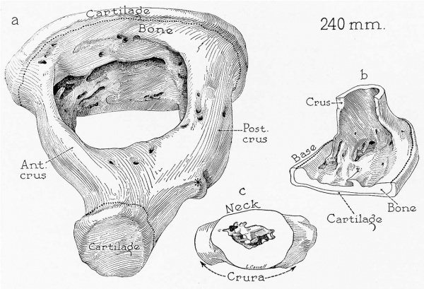
(27 weeks; series 112) — The stapes has undergone more complete excavation of the base and complete destruction of the obturator wall (fig. 14 a). However, large, irregular fragments of endochondral bone persist on the obturator aspect of the neck and within its excavated portion; a small amount of marrow is also retained in the capital space. The general proportions of the ossicle are massive (fig. 14 b) ; the bone of the crura and of the head is thick (fig. 14 C). The heavier bridges of bone have been removed from the base; only irregular, flattened trabeculae remain on the lateral (tympanic) Surface of the base and in the area between the base and the adjacent portion of each crus (fig. 14 Z7).
246 mm Fetus
(27.5; weeks; series 30, 15) —The stapes of fetus 30 is generally similar to that of the 230 mm. specimen (series 2) , whereas in fetus 15 it is advanced beyond the stage shown in the latter. In fetus 15 endochondral bone has covered the basal plate of cartilage at the periphery in such a way as to form osseous channels, through which the blood vessels course.
Fig. 14. Drawings of a reconstruction of the stapes in a 240 mm. (27 week) fetus (Wisconsin series 112); x 7: (a) reconstruction pictured entire and viewed from a superolateral position; (b) the basal portion of the anterior crus and the adjacent anterior third of the base seen as though from the obturator space; (c) the neck of the stapes (capital portion removed) and the adjacent portions of the two crura, in lateral view. In a are shown the limits of the portions shown in b and c.
The crura are now channeled columns (a and b) and are foraminous in certain areas (a). On the base of the ossicle some of the persistent perichondrial bone has formed an incomplete crista stapedis (a and 12); under arrow in (c). The neck and head are hollowed; yet some endochondral bone, in the form of spicules, remains to close the capital space (c). The stapes has assumed typical adult form: The anterior crus is the shorter and thinner of the two; a pronounced tubercle appears on the posterior crus, for attachment of the tendon of the stapedius muscle, and the head is foveate on its articular surface (at). The osseous lamina of the base is composed of endochondral bone, with which have fused remnants of the original perichondrial bone from the once complete obturator wall of the base (a and b). As is regularly the case, the developmental changes affecting the head of the stapes are retarded; part of the obturator wall here remains to close the capital space medially (c). Such bone sometimes persists in the stapes of adults, either in the form of a perforate plate or in that of osseous bridges.
265 mm Fetus
(29 weeks; series 69) — The process of canal formation around the periphery of the stapedial base continues, with‘ the intrachondrial bone taking part by further walling off these channels. Endochondral bone is covering the now extremely thin layer of basal cartilage.
The fissula is of unusual form in that its tympanic extremity is wider than the vestibular orifice. There is no fenestral opening, although the connective tissue within the fissular channel approaches the fenestral cartilage. At the tympanic opening the tissue is of such concentrated type as to suggest a perichondrium.
275 mm Fetus
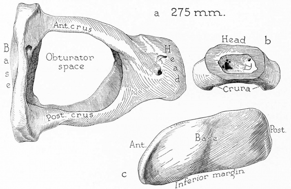
(30 Weeks; series 4) —The stapes is essentially of adult form (fig. 15 a). The head is now an osseous cylinder capped by a plate of articular cartilage. Its surface is irregular outside and inside, and in the latter situation the irregularities represent openings of vascular spaces, some of which are continued into the crura. The medial (obturator) end of the capital cylinder is crossed by a ledge of bone (figs. 7 c, 15 b) similar to that seen not only in some early post— natal stages but also in adult specimens. The crura are channeled, thinned and bowed and are now invested on all aspects with mucous membrane. The base is bilaminar and is canalized peripherally; the vessels within the canals communicate with those of the newly formed connective tissue. Marrow tissue has not been entirely removed, however; some remains in the head and neck. In general, the irregularities characteristic of the stapes in earlier stages of development have been smoothed out. There is further flattening of the trabeculae 011 the tympanic (lateral) aspect of the base. The vestibular (medial) surface of the base is smooth and reniform (fig. 15 c).
290 mm Fetus
(32 weeks; series 66) — A slight amount of marrow persists in the head of the stapes despite the fact that a ledge, such as that described in the preceding stage, is wanting in this specimen. Bone is present on the medial (internal, or obturator) aspect of the articular cartilage (fig. 7 d). The channeled crura are thinned and are _invested with mucous membrane. The base is thin and bilaminar. The marginal spaces, formed by the fusion application of the persistent portion of the periosteal bone with the adjacent endosteal bone, are striking morphologic features (figs. 1 1‘, 7d). In being less massive than the stapes in the 275 mm. stage (series 4), and in the absence of the cervical obturator plate (to leave an open, marrow-free capital extremity), the ossicle of the 290 mm. fetus has attained adult form.
The presence of marrow in the capital part represents the sole fetal feature.
Bone has filled the spaces which formerly existed in the area bounded by tympanic, cochlear and vestibular walls, a circumstance which renders the fissular tract inconspicuous. In this way the fissular region of the capsule assumes the appearance of compactness; consequently, it may, for the first time, be described as petrous. The fissula is of simplest type; it is short, slightly broadened in the part between two orifices, and contains a fibrous core around which cartilage forms an incomplete shell. The latter is thin Where present, and the bordering bone is largely of endochondral type. A fenestral orifice is represented by a shallow cleft which opens on the anterior surface of the vestibular window. This orifice is surrounded by an irregular mass of cartilage which is continuous with that of the articular margin (fig. 8c).
Fig. 15. Drawings of a reconstruction of the stapes in a 275 mm. (30 week) fetus (Wisconsin series 4) ; X 7: (a) the reconstruction entire, viewed from above (superior, or cranial, aspect); (b) the neck and adjacent portions of the crura, in lateral (tympanic) view; (c) the base of the ossicle seen in medial (vestibular) view.
The stapes has attained adult form; of the crura, now deeply channeled, the posterior is longer and larger (as); the base is flatter on the posterior than on the anterior margin (c) ; the head and neck are hollowed (b). In this fetal specimen, as in some adult ossicles, a ledge of bone partially closes the space of the neck toward the obturator foramen.
295 mm Fetus
(33 weeks; series 20) —The stapes is now snugly invested with mucous membrane, continuous with that on the adjacent tympanic wall. The abundant blood vessels in the lamina propria are unusually clear.
The connective tissue which fills the fissular channel is slight in amount and contains but few small vessels; the vertical distance between the cranial border of the vestibular orifice and the small tympanic opening is slight. The immediately investing wall is composed of cartilage and perichondrium, external to which are situated areas of endochondral and intrachondrial bone. This is equivalent to saying that the fissula and the related area are, respectively, of adult form and of mature histologic structure. '
305 mm Fetus
(34 weeks; series 60 A) —The stapedial structure is substantially that of the ossicle in the term fetus (to be described later).
345 mm Fetus
(38 weeks; series 61) —The stapes is indistinguishable from that of the postnatal stages (fig. 7 e). Bone covers the inner surface of both capital and basal cartilages. The base is thinner than it was at any preceding stage of development; cartilage comprises approximately two thirds, and bone one third, of the thickness. The mucous membrane, which has replaced marrow, extends inward to‘ invest the former endosteal surface of the head.
The fissular part of the capsule is similar to that encountered in the ear of an adult. An auxiliary (fenestral) orifice is present. The vestibular orifice of the fissula is a narrow cleft; the tympanic opening is small and virtually circular. The channel retains a thin wall of cartilage (fig. 8 d). The capsular bone is of dense, compact type; spaces are few and small.
363 mm Fetus
(40 weeks; series 16) —The stapes is of adult form. The thin, bilaminar base is vascularized circurnferentially. The head is hollowed, and its articular portion is bilaminar. The crura are exceedingly thin.
The small fissula is of typical form in its lower part but is aberrant cranially in lacking a tympanic opening. The vestibular opening crosses obliquely to the window, producing an uninterrupted vestibuletympanic cleft.
Term Fetuses
(series 67, 127, 95, 102 and 115) — In fetus 67 the internal surface of the head of the stapes is extremely irregular, being sulcate for the transmission of blood vessels. The external surface is also somewhat irregular. The crura are strikingly thinned; as a consequence, they are indistinguishable from those of an adult ossicle. The base, like the head of the ossicle, is bilaminar and thin.
In sections, the fissula appears as a narrow fibrous seam, clearly traceable from the vestibular to the fenestral opening. However, near the tympanic orifice the fibrous tissue passes into the surrounding cartilage by such imperceptible gradation that the former tissue seems to be wanting. As a result, the tympanic opening of the fissular channel is inconspicuous.
Beneath the thin periosteal shell the capsule is well filled with endochondral bone; marrow spaces occupy less than one fourth of the entire fissular portion of the otic capsule.
In fetus 127 the stapes is of adult form. The base, crura and head have been excavated and hollowed to striking thinness. Submucosal vessels are now prominent; marrow has disappeared.
Cartilage is minimal in amount on the fissular wall, being present as a distinct layer only at the vestibular end. The surrounding bone is chiefly endochondral, with some intrachondrial bone deeply embedded in its substance. There is no fenestral orifice. The small tympanic opening of the fissula ante fenestram is situated posterior to the developing semicanal of the tensor tympani muscle. Altogether, the fissula may be said to be of the kind previously described as typical.
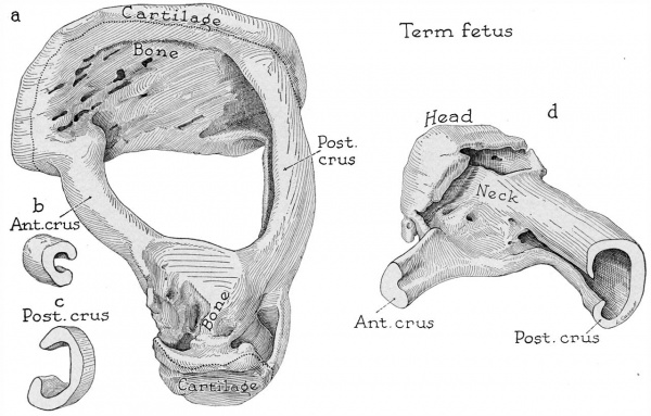
Fig. 16. Drawings of a reconstruction of the stapes in a fetus at term (Wisconsin series 102) ; x 7: (a) the reconstruction entire, seen from a superolateral position; (b) segment of anterior crus; (c) segment of posterior crus; (d) lateral part of the reconstruction, in posteromedial view. In a are indicated the limits of the segments shown in b and c.
The cartilage which covers the vestibular (medial) surface of the base is carried over the fenestral margin as a circumferential lip, while the tympanic (lateral) surface is formed by a plate of irregular bone which is composed of both endochondral and perichondrial bone (:2; cf. figs. 13a and 14 a). The crura are deeply channeled (16a to c). The head and neck of the ossicle are strikingly eroded, despite the fact that the cavity of the capital portion is crossed by a plate of bone (a and d). This feature of sculpturing is persistent, being encountered in ossicles from adults (specimens from a 57 year old person and from other adults). The anterior crus, much the slenderer and shorter of the crura, is implanted in the base at the inferior margin of the crus (a); at the point of continuity with the head, the crus flattens to meet the intercrural plate (d). The bulkier posterior crus is implanted widely into its portion of the base (a); its cavity, relatively capacious (c), opens into that of the head and neck by a small orifice (d). Cartilage covers the articular surface of the head as a layer of restricted extent (a).
There is a sharp line of demarcation between the bone of the original capsule and that added in later development. Marrow spaces of fair size occur only in the area midway between the tympanic and the cochlear wall. This is the site of the space which originally contained only intrachondrial bone and marrow (compare the 230 mm. fetus, series 2‘).
In fetus 95 the stapes is again of adult type. The vestibular orifice of the fissula is of conventional form. Its connective tissue core is uninterrupted. The fibrous tissue extends into a small, but definite, fenestral orifice, whose walls are formed by the cartilaginous tissue of the fenestral rim. The tympanic extremity is aberrant in being occupied by a cartilaginous nodule without fibrous content. The nodule touches, but remains covered with, a thin part of the periosteal tympanic wall; the area on which it tends to open is that of the semicanal.
In fetus 102 the stapes in all respects is, again, an adult ossicle (fig. 16a to d ). The vestibular and fenestral orifices of the fissula are of typical form and size. The tympanic orifice is indistinct, since in the cranial part of its extent the meager connective tissue is displaced by a concentrated tissue which resembles a perichondrium. The occurrence of such tissue is of interest in connection with the formation of cartilaginous nodules in the cranial part of the fissular tract.[4] It is evident that new cartilage, formed from the usually dormant lining of the fissula, may spread to obliterate the normal fibrous tissue of the fissular canal.[5] Subsequently, the cartilage is likely to ossify. In the current specimen such a change was doubtless in progress, since the peripheral part of the perichondrium-like tissue gradually merges with the bone.
In fetus 115 the stapes is of an adult form except for the presence of a ledge on the interior of the head. The ledge corresponds to the partition seen in the 275 mm. specimen (series 4). In certain portions of the base the osseous lamina is thicker than the cartilaginous, a proportion which We formerly believed obtained only in adult stages.
The fissula is of the typical variety. It is thin mediolaterally; its content is fibrous. The vestibular and tympanic orifices are narrow; an auxiliary (fenestral) opening is wanting. The capsule is well filled in with endochondral bone, marrow spaces being large only in a line corresponding to that of the junction of the early capsule and the newly added shell of periosteal bone.
In general, it may be said that the stapes of the fetus at term is extensively excavated throughout its obturator aspect. The superior portion of the anterior basocrural junction is decidedly attenuated. The diameter of the anterior crus is approximately one-half that of the posterior crus. The superior surface of the crurocervical region is likely to be more irregular than the inferior. Ledges of bone are likely to persist on the inner surface of the head and base.
Newborn
(series 315) — In the newborn the stapes is of fully mature form and structure (fig. 8e).[6] The fissula ante fenestram extends toward the fenestral margin, but does not communicate with the vestibular window (compare 345 mm. stage, series 61). The capsule is compact, its relatively petrous nature suggesting the texture of the adult capsule.
Summary
Ossification has been initiated in the stapes of the 150 mm. fetus. The process begins in a solitary center on the obturator surface of the base. However, in a fetus at the 160 mm. stage, bone development is relatively tardy. Encroachment from the single center to the crurobasal cartilage is in evidence in the 163 and 175 mm. stages; erosion and calcification of cartilage is well advanced in the 160 mm. stage. In the 170 mm. fetus periosteal bone surrounds the anterior crus, extending from the base to the future neck of the stapes. The crura are converted into hollowed cylinders in the 190 and 202 mm. fetuses; concurrently, calcification of cartilage in the neck is under way, in anticipation of the spread of bone across the crural junction. In the 180 mm. fetus, and its developmental counterpart, the 205 mm. stage, this junction has been effected; through this fusion the obturator wall is formed circumferentially of foraminous periosteal bone; spicules of calcified cartilage and intrachondrial bone persist in the cervical and basal portions. Erosion of the capital cartilage is advanced in the 215 mm. stage; absorption of the basal obturator plate has progressed further, thereby enlarging the irregularly distributed foramens. This process is approaching completion at the 222 and 210 mm. stages, leaving articular cartilage at the capital extremity and an irregularly thickened basal cartilage. While resorption of the periosteal bone on the obturator wall of the base is progressing, this “roof” of the basal space is gradually approximated to the tympanic portion, or “floor.” Concornitantly, resorption of the corresponding portions of the crural Wall has produced incomplete channeling of the limbs, a change which suggests the adult form. At this stage, bone is generally‘ of coarse fibrillar type; however, maturation is evident at the capitocrural junction.
The entire obturator portion of the foraminous periosteal bone has been removed in the head and the crura of the stapes at the 245 mm. stage; in the base the original periosteal bone persists only as marginal elevations and as an inconstant central crista. The articular surface of the head is now bilaminar, since the cartilaginous plate has been completely lined with endochondral bone. The stapes at the 275 mm. stage is of massive proportions, with the persistence of a bony ledge on the obturator surface of the neck. At the 290 mm. stage the osseous stapes is approaching definitive proportions. Further modeling contributes to the stapes at the 345 mm. stage the histologic structure and general conformation of the postnatal ossicle. The stapes of the fetus at term is indistinguishable from that of an adult.
The transition of a fibrous seam in the primordial fissula ante fenestrarn into cartilage with realteration of the surrounding capsular tissue advances on a variable growth gradient, now showing a mature fissular zone at the early fetal stage, again persisting as a primitive stripe late into fetal life. An auxiliary orifice occasionally opens on the fenestral margins, and in some cases the fissular orifice is an open cleft extending continuously from the tympanic, through the fenestral, to the vestibular surface of the otic capsule. Rarely is the tympanic orifice lacking.
At the 160 and 179 mm. stage the fissula is merely a fibrous seam in a cartilaginous capsular mass. Broadening of the fibrous component, with early vascularization, occurs in a 170 mm. fetus. Gradual erosion of the surrounding cartilaginous mass leads to isolation of the fissula from the cochlear portion of the capsule‘; this process can be traced successively through the 170, 175, 202 and 167 mm stages. Persistent fissular cartilage undergoes transitional change into calcified cartilage (.170 mm), then into intrachondrial bone (167, 190 and 193 mm.) and, finally, into endochondral bone (180 and 205 mm), with concurrent reduction of adjacent primitive marrow spaces. At the 210 mm. stage the fibrocartilaginous fissula is invested with endochondral bone, cartilage persisting only at the vestibular orifice. The fissula of the 230 mm. fetus resembles that of the term fetus; it will undergo little intrinsic reorganization; subsequent changes will be predominantly extrafissular, serving to remodel the adjacent capsule. At the 265 mm. stage, the fibrous fissula is bordered chiefly by intrachondrial and endochondral bone. These osseous elements become abundant enough to render the fissula at the 290 mm. stage an inconspicuous stripe. Thereafter, at the 295, 345, 363 and 370 mm. stages the fissula is of stabilized, “adult” type, resembling that of the infant at term.
The perifissular cartilaginous mass, continuous with that of the cochlea anteriorly and the fenestral rim posteriorly, undergoes gradual resorption and replacement by a series of intermediate tissues. Calcification and formation of intrachondrial and endochondral bone occur on the lateral and medial aspects of the fissula, leaving cartilaginous continuity with the tympanic and vestibular orifices, respectively. Subsequently, separation from the cochlear cartilage is effected. Continuity with the margin of the vestibular (oval) window persists, especially in the presence of a separate auxiliary (or fenestral) fissular orifice.
At the 180 mm stage, cartilaginous orifices are in Contact with capsular periosteal bone, the latter gradually encroaching on the vestibular extremity (193 mm.). Ossification of the orifice is effected at the 230 mm. stage, isolating the fissula from cartilaginous contact with the fenestral margin. Periosteal bone then enters the fissular orifices to merge with the endochondral layer surrounding the fissular cartilage (240 mm). Thereafter, similar osseous relations are preserved to the postnatal stage, with the exception of those stages at which the fissular continuity is maintained with the fenestral margin. In such instances the endochondral investment of the fenestral orifice establishes contact with the periosteal bone adjacent to the fenestral articular rim.
References
- ↑ Anson, B. J.; Karabin, J. E., and Martin, J.; Stapes, Fissula Ante Fenestram and Associated Structures in Man: I. From Embryo of Seven Weeks to That of Twenty-One Weeks, Arch. Otolaryng. 28:676—697 (Nov.) 1938; II. From Fetus at Term to Adult of Seventy, ibid. 29:939-973 (June) 1939.
- ↑ 2.0 2.1 Cauldwell, E. W., and Anson, B. J.: Stapes, Fissula Ante Fenestram and Associated Structures in Man: 111. From Embryos 6.7 to 50 Mm. in Length, ibid. 36:89l-925 (Dec.) 1942. Anson, B. J., and Cauldwell, E. W.: Stapes, Fissula Ante Fenestram and Associated Structures in Man: IV. From Fetuses 75 to 150 Mm. in Length, ibid. 37:650-671 (May) 1943.
- ↑ The malleus, on the contrary, is now composed partially of endochondral bone. Except for the articular surface, the articular end is bony, while the tympanic aspect of the base is still cartilaginous. The incus is osseous throughout the greater part of its length. In the reconstruction of the 161 mm. stage (Bast, T. H.: Contrib. Embryol. [no. 121] 21:53-82, 1930), these differences are clearly evident.
- ↑ Bast, T. H.: III. Development of Otic Capsule: Fetal and Infantile Changes in Fissular Region and Their Probable Relationship to Formation of Otosclerotic Foci, Arch. Otolaryng. 23:509—525 (May) 1936.
- ↑ Anson, B. J.; Cauldwell, E. VV., and Bast, T. H.: Ann. Otol., Rhin. & Laryng. 57:l03-128, 1948.
- ↑ For the reader who may be interested in the form of the adult stapes and in that of embryonic, fetal and infantile stages in its morphogenesis, figures of reconstructions are available in three of the four antecedent contributions published in this journal (Anson, Karabin and Martin.1 Anson, B. J., and Cauldwell, E. W.: Stapes, Fissula Ante Fenestram and Associated Structures in Man: IV. From Fetuses 75 to 150 Mm. in Length, ibid. 37:650-671 May 1943). These stages, in the order of age, are as follows: 22.8 mm. (7.7 weeks) ; 25 mm. (8 weeks) ; 29 mm. (8.5 weeks); 40 mm. (10 weeks); 50 mm. (11 weeks); 78 mm. (13 weeks); 100 mm. (15.5 weeks); 111 mm. (16.5 weeks); 126 mm. (18 weeks); 135 mm. (17 weeks) ; 150 mm. (20 weeks) ; 161 mm. (19.5 weeks) ; 183 mm. (21 weeks) ; term fetus; infant of 3 months: child of 3 years; adult of 18 years; adult of 57 years; adult of 70 years. These may be interpolated in, or added to, the group of reconstructions pictured in the current article.
Cite this page: Hill, M.A. (2024, April 28) Embryology Paper - Stapes, fissula ante fenestram and associated structures in man 5. Retrieved from https://embryology.med.unsw.edu.au/embryology/index.php/Paper_-_Stapes,_fissula_ante_fenestram_and_associated_structures_in_man_5
- © Dr Mark Hill 2024, UNSW Embryology ISBN: 978 0 7334 2609 4 - UNSW CRICOS Provider Code No. 00098G


