Paper - Radioangiographic studies of circulation in the maternal placenta of the rhesus monkey
| Embryology - 27 Apr 2024 |
|---|
| Google Translate - select your language from the list shown below (this will open a new external page) |
|
العربية | català | 中文 | 中國傳統的 | français | Deutsche | עִברִית | हिंदी | bahasa Indonesia | italiano | 日本語 | 한국어 | မြန်မာ | Pilipino | Polskie | português | ਪੰਜਾਬੀ ਦੇ | Română | русский | Español | Swahili | Svensk | ไทย | Türkçe | اردو | ייִדיש | Tiếng Việt These external translations are automated and may not be accurate. (More? About Translations) |
Ramsey EM. Corner GW. Jr. Donner MW. and Stran HM. Radioangiographic studies of circulation in the maternal placenta of the rhesus monkey: preliminary report. (1960) Proc. Natl. Acad. Sci. U.S.A., 46(7): 1003-8 PMID 16590693
| Historic Disclaimer - information about historic embryology pages |
|---|
 |
Radioangiographic studies of circulation in the maternal placenta of the rhesus monkey: preliminary report
By Elizabeth M. Ramsey, George W. Corner, Jr., Martin W. Donner, And Herbert M. Stran
Department Of Embryology, Carnegie Institution Of Washington, And The Departments Of Obstetrics And Radiology, Johns Hopkins University And Hospital
Communicated by R. K. Burns, May 2, 1960
It has been the consistent testimony of recent anatomical studies of the maternal placenta of primates that the vis a tergo of maternal arterial blood pressure is the controlling factor effecting circulation of blood through the placenta.”
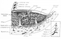
The primate placenta is of the hemochorial type in which clusters of fetal chorionic villi containing the fetal blood vessels hang free in a pool of maternal blood, the intervillous space. Maternal blood is supplied to this pool through the base of the placenta by endometrial branches of the uterine arteries. Blood is drained from the intervillous space by endometrial branches of the uterine veins (Fig. 1). Exchange of metabolites between mother and fetus takes place across the tissue of the villi in Whose fibrous core the fetal blood vessels are embedded.

Fig. 1. — Diagrammatic representation of a portion of the primate (hemochorial) placenta attached to the uterine wall. The inset shows, at higher magnification, the components of a single arterial stem. (From Ramsey,‘ in Report of 15th M & R Pediatric Research Conference 1955.)
Experimental procedures employed in the Carnegie Department of Embryology to study the circulation in the primate placenta have been as follows.
In rhesus monkeys studied at close intervals throughout pregnancy one or another of three techniques was used:
- Intra—aortic injection of India ink at laparotomy with subsequent removal of the uterus and serial sectioning of all or part of the placenta in situ and the full thickness of the adjacent uterine wall.3
- Injection of nontoxic colloidal mercuric sulfide into the femoral vein with subsequent laparotomy and removal of the uterus when the dye has been fully distributed through the systemic vascular bed. The placenta was sectioned as in the first technique.‘
- An “auto—injection” procedure whereby, at laparotomy, the in situ uterus was immersed in copious amounts of Bouin’s solution for periods of from 4-7 min. The uterine ligaments and their vessels were then clamped simultaneously and

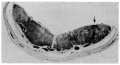
Fig. 2. — Mercuric sulfide injection of a monkey placenta. The arrow indicates the location of a spurt of dye high into the intervillous space from the mouth of an endometrial arteriole. Monkey C-747. 121 days pregnant. Primary placenta. Section B 17. x 2.
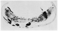
the uterus was removed for serial sectioning. Contact between the Bouin’s solution and the blood, which was still circulating during the interval before clamping, so changed the color of the blood that its course through the placenta could be traced in the sections.
In human uteri removed at operation or at autopsy at various stages of pregnancy from the 8th week to term, injections of India ink were performed. Canulas were inserted in one or both uterine arteries immediately after removal of the specimen and the vascular bed was flushed at pressures below diastolic arterial pressure with an isotonic saline solution to which 1 per cent sodium nitrate and 0.1 per cent histamine had been added. This perfusion was followed by India ink injection at the same pressure. The placenta was studied by serial sections a.s in the monkey experiments.‘
Plastic sheet reconstructions of human and rhesus material were prepared to show the changing character of the uterine vascular bed during pregnancy.
Whenever the point of connection of a maternal artery with the intervillous space was encountered in the sections it was noted that the maternal blood (or injection material) entered the placenta in the form of a “spurt” or fountain-like “jet” which carried the blood in a discrete mass far toward the chorion before lateral dispersion occurred (figs. 2 and 3). Repeated observation of this phenomenon suggested interpretation of the circulatory mechanism in terms of the following “physiological concept”:
“Arterial blood enters the placenta from the endometrial arteries under a head of pressure sufficiently higher than that prevailing in the vast, amorphous lake of the intervillous space that the incoming stream is driven high up toward the chorion. Gradually this force is spent and lateral dispersion occurs, aided by the villi which, acting as bafiles, promote mixing and slowing and by their own pulsation effect a mild stirring. Eventually the blood in the intervillous space falls back upon the orifices in the basal plate which connect with maternal veins, and since there is an additional fall in pressure between the intervillous space and the endometrial veins, drainage is accomplished.” This circulatory progress is enhanced by intermittent myometrial contractions throughout pregnancy.

Fig. 3. — Autoinjection of a monkey placenta. Bouin’s bath of in situ uterus. Maternal blood, colored by contact with Bouin’s solution, spurts high into the intervillous space at the point marked by the arrow. Monkey C-750. 123 days pregnant. Secondary placenta. Section 100. x2.
Physiological studies to test this working hypothesis have been carried out in the monkey. Pressure transducer systems have been introduced simultaneously into the femoral artery and femoral vein, the intervillous space of the placenta and the amnionic cavity in early, mid, and late pregnancy. Although pressures in the femoral artery and femoral vein are only roughly representative of the effective arterial and venous pressures within the uterine wall at the placental base, the data obtained have confirmed the pressure gradients assumed in the physiological concept and established the existence, character, and effect of the myometrial contractions
Highly suggestive as these results appear to be, they must be regarded as indirect evidence only, because of the artificial conditions introduced into the experiments. In the search for a more physiological procedure we have gratefully adopted a suggestion of Dr. Louis M. Hellman who proposed that we employ the technique of radioangiography with which he had performed some preliminary
studies in the human. This procedure, originally worked out by Fernstr6m° and Borell,7 has limited usefulness for prolonged or repeated investigations in humans because of the associated risk of overexposure to radiation.

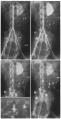
Fig. 4. — Photographs of four of a series of radiograms made at 3.5, 4, 5, and 6 sec, respectively, following injection of a radiopaque dye into the right femoral artery of Monkey 12B, 111 days pregnant. The insert in the third photograph (lower left) is a magnification (x4) of the area inclosed in the box. The arrows indicate spurts of dye into the intervillous space. RA, renal artery; SA, spiral artery of endometrium; HA, hypogastric artery; UA, uterine artery.
The pregnant monkey is anesthetized with intravenous pentobarbital sodium and one femoral artery is exposed under sterile conditions. A 20-gauge needle is then inserted into the artery counter to the flow of blood. The needle is connected, by means of a polyethylene catheter, to an automatic pressure syringe containing 15 cc of a 90 per cent solution of diatrizoate sodium (Hypaque), a well-tolerated tri-iodine, radiopaque dye. Under 90 lbs. of pressure delivered from a cylinder of compressed nitrogen the dye is instantaneously injected into the artery and at the same time rapid serial radiography is commenced. X-ray tubes fixed at right angles to each other permit simultaneous anteroposterior and lateral views to be taken. Exposures of 80 kv, 500 ma at 1/20 sec for the AP view and 95 kv for the lateral have been found to be optimum for a 5,000 to 6,000 gm monkey in mid-pregnancy. The X-ray films are contained in an automatic film changer (Schonander) set to deliver two films per second foratotal of 13 seconds. Single preliminary and post-experimental films are made to verify the stage of pregnancy and to check the time of clearance of the dye by the kidneys.
The representative X-rays reproduced in figure 4 illustrate the manner in which the retrograde progression of the dye may be followed from the point of injection. The dye courses up the femoral artery under pressure into the aorta and reaches the level of the left renal artery just as the applied pressure is spent. It then returns under the force of arterial pressure and enters the hypogastric arteries, the uterine arteries and finally the intervillous space. The characteristic spurts appear as the dye enters the placenta.
Discussion
The artificial conditions imposed by barbiturate anesthesia and incision into the femoral triangle do not alter the uterine circulation. Confirmation of this lies in the similarity of the circulatory pattern observed in the monkey with that in the unanesthetized human patients in whom injection was made through the intact skin.“ The high injection pressure is an essential part of the technique in order to avoid dilution and dispersion of the dye before it reaches the uterine arteries. As mentioned above, the excess pressure is dissipated before the dye enters the uterine vessels. Further evidence that the spurts have not been caused artificially lies in the fact that identical formations were obtained in the second type of anatomical study described above in which maternal pressure was the only propulsive force (Fig. 2).
Studies currently in progress are directed toward visualization of the venous drainage of the intervillous space and toward establishment of the circulatory pattern at various stages of pregnancy and under different conditions of myometrial activity.
It is concluded that radioangiography affords a physiological method for study of the circulation within the maternal placenta. Use of this technique in pregnant monkeys has provided results in full harmony with those obtained by anatomical studies and confirmed by pressure recordings. The uniformity of the results obtained by at least six different methods of study confirms the “physiological concept” that the circulation of blood in the maternal placenta is controlled by the head of pressure in the maternal arterial system.
References
1 Ramsey, E. M., Am. J. Anat., 98, 159 (1956).
2 Ramsey, E. M., Ann. New York Acad. Sci., 75, 726 (1959).
3 Ramsey, E. M., Contrib. Embryol., 35, 151 (1954).
4 Ramsey, E. M., in Proe. 3rd Conference on M icrocirculatory Physiology and Pathology, Amer. Physiol. Soc., (Baltimore: Waverly Press, Inc., 1958)
5 Ramsey, E. M., G. W. Corner, Jr., W. N. Long, Jr., and H. M. Stran, Am. J. Obst. and Gynee., 77, 1016 (1959).
6 Fernstrom, I., Acta rad2'ol., Stockholm, Suppl., 122, 1 (1955).
7 Borell, U., Geb. u. Frauenh., 18, 1 (1958).
8 Ramsey, E. M., “Uterine and Placental Circulation,” in Respiratory Problems in the Premature Infant, Report of the fifteenth M & R Pediatric Research Conference (1955).
Figures
1960 haemochorial placenta
Copyright
Proceedings National Academy of Sciences (PNAS) Liberalization of PNAS copyright policy: Noncommercial use freely allowed Note original Author should be contacted for permission to reuse for Educational purposes. See also PNAS Author Rights and Permission FAQs
- Cozzarelli NR, Fulton KR, Sullenberger DM. Liberalization of PNAS copyright policy: noncommercial use freely allowed. Proc Natl Acad Sci U S A. 2004 Aug 24;101(34):12399. PMID15314225 "Our guiding principle is that, while PNAS retains copyright, anyone can make noncommercial use of work in PNAS without asking our permission, provided that the original source is cited."
Cite this page: Hill, M.A. (2024, April 27) Embryology Paper - Radioangiographic studies of circulation in the maternal placenta of the rhesus monkey. Retrieved from https://embryology.med.unsw.edu.au/embryology/index.php/Paper_-_Radioangiographic_studies_of_circulation_in_the_maternal_placenta_of_the_rhesus_monkey
- © Dr Mark Hill 2024, UNSW Embryology ISBN: 978 0 7334 2609 4 - UNSW CRICOS Provider Code No. 00098G