Introduction
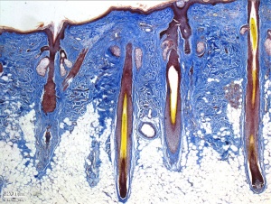
Adult skin histology showing epidermis, dermis and hypodermis as well as specializations, such as hair follicles and sweat glands
The skin provides a barrier between ourselves and our environment (temperature, water, UV), and contains specializations in different regions including hair, nails, teeth, glands and sensory receptors. In other species there are also specializations of beaks, scales and feathers.
The two major tissue organizations of epithelial (ectoderm, epidermis) and mesenchyme (mesoderm connective tissue, dermis and hypodermis) are shown within skin. In addition, we have also have extensive populating by melanocytes (neural crest) and sensory nerve endings.
Possibly the first epithelial tissue specialization from which arose other epithelial specializations now located inside the body. The external skin associated structures have many different roles and functions. This system is also an excellent model for distribution or "pattern" and adult stem cells.
Integumentary Lecture PDF
Shown below on this page is for only background information for this topic.
Textbooks
| References
|
| Hill, M.A. (2020). UNSW Embryology (20th ed.) Retrieved Mayıs 4, 2026, from https://embryology.med.unsw.edu.au
|
2015 Integumentary Lecture Slides PDF | 2014 PDF | 2010 Lecture
|
|
Moore, K.L., Persaud, T.V.N. & Torchia, M.G. (2015). The developing human: clinically oriented embryology (10th ed.). Philadelphia: Saunders. (links available to UNSW students)
|

|
Schoenwolf, G.C., Bleyl, S.B., Brauer, P.R., Francis-West, P.H. & Philippa H. (2015). Larsen's human embryology (5th ed.). New York; Edinburgh: Churchill Livingstone.
The following chapter links only work with a UNSW Library connection.
|
| Links: Embryology Textbooks
|
Objectives
- Understand the embryonic origin and differentiation of the epidermis and dermis.
- Understand the formation of hair and nails.
- Understand the formation of sweat glands, mammary glands.
- Understand the formation of teeth.
- Brief understanding of associated abnormalities.
|
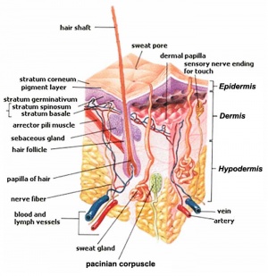
Skin structure cartoon
|
Online Textbooks
External Links
External Links Notice - The dynamic nature of the internet may mean that some of these listed links may no longer function. If the link no longer works search the web with the link text or name. Links to any external commercial sites are provided for information purposes only and should never be considered an endorsement. UNSW Embryology is provided as an educational resource with no clinical information or commercial affiliation.
Duverger O & Morasso MI. (2008). Role of homeobox genes in the patterning, specification, and differentiation of ectodermal appendages in mammals. J. Cell. Physiol. , 216, 337-46. PMID: 18459147 DOI.
Figure 1 Key steps in the development of three major ectodermal appendages
Terms
| Integumentary Terms
|
Integumentary Development
- acrosyringium - coiled intra-epidermal region of the eccrine gland sweat duct.
- apocrine gland - (sweat gland) proteinaceous secretion associated with hair (axilla, areola, genital and anal regions). Additional glands associated with eyelashes are called the glands of Moll (ciliary gland). (More? image - apocrine secretion)
- arrector pili muscle - bundle of smooth muscle associated with hair follicle, inserts into the papillary layer of the dermis and attaches to the dermal sheath of the hair follicle. (More? image - arrector pili muscle)
- Blaschko lines - (lines of Blaschko) may represent pathways of epidermal cell migration and proliferation during development. Specific type of lupus erythematosus shows this distinctive pattern. Named after Alfred Blaschko a German dermatologist who first described the feature in 1901. (More? PMID 21396561 | Historic Terminology)
- bulb - the hair follicle enlargement located at its deepest end, dividing cells form the hair and the root sheath.
- café-aut-lait macule - (French, cafe-au-lait = coffee with milk; birthmark) describes the characteristic colour of the skin hyperpigmented patch present at birth (congenital) or appearing in early infancy. Common single feature, multiple are associated with various genetic syndromes including Neurofibromatosis type 1 and 2.
- corneocytes - terminally differentiated keratinocytes forming the stratum corneum.
- cutis - alternative term for the epidermis and the dermis layers of the skin.
- dermal papillae - interdigitation of the dermis with the epidermis.
- dermatoglyphic patterns - (Greek, derma = "skin", glyph = "carving") fingers, palms, toes, and soles skin patterns.
- dermis - connective tissue middle layer of the skin, consists of two sublayers (papillary and reticular layers) that do not have a clear boundary. Embryologically derived from the somite dermatome.
- dermomyotome - Early embryonic dorsolateral half of the somite that will later divide to form both the dermatome and myotome. The dermatome will contribute the dermis and hypodermis of the skin. The myotome will contribute the skeletal muscle of muscoloskeletal system. Development sequence: mesoderm to paraxial mesoderm to somite to "dermomyotome" then dermatome and myotome. (More? Somitogenesis | Musculoskeletal System Development | Integumentary System Development)
- eccrine gland (Greek, ekkrinein = "secrete"; merocrine glands) sweat glands unique to some primates and used in humans for thermoregulation. Adult body has 2 to 4 million sweat glands with concentrations (700/cm) on the palms of the hand, soles of the feet and forehead. Secretion is timulated by sympathetic nervous system, post-ganglionic cholinergic branch, and other stimuli
- ephilis - (pl., ephilides; freckle) Clinical term describing a "freckle", that is a small brown or tan mark on the skin. These inherited features result from a copy of variant Melanocortin 1 Receptor (MC1R) gene and are common on fair skinned Celtic children. Melanocytes produce locally more melanin, this can also increase following exposure to ultraviolet radiation in sunlight. (More? Integumentary | Neural Crest | OMIM MC1R)
- epidermis - Histological term describing the external cellular epithelial layer of the integumentary (skin) covering the entire body. This surface layer of keratinocytes is ectoderm in origin, while the underlying connective tissue layers of dermis and hypodermis are mesoderm in origin. (More? Integumentary Development)
- epidermal differentiation complex - (EDC) human chromosome (1q2) containing linked 63 genes within four gene families that are molecular markers for stratified epidermis terminal differentiation.
- epidermal growth factor receptor - expressed on cells in the epidermis basal layer, signaling stimulates both epidermal growth and wound healing and also mediates an inhibition of differentiation.
- glabrous skin - skin without hair, refers to the palms of hands and soles of feet.
- hair - (pili) in humans consists of vellus and terminal hairs.
- holocrine - form of gland secretion where the secretory cells eventually lyse (rupture) and are lost. On the skin, these cells release sebum consisting mainly of lipid. (More? image - holocrine secretion)
- hypodermis - (subcutis, subcutaneous adipose) a connective tissue ilower layer of the skin that binds it to underlying structures.
- integumentary - term for the skin and its appendages.
- involucrin - protein that binds loricrin in the development of the cell envelope protecting corneocytes in the skin.
- keratinocyte - the main cell type forming the layers of the epidermis, derived from ectoderm.
- keratohyalin granule - found in the stratum granulosum consist of profilaggrin and loricrin.
- Langerhans cell - skin dendritic cell (antigen presenting cell) develops initially from fetal liver monocytes and yolk sac macrophages. May, depending on the immunological setting, elicit immunity or tolerance. Named after Paul Langerhans.
- Langer's lines - (skin cleavage lines, cleavage lines) Clinical term for the orientation of reticular dermis collagen bundles causing tensions on skin and subcutaneous tissues. Lines tend to be horizontal in the trunk and neck, and longitudinal in the skin and limbs. (More? PMID 15791423)
- Meissner corpuscle - sensory structure acting as a rapidly-adapting mechanoreceptor mainly in the dermal papillae of (digital) skin. (More?Touch
- melanin - (Greek, melanos = black) The pigment produced by melanocytes that provides photoprotection, preventing cellular DNA damage, and colouring of the basal epithelial cells that absorb the pigment.
- melanodermia - hyperpigmentation causing abnormal darkening (brown/black) of the skin due to excess melanin or by metallic substances. See also the abnormality ceruloderma (blue/grey). (More? PMID 23522626)
- melanocyte - (Greek, melanos = black) A pigmented cell, neural crest in origin, differentiating from melanoblasts located in the skin and other tissues that produces melanin. The melanocytes within the integument (skin) transfer melanin to keratinocytes to give skin colour and to the hair follicle to give hair colour. Melanocytes are also located within "non-cutaneous" tissues in the eye (for eye colour), harderian gland and inner ear. This is the cell type that proliferates in the cancer melanoma. (More? Neural Crest Development | Integumentary System Development)
- Merkel cell - An epidermal-derived cell in touch-sensitive area of the epidermis and mediate mechanotransduction in the skin. Previously thought to be neural crest in origin, but recently shown to arise from the embryonic epithelium. The cells are named after Friedrich Sigmund Merkel, a German anatomist who was the first to describe them in 1875. (More? Touch | Lecture - Integumentary Development | PMID 19786578 | PMID 3782861)
- merocrine gland - (sweat gland, eccrine sweat) simple tubular glands located at the border between the dermis and hypodermis. These glands regulate the body temperature. (More? image - merocrine secretion)
- nestin - (neuroectodermal stem cell marker) an intermediate filament protein (type VI) expressed in stem cells and transiently during development, and in cells within hair follicles, sebaceous and sweat glands.
- papillary layer - dermis sublayer that appears less dense and contains more cells lying close beneath the epidermis. (More? image)
- pilosebaceous unit - term used to describe a hair and its associated structures: hair follicle, arrector pili muscle and sebaceous gland.
- rete ridge - the extensions of the epidermis into the dermis. These epidermal surface thickenings extend downward between underlying connective tissue dermal papillae. This is also the site of initial eccrine gland differentiation.
- reticular layer - dermis sublayer that appears denser and contains fewer cells with thick collagen bundles lying beneath the papillary layer parallel to the skin surface. (More? image)
- root sheath - cell layers that surround the hair.
- sebaceous gland - holocrine gland associated with both the hair follicle and hairless parts of the skin (lips, cheek oral surface and external genitalia). Embedded in the dermis and are sites of infections (acne).
- simple - consisting of a single cell layer.
- terminal hairs - hair seen in obviously hairy parts of the body.
- thick skin - refers to the skin histology found on the palms of the hand and soles of the feet, does not contain hair. Note that this is used as a histological term not a measurement of overall skin thickness.
- thin skin - refers to the skin histology found on skin on all body regions, other than palms and soles (thick skin).
- vellus hairs - fine short hairs only lightly pigmented covering the body.
- vernix caseosa - (vernix, Latin, "caseosa" = cheese-like) a fetal protective coating consisting of sebum, skin cells and lanugo hair. Forming late in fetal development in a rostra-caudal sequence associated with epithelium differentiation.
- Voigt's lines - clinical term to describe the skin borders between areas of innervations by specific peripheral cutaneous nerves. (More? Sensory Touch | Historic Terminology)
|
|
|


