Book - Manual of Human Embryology 11D
| Embryology - 26 Jul 2026 |
|---|
| Google Translate - select your language from the list shown below (this will open a new external page) |
|
العربية | català | 中文 | 中國傳統的 | français | Deutsche | עִברִית | हिंदी | bahasa Indonesia | italiano | 日本語 | 한국어 | မြန်မာ | Pilipino | Polskie | português | ਪੰਜਾਬੀ ਦੇ | Română | русский | Español | Swahili | Svensk | ไทย | Türkçe | اردو | ייִדיש | Tiếng Việt These external translations are automated and may not be accurate. (More? About Translations) |
Bardeen CR. XI. Development of the Skeleton and of the Connective Tissues in Keibel F. and Mall FP. Manual of Human Embryology I. (1910) J. B. Lippincott Company, Philadelphia.
| Historic Disclaimer - information about historic embryology pages |
|---|
 |
XI. Development of the Skeleton and of the Connective Tissues
By Charles R. Bardeen, Madison, Wis.
- Skeleton and Connective Tissues: Connective Tissue Histogenesis | Skeletal Morphogenesis | Chorda Dorsalis | Vertebral Column and Thorax | Limb Skeleton | Skull Hyoid Bone Larynx
D. Skeleton of the Limbs
One of the most studied subjects in morphology has been the development of the vertebrate limbs. Since, fortunately, critical summaries of its literature have recently been given by several noted investigators, among whom may be mentioned Wiedersheim (1892), Mollier (1893, 1895, 1897), Gegenbaur (1898), Rabl (1901), Fiirbringer (1902), Ruge(1902), and Braus (1904), no attempt will be made here to review this work except so far as it deals directly with the development of the human limb.
During the third week of embryonic life the limb buds become filled with a vascular mesenchyme. The source of this tissue is uncertain. In part it may come from the primitive body-segments, but it seems probable that in the main it comes from the parietal layer of the unsegmented mesoblast. Toward the end of the fourth week a slight condensation of the mesenchyme can be seen at the centre of the arm bud, and early in the fifth week a similar condensation may be noted in the leg bud. This condensation represents the first rudiment of the skeleton of the limb. The tissue composing it may therefore be called * * scleroblastema. " From the scleroblastema there is developed a membranous skeleton. In this a cartilaginous skeleton is differentiated, and this in turn is replaced by the permanent osseous skeleton. We may thus distinguish three overlapping periods, a blastemal, a chondrogenous, and an osseogenous. We shall first consider in some detail the development of the skeleton of the inferior extremity and then more briefly that of the superior extremity.
Inferior Extremity
Blastemal Period
At the time when the condensation takes place in the leg bud the latter has the general form shown in outline in Fig. 274. The bud projects considerably from the body, but shows no definite resemblance to the limb to which it is to give rise. The condensed tissue, scleroblastema, is not sharply outlined. It represents the region of the acetabulum and the proximal end of the femur.
Once begun, skeleton differentiation proceeds rapidly. In an embryo 11 mm. long (Fig. 275) it may be seen that from the original centre of skeletal formation the condensation of tissue has extended both distally and proximally, but much more freely in the distal direction. Distally the scleroblastema shows femur, tibia, fibula, and a foot-plate; proximally, an iliac, a pubic, and an ischial process. A series of sections through the skeletal mass shows that in the femur, tibia, and fibula chondrification has begun. At centres in the blastema of the ilium, ischium, and pubis a still earlier stage of chondrification has made its appearance. The leg of this embryo, therefore, represents a stage of transition from the blastemal to the chondrogenous stage of development.
Chondrogenous Period
The further development of the skeleton of the limb during the second and third months of intra-uterine life may be followed in Figs. 276, 277, and 278. For the sake of convenience the development of the several parts of the skeleton will be taken up as follows: (a) the os coxae; (6) femur, hip-joint, tibia and fibula, and knee-joint; (c) ankle and foot.
(a) The Os Coxm. — The pelvic scleroblastema of embryos of the stage illustrated in Fig. 275 undergoes a rapid development. Its iliac portion extends in a dorsal direction toward the vertebrae which are to give it support. The costal processes of the latter at the same time become fused into a dense mass of tissue which enters into close association with the iliac blastema (Fig. 276), although for some time separated from this by a narrow band of tissue staining less densely than the blastema. Cranialwards the iliac blastema extends toward the abdominal musculature, to which it finally gives attachment.

Fig. 274-278. — (After Bardeen, Amer. Journ, of Anat.. 1905.) Lateral view of models to illustrate the development of the distal part of the spinal column and of the inferior extremity of embryos 9-50 mm. long, In Figs. 274. 275, and 276 the scleroblastema is shown, and this is in Figs. 275 and 276 the centres of chondrification. In Figs. 277 and 278 the cartilaginous skeleton is shown, and in this in F 27.
Length of embryo, mm. Fig. 275. Iength of embryo. 11 mm. Fig. 276. Length of e bryo. 14 mm. Fig. 277. Length of emhryo. 20 mm. Fig. 278. Length of fetus. 5O mm. Chd,. ehoi dorsalis; Co", first coccygeal vertebra; Cnifa /*, iwcHih rib: Fi, fibular F.o., foramen obturatum; . ilium; L.i., ligamentum inguinale: M.id., membnns interdorsalin: P.. pubis; Pr.a.o.. proee.wus artii processus tmnaverBUs ; Ti, tibia.
While the blastemal ilium is thus becoming differentiated the pubic and ischial processes of the pelvic blastema extend rapidly forward. Ventral to the obturator nerve they become united by condensed tissue, which completes the boundary of the obturator foramen. Betwen the crest of the ilium and the ventral extremity of the pubis dense tissue is formed to give attachment to the oblique abdominal musculature. This represents the embryonic inguinal ligament and completes a femoral canal (Fig. 276).
While the blastemal pelvis is being completed the three centres of chondrification, barely visible in the 11 mm. embryo, give rise respectively to iliac, pubic, and ischial cartilages in which the adult form becomes gradually more distinct. (Compare Figs. 276, 277, and 278.) In embryos between 15 and 20 mm. long each of the three cartilages gives rise to a plate-like process over the head of the femur. These processes fuse with one another and give rise to a shallow acetabulum (Fig. 277), which during the third month gradually becomes deeper (Fig. 278). The iliac and ischial cartilages furnish a greater part of the floor of the acetabulum than the pubic cartilage and unite with one another before being joined by the pubic cartilage.
Toward the end of the second month and the beginning of the third month of development the symphysis pubis is formed. This is at first composed of dense blastemal tissue. In this tissue first hyaline and then fibrocartilage become differentiated. At the centre of the joint a slight fissure may appear in adult life (Farabeuf, 1895).
(b) Femur and Hipjoint. Tibia, Fibula, and Knee-joint. — The rapid development of the blastemal skeleton of the lower limb has been briefly described above. Soon after the anlage of the femur makes its appearance condensation of tissue marks out the anlages of the tibia and fibula and the skeleton of the foot. This last seems to be at first a somewhat irregular continuous sheet of tissue. It is not clear whether or not the anlages of the tibia and fibula also begin as a continuous sheet which becomes divided, by ingrowth of blood-vessels, into tibial and fibular portions. The incomplete development of the interosseous fissure in an 11 mm. embryo suggests this (Fig. 275). The blastemal anlages of the tibia and fibula are here very incompletely separated.
Within the blastema of the femur, tibia, and fibula chondrification begins as soon as the outlines of the blastemal skeleton are fairly complete (Fig. 275). The embryonic cartilage appears slightly kneewards from the centre of the shaft of each bone and then eKtends toward the ends. The cartilage of the femur consists of a bar largest at the knee, whence it tapers off toward the hip. The cartilages of the lower leg lie nearly in a common plane. That of the tibia is larger than that of the fibula and toward the knee it broadens out considerably. At this stage the joints consist of a solid mass of mesenchyme (Figs. 279 and 280). The tissue uniting the femur and tibia has temporarily somewhat the appearance of precartilage (Fig. 283). From this period onwards the development of the individual bones and joints is rapid.

Fig. 279 to Fig. 284.
The cartilaginous femur expands at the expense of the surrounding blastemal perichondrium and at the same time acquires adult characteristics (Figs. 276, 277, and 278). the hip-joint is at first completely filled with a dense blastemal tissue (Fig. 280). While the embryo is growing from 20 to 30 mm. in length, cavity formation begins in the tissue lying between the cartilaginous floor of the acetabulum and the head of the femur. The first stage in the process is marked by a condensation of the capsular tissue immediately bordering upon the joint and of the perichondral tissue which at this stage covers the cartilages on tiieir articular surfaces as well as elsewhere. In the region of the ligamentum teres a fibrous band is likewise differentiated from the blastema of the joint. The rest of the tissue becomes looser in texture and ultimately is absorbed (Fig. 284). Henke and Eeyher (1874) gave a good account of the development of the hipjoint. Moser (1893) has described that of the ligamentum teres. Schulin (1879) has given a good account of the later development of the joint cavity in its relations to the head and neck of the femur (see Figs. 286-288). It is to be noted that in the fetus 25 cm. long (Fig. 286) the joint cavity extends about the neck of the femur in a pocket lined on one side by perichondrium, on the other by the capsule of the joint, and that later the peridiondral lining becomes periosteum (Fig. 288).

Fig. 285 to Fig. 288. (After Schulin, Archiv f. Asatoici*. 1879.) Fig. 285. Median section through the knee-joint of a fetus 13 cm long. a, patella; b. connective tissue over patella; c, lig
Fig. 286-288. Hip-joint of a male fetus 25 cm. long, of a female child six years old, and of a male adult. a. region of insertion of joint capsule; b, intracapsular connective-tissue surface; c, limit of endochondral ossification; d, epiphyseal osseous nucleus; e, ligamentum teres.
The tibia and fibula at first lie nearly in the same plane (Fig. 275). As the head of the tibia enlarges toward the knee-joint it comes to lie ventral to the proximal extremity of the fibnla. This may be seen in Figs. 276 and 277.
The development of the knee-joint in man has been studied by a number of competent observers. Bernays (1878) gave a good review of the previous work of von Baer, Bruch, Henke, and Reyher, and an accurate description of the processes which take place. Of the more recent articles those of Schulin (1879), Kazzander (1894), and Lucien (1904) deserve mention.
Until the embryo reaches a length of about 17 mm. the kneejoint is marked by a dense mass of tissue (Fig. 279). The medullary tissue at the knee, like that at the hip and other joints, is less dense than the surrounding cortical substance, so that when the cartilages of the femur, tibia, and fibula are first differentiated they seem to be connected by a tissue which, in some respects, resembles the cartilage of which they are composed (Fig. 283) ; but as the cartilages become more definite the apparent continuity disappears. As the musculature becomes differentiated a dense tendon for the quadriceps is formed in front of the knee-joint. At this period the joint is flexed at nearly a right angle.
In embryos of about 20 mm. the tissue immediately surrounding the cartilages becomes greatly condensed into a definite perichondrium. The peripheral blastemal tissue at the joint becomes transformed into a capsular ligament, strengthened in front by the tendon of the quadriceps. Within the joint most of the tissue begins to show signs of becoming less dense, but the menisci and the crucial ligaments, like the ligaments of the capsule, are differentiated directly from the blastema (Figs. 281 and 282). In the differentiation of the articular blastema the menisci first become distinct, then the capsule, then the crucial ligaments, the patella, and the lig. mucosum.
A knee-joint cavity first appears, in embryos about 30 mm. long, between the patella and the femur. according to Lucien, two other cavities somewhat later appear between the condyles of the femur and the menisci. These cavities secondarily communicate with the retropatellar cavity and with cavities formed between the menisci and the tibia. The cavity of the knee-joint is primitively partly divided into two parts by a median septum (lig. mucosum), which becomes greatly reduced in fetuses 10-12 cm. long (Fig. 285, C) and in the adult is replaced by a fat pad.
The shafts of the tibia and fibula are incompletely separated in the blastemal stage. The cartilages which arise in the scleroblastema are, on the other hand, separated by a distinct interval (Fig. 279). At first short and thick, the shafts become gradually more slender in proportion to their length. The fibula, at all times smaller, becomes increasingly more slender in comparison with the tibia. In fetases 50 mm. long (Fig. 278) both bones, and especially the fibula, are still relatively thick compared with the adult bones.
During a period of rapid development, in embryos of 15 to 20 mm., the tibia and fibala, like the femur, may extend so rapidly in length as to become temporarily distorted by resistance at the ends. This is often especially marked in hardened specimens. Holl (1891), Sohomburg (1900), and others have called attention to this distortion.

Fig. 289. — (After R. Quain. Quain's Anatomy, 10th ed„ vol. ii. Pt. I. Figs. 157 and 158) A. Bone at birth. B. Child under six years of age. C. Child two to three years older than B. D. Person of about twenty years. E. Acetabular region of hip-bone at fourteen years of age. 1. ilium; 2, ischium: 3, pubis; 4. os acetabuli; 5. bony nodules between ilium and ischium: 6 and 7, epiphyseal laminae on ilium and ischium; 8, 9, 10. 11, epiphyses of anterior inferior iliac spine, iliac crest ischial tuberosity, and symphysis pubis.
(c) Ankle and Foot. — Of the papers dealing with the early development of the skeleton of the human foot the more important are those of Henke and Reyher (1874), Leboucq (1882), v. Bardeleben (188.3, 1885), Lazarus (1896), and Schomburg (1900).
During the fifth week of embryonic development the free extremity of the limb bud becomes flattened and differentiated into a foot-plate (Fig. 275). Toward the end of the fifth week the anlages of the individual bones of the ankle and foot begin to become marked by specific condensation of the blastemal tissue. Within these anlages precartilage soon appears. The digital rays are marked at first by condensed bars of tissue, in which segmentation into metatarsals and phalanges appears during the period of chondrification (Fig. 276). The metatarsal cartilages become differentiated before the tarsal cartilages. The phalangeal cartilages appear relatively late.

Fig. 290. — (After R. Quain, Quain's Anatomy, 10th ed., vol. u, Pt. I, Fig. 169.) Ossification of the femur. A. Before the eighth month. B. At birth. C. About a year old. D. At about the fifth year. E. Near the age of puberty. 1, diaphysis; 2, distal epiphysis; 3, head; 4, great trochanter; 5, small trochanter.

Fig. 291. — (After R. Quain, Quain's Anatomy, 10th ed., vol. ii, Pt. I. Fig. 160.) Ossification of the tibia. A. Some weeks before birth. B. At birth. C. At the third year. D. Betweoi the eighteenth and twentieth years. E. Example of separate centre for tuberosity. 1, diaphysis ; 2, proximal epiphysis ; 2*, epiphysis of tuberosity; 3, distal epiphysis. The earliest appearance of the tarsal cartilages is found in an embryo about 14 mm. long (Fig. 276). Toward the end of the second month these cartilages become much more distinct (Fig. 277). By the middle of the third month the cartilages of the foot have a form distinctly corresponding to the adult. The similarity is still better marked at the end of the third month (Fig. 278).
The joint cavities begin to develop while the embryo is growing from 25 to 30 mm. in length. As in other cases, so here the blastemal tissue in which the cartilages are developed becomes condensed at their articulating ends and about the joint, while in the region of the joint the tissue becomes less dense and finally disappears, leaving a joint cavity. In embryos of about 30 mm. the joint cavities of the foot are filled with a loose fibrous tissue ; in fetuses of 50 nun. definite cavities are to be made out. During the progress of form differentiation above described, the shape of the foot is markedly altered. At the beginning of the development of the foot the tarsal and metatarsal bones lie nearly, though not quite, in the same plane as the bones of the leg. They are so arranged, however, that the foot is convex on its dorsal surface and concave on the plantar, and the projections of the calcaneus and talus serve to deepen the plantar fossa. The metacarpals spread widely apart. As differentiation proceeds, the metatarsals come to lie more nearly parallel to one another, and the tarsal elements become compacted in such a way as to give rise to the tarsal arch. The foot at the same time is dorsally flexed at the ankle and slightly everted. The toes are flexed. In the further development of the skeleton of the foot the various constituent structures are elaborated, and the foot gradually becomes more flexed dorsally and turned toward the fibular side.

Fig. 292. — (After R. Quain, Quain's Anatomy, 10th ed., vol. ii, Pt. I, Fig. 161.) Oaflification of the fibula. A. At birth. B. At about two yean. C. At about four years. D. At about twenty years. 1, diaphysis; 2, distal epiphysis; 3 proximal epiphysis.
Period of Ossification
The hip-bone is ossified from three primary centres, one for each of its constituent parts, the ilium, ischium, and pubis, and from several epiphyses. There is one primary centre of ossification for each of the other bones of the inferior extremity, and in addition most of the bones have one or more epiphyses. In the tarsus the calcaneus alone regularly has an epiphysis. The patella has no epiphysis. With the exception of those of the tarsal bones and of the various sesamoid bones the primary centres of ossification appear relatively early in intra-uterine life. At birth there are usually centres of ossification present in the calcaneus, talus, and cuboid, but not in the other tarsal bones. Ossification in these latter tarsal bones and in the sesamoid bones and the various epiphyses appears after birth. In the talus, according to Sewell (1906), dark-staining regions in the hyaline cartilage of which it is composed in the sixth fetal month indicate structural features characteristic of the adult bone. The following tables and the accompanying figures illustrate the process of ossification in the inferior extremity. Authors differ in the data which they give concerning the time of ossification of the various bones. When not otherwise indicated the tenth edition of Quain 's Anatomy is followed in the tables.

Fig. 203. — (AfMr R. Quain, Quain's the bona of (he foot. A. Right foot or a digital phlange< an owHlied. The Uraus i caneus. B. Fetus o( 7-8 monthe. Nurleit y«r. Nucleus in third cuDeiform. E. Ii G. About the age ol puberty, calcaueua. Epiphyees of metacaneusi 1', in G, the epiphyMs of the caleaneu
nueloua o( the talun; 3, of the euboid : . . . . uLar; 7. of thosecond ouneiform; S, metAlanal bones; 8'. distal epiphysis of llie senmd metatarsal bone; S" p^o^i^lBl etiiphysis of the first metBtanal bone ; 9, first phalanx of Ihe second toe; 9', proximal epiphysifi of this phalanx; 9*, (hat of the first phalanx of th« ereiit U>9 ; 10. second phalanx; 10'. the ^pbysis of tliie phalanx; 10*, epiphysia of the terminal phalanx of the great toe : 11. terroiDal phalanx; 11 ', iu epiphysis.
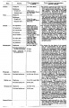
Table of Ossification of the Bones of the Inferior Extremity
| Bone | Centres | Time of appearance of centre | Time of fusion: general remarks |
|---|---|---|---|
| Os coxae | Os ilium | 56th day (Mall) | The rami of the ischium and the pubis are united by bone in the 7th or 8th year (Quain) ( 12-14 year Sappey). In the acetabulum the three hip bones are separated by a Y-shaped cartilage until after puberty. In this cartilage between the ilium and pubis the "os acetabuli" appears between the ninth and twelfth years. This bone, variable in size, forms a greater or less part of the pubic portion of the articular cavity. Leche (1884). Krause (1885), and many others consider it primarily an independent bone. About puberty between the ilium and ischium and over the acetabular surfaces of these bones small irregular epiphyseal centres appear. The os acetabuli becomes imited to the pubic bone about puberty and soon afterwards the acetabular portions of the ilium and ischium and the ischium and pubis begin to become united by bone. The acetabular portions of the pubis and ilium are unite a little later. Osseous union takes place earlier on the pelvic than on the articular surface of the acetabulum. The union of the several primary centres and the epiphyses is usually completed about the twentieth year. |
| Os ischii | 105th day (Mall) | ||
| Os pubis | 4th to 5th fetal month | ||
| Os acetabuli. | 9th to 12th year | ||
| Epiphyses:
Those of the acetabulum |
Soon after puberty | ||
| Crest of ilium | Soon after puberty | Fuses with main bone 20th to 25th year | |
| Tuberosity of ischium | Soon after puberty | Fusion begins in the 17th year and is completed between the 20th and 24th years (Sappey) | |
| Ischial spine | Soon after puberty | 18th to 20th year (Poirier). | |
| Ant. inf. spine of ilium | Soon after puberty | 18th to 20th year (Poirier) | |
| Symphysis end of os pubis (1 or 2 centres) | 18th to 20th year (Sappey) | After the 20th year | |
| Femur | Diaphysis | 43d day (Mall) | |
| Epiphyses:
Distal end |
Shortly before birth1 | 20th to 24th year | |
| Head | 1st year | 18th to 19th year | |
| Great trochanter | 3d to 4th year (Osseous granules soon after birth, (Poirier) | 18th year | |
| Small trochanter | 13th to 14th year
8th year (Sappey) |
17th year (Quain)
Proximal epiphysis 18th to 22d year (Poirier) | |
| Patella | 3d to 5th year | The osseous patella reaches its definitive form soon before puberty | |
| Tibia | Diaphysis | 44th day (Mall) | |
| Epiphyses:
Proximal end |
About birth | 19th to 24th year (Sappey) | |
| Distal end | 2d year | 16th to 19th year | |
| Tubercle (occas.) | 13th year | Fuses with epiphysis of the proximal end and then with this to the diaphysis | |
| Fibula | Diaphysis | 55th day (Mall). | |
| Epiphyses:
Distal end |
2d year | 20th to 22d year | |
| Proximal end | 3d to 5th year | 22d to 24th year | |
| Calcaneus | Chief centre | 6th fetal month | The chief nucleus is endochondral. A periosteal nucleus appears frequently in the 4-5 fetal month (Hasselwander) |
| Epiphysis (distal end) | 10th year (Quain)
7th-8th year ( Sappey) |
15th-16th year (Quain)
16th-18th year (Poirier) M 17-21, average 20 years F 13-17, average 16 years (Hasselwander) | |
| Talus | 6th fetal month (Hasselwander) | In the 7th-8th year the posterior part of the talus, the os trigonum, is frequently ossified from a special centre (v. Bardeleben). It fuses about the 18th year. | |
| Cuboid | About birth | ||
| Cuneiform III | 1st year | ||
| Cuneiform I | 2d-3d year | ||
| Cuneiform II | 3d-4th year | ||
| Navicular | 4th-5th year | ||
| Metatarsals | Diaphyses | 8th-10th week | According to v. Bardeleben a second centre of ossincation appears much later than the primary in the navicular, and finally about the time of puberty a medial epiphyseal centre arises. |
| Epiphyses | 3d-8th year | The centre for the 2d metatarsal usually appears first, then come the 3rd, 4th, 1st and 5th. The epiphysis of the 1st metatarsal appears at the proximal end of the bone: the other epiphyses arise at the distal ends of the metatarsals. There may be a distal epiphysis in the first metatarsal also.2 In some instances a proximal epiphysis is formed on the tuberosity of the fifth metatarsal (Gruber). The epiphyses unite with the shafts in the 17-21 year in males and in the 14-19 year in females. (Hasselwander). | |
| Phalanges: | |||
| Terminal row | Diaphyses | 58th day (Mall) | |
| Epiphyses (distal) | 4th year | M 13-23, average 16-21 year.
F 13-17, average 14-17 year (Hasselwander). | |
| Middle row | Diaphyses | 4th-10th fetal month | |
| Epiphyses | 3d year | M 15-19 year
F 13-16 year (Hasselwander) | |
| Proximal row | Diaphyses | 3d fetal month | |
| Epiphyses | 3d year | M 15-17 year.
F 14-15 year (Hasselwander) The centres for the shafts of the phalanges often appear double, one for the dorsal and one for the plantar surface. The centres for the medial phalanges in each row usually appear before the more laterally placed centres. The centre for the 5th terminal phalanx appears much later than the other centres in this row (Mall). According to Rambaud and Renault the epiphyses arise each from two centres which fuse together. In the terminal phalanx of the great toe the ossification centre of the epiphysis often appears as early as the second or even the first year. (Hasselwander) | |
| Sesamoid bones of the great toe | M 14th year
F 12th-13th year |
Ossification may begin in the 8th year in females, in the 11th in males (Hasselwander). | |
| |||
| Days and weeks refer to the prenatal, years to the postnatal period. M = male F = female. According to Poirier, Traite d'Anatomie, p. 138, two centres appear in the eighth week, and unite in the third month to form a centre of ossification for the body of the scapula. | |||
| Links: limb | bone | upper limb ossification timeline | lower limb ossification timeline | Historic - Chapter 11 Development of the Skeleton | timeline | Category:Timeline Table Data Reference[1] | |||
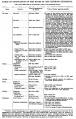
Part 1 Table as picture.
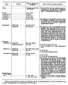
Part 2 Table as picture.


Infantile Characteristics of the Skeleton of the Inferior Extremity. — In the infant the pelvis is small in proportion to the size of the body and contains a smaller proportion of the abdomino-pelvic viscera than in the adult. The cavity of the infantile pelvis is cone-shaped and diminishes in diameter from the entrance to the outlet (Fehling, 1876, Hennig, 1880). The blades of the ilium are relatively slightly developed. In the first half of fetal life the sacropelvic angle is similar to that of quadrupeds, but during the latter half and after birth the angle becomes greater, expanding from 55** to 90-110" in the adult (Le Damany)."
The acetabulum is relatively shallow in the new-bom as compared with the adult. The shafts of the long bones are relatively shorter and thicker. The neck of the femur is but slightly developed at birth. The infantile foot has certain ape-like characteristics and is strongly flexed and inverted. The head of the talus is directed more medialwards than in the adult, the first metatarsal is relatively short and inclined medialwards by the oblique articular surface of the first cuneiform (Leboucq, 1882).
Superior Extremity
Blastemal and Chondrogenous Periods
In general the development of the superior extremity resembles that of the inferior extremity. The various stages of differentiation begin in the former a little earlier than in the latter. W. H. Lewis (1902) has described the earlier stages in the development of the arm. His description is closely followed here.

Fig. 294. —
In an embryo at the end of the fourth week the scleroblastema of the limb bud is marked by a slight condensation of the tissue near the future head of the humerus. Early in the fifth week this condensation has extended to the distal part of the limb bud and the anlages of the scapula, humerus, radius, and ulna are distinguishable (see Fig. 294). The skeleton of the wrist and hand is marked by a plate of condensed tissue. There are no distinct centres of chondrification at this stage.
In an 11 mm. embryo marked alterations have taken place in the skeleton of the superior extremity (Fig. 295). Centres of chondrification appear.

Fig. 295. —
The scapula is composed of precartilage surrounded by a dense blastema. It lies opposite the lower four cervical and the first one or two thoracic vertebrae. From the superior border there springs a large curved acromion process. On the medial (costal) surface, at the junction of the humerus with the scapula, arises a large hooked coraccid process. A slight ridge on the medial surface marks the future anterior border. The perichondrium is well marked onlv on the medial surface.
The clavicle is an ill-defined mass of condensed tissue which extends from the acromion about a third of the distance to the tip of the first rib. The coraccclavicular ligament is partially differentiated.
For recent accounts of the development of the pelvis, see Merkel (1902) and Falk (1908). Fehling recognized sexual differences in the pelvis early in fetal life.
The humerus is short and thick. The shaft is composed of embryonic cartilage surrounded by a dense layer of perichondrium. Towards each end of the shaft the central tissue is precartilaginous in character. The surrounding perichondrium is continued directly into the dense tissue of the neighboring skeletal parts.
There is more flexion at the elbow than during the preceding stage. The forearm is midway between supination and pronation. The core of the shaft of each bone is composed of hyaline cartilage.
The hand-plate is composed of condensed mesenchyme. There are several centres of increased condensation which probably correspond to the carpal bones. The digits are marked by condensed tissue in which no segmentation into metacarpals and phalanges is visible.

Fig. 296. —
In an embryo of 14 mm. the skeleton of the superior extremity is well advanced in development (Fig. 296). The form of the scapula is shown in this figure. It is composed mainly of cartilage, covered by a thick layer of perichondrium. It has migrated caudalwards so that less than one-half of it lies anterior to the level of the first rib. The clavicle is a rod composed of dense tissue. It extends from the acromion to the tip of the first rib, where it is continued into the sternal anlage. It contains a small core of a peculiar precartilaginous tissue. The acromioclavicular ligament is distinct. The humerus is larger and more slender than at the preceding stage and has expanded at each end. It is composed chiefly of cartilage surrounded by a thick perichondrium which is continuous with that of the lateral angle of the scapula. There are no signs of a joint cavity at the shoulder.
The ulna and radius are likewise composed of cartilage surrounded by a thick perichondrium continuous at one end with that of the humerus and at the other with that of the wrist. There are no joint cavities at the elbow.
The carpus is composed of a dense tissue in which are embedded cartilages which represent the bones of the wrist with the exception of the lunar and the pisiform. These are still composed of condensed tissue.
The metacarpals are represented by five slender cartilages surrounded by a dense perichondrium. The first metacarpal is only about half the length of the others.
The phalanges of the first row, with the exception of that of the thumb, have cartilaginous cores. The basal phalanx of the thumb is composed of condensed tissue. At the tip of each digit is a mass of condensed tissue. There are no joint cavities present in the hand.

Fig. 297. —
In an embryo 20 mm. long the cartilaginons anlages of various bones of the superior extremity are all well marked, except those of the distal row of the phalanges of the fingers. The clavicle extends from the acromion to the sternum. It is composed of a peculiar kind of precartitaginous tissue. The general shape of the other cartilages may be seen from Fig. 297. The spine of the scapula is not yet distinct. There are distinct coraccclavicular, costoclavicular, and interclavicular ligaments. There is no joint cavity at the shoulder, but a capsular and a coraeo-humeral ligament may be distinguished. The humerus has well-marked tuberosities and condyles. The ulna and radius are larger and longer than at the preceding stage. The olecranon, coraccid, and styloid processes are composed of cartilage and condensed tissue. The perichondrium about the ulna and radius is quite thick. The capsular and annular ligaments are present, but there are no joint cavities.
Magn. R : 1. Flu. 2Bn.— (AfU im. emiir.vo. Magn. about 13 :
All the bones of the carpus have cartilaginous centres. There are no joint cavities in the hand.
During the third month of development the cartilages of the superior extremity assume more and more the form characteristic of the adult bones ; in several ossification begins ; the joint cavities appear at this time.
The early development of the bones of the forearm and hand, and especially those of the wrist, has engaged the attention of several investigators. The following details are based upon the recent paper of Graefenberg (1906).
Forearm
The form and relations of the cartilaginous radius and ulna in the fifth, sixth, and the seventh weeks of embryonic life are shown in Figs. 298, 299, 300. The two cartilages are at first some distance from one another. The proc. styloideus of the ulna begins to develop during the latter part of the second month. It extends at first to the dorsal side of the triquetrum and becomes relatively large. Later the process becomes smaller, and is carried proximally and volarwards.
The discus articularis arises from a mass of tissue which lies between the radius and the styloid process of the ulna. This mass of tissue gives rise to a special centre of chondrification, which by some is supposed to represent the os intermedium antebrachii of the lower vertebrates.
The Carpus
Os Centrale. — Most of those who have studied the development of the human carpus have described a cartilage which is homologous with the os centrale of the carpus of lower vertebrates. The position of this element is shown in Figs. 298, 299, and 300. It later disappears. In the process of retrograde metamorphosis it may become divided into several parts. according to Graefenberg it does not fuse with any of the other carpal.
The Proximal Row or Bones. — The navicular arises from two centres of chondrification. It is homologous with the radiale of lower forms. The lunar is the last of the carpalia to be differentiated. according to Gegengaur and some other investigators, it is homologous with the os intermedium of the lower vertebrates. In man, however, there are no indications of its wandering from the forearm into the wrist. The triquetrum is relatively small when first differentiated, but grows rapidly in size (Figs. 298, 299 and 300). Perna has found it arising from two centres of chondrification. The pisiform is relatively large in embryonic stages. It is a canonic carpal element and not a sesamoid bone. It arises later than the triquetrum. During its development it wanders from the ulnar margin to the volar surface of the triquetrum.
The Distal Row. — The cartilages of the distal row are at first relatively large compared with those of the proximal row

Fig. 298-299. —
J. AnBlomiMhe Hefte, IBOO, Fi«. 1.) Dorml view of ■ model of th» le forearm and hand of * Gve-weeks human onbryo. Magn. TO : I. jf ibe skeleton of the forcann and hand of a
liniu; the cmtmla betwe
atum; »un.. humerus; M.mo)'., multaii(ulun ., pare radialit, ubt., i»r« ulnaris : P., pisiforme ; The oafwtatum Um betweeo tha bunatum and the muKanpi

Fig. 300. — A. (After Grfenberc, 1906. fig. G.) Skeleton of the right hand of a ten-weeks human fetus.
(Figs. 298 and 300). The capitatum is the largest, next comes the hamatum. The multangalar carpalia are small; the M. majus is for a time considerably smaller than the M. minus. The capitatum and hamatum are the first elements of the carpus to undergo chondrification. The hamulus ossis hamati is differentiated from a special centre of chondrification.
urn tuid hsmBtum. B. Elbow-joint of a fetui c. centnl udLdq: d, perichoadr*! put of Juia anoa. D, £, F. Should er-joiat o[ a fetua 13 m of mpiiulei t. intracepsuUr eonnwtive tiMi
Anat. Abt., 1879.) Fir diocarpflj ioiut in thrc«
Metocarpalia. — These are at first relatively large. The first metacarpal, according to some investigators, represents a basal phalanx. Galen was the first to express the view that the metacarpal of the thumb is not present. Others have thought that a phalanx is missing from the thumb, but Graefenberg accepts the view of Galen. The other four cartilaginous metacarpals arise at some distance from one another (Fig. 298). They spread apart distally. The bases are first brought into contact with one another, and later the distal ends. The fifth metacarpal articulates at first with the triquetrum (Fig. 298) and later with the hamatum (Fig. 299).
The phalanges are differentiated in serial order, the basal row appearing first, the terminal row last. according to Graefenberg, the terminal phalanges show evidence of being composed of two elements, a proximal and a^ distal. The latter is composed of cartilage, the cells of which rapidly enlarge. It may represent a fourth phalanx. Primitively the digits were probably composed of many phalanges. The terminal phalanges are at first smaller than those of the middle row, but then develop faster so as to exceed them in length. After a time retrograde metamorphosis overtakes the distal ends of the terminal phalanges, so that the middle row once more exceeds the terminal in length. The tuberositas unguicularis is composed of fibrous tissue which becomes transformed directly into bone.
The sesamoid bones, according to Thilenius, are more numerous in the fetus than in the adult. The following table, after Pfitzner and cited by Dwight (1907) illustrates this. This table is based on a study by Thilenius of 30 hands of fetuses of the fourth month, and of 1440 hands by Pfitzner of individuals from fourteen to eighty-nine years of age. The Roman numerals indicate the metacarpophalangeal joints opposite which sesamoid bones were found, and the Arabic numerals represent the percentage of frequency with which the bones were found.

Fig. 302. (After R. Qiiain, Qtiain's Anatomy, 10th ed., vol. ii, Pt. 1, Fig. 1 15.) Ossification of the clavicle. A. Clavicle at birth. B. At about the twenty-third year. 1, shaft; 2, epiphyma.

Joints
Schulin (1879) has given some account of the development of the joints of the upper extremity.

Fig. 301. —
At the shoulderjoint (Fig. 301, D, E, F) a joint fissure arises in the periphery of the intermediate zone and thence extends inwards between the head of the humerus and the glenoid fossa (Fig. 301, D). The joint cavity extends into the perichondrium for some distance on each side of the head of the humerus, so that there is from a very early period a well-developed layer of intracapsular connective tissue (6). The labrum glenoidale is differentiated at an early period. After the joint cavity appears the head of the humerus undergoes considerable development. (Compare D, E, F, Fig. 301.) During fetal development the tendon of the long head of the biceps sinks in through the capsule of the joint. For a time it is covered by a layer of synovial membrane which attaches it to the capsule, but in the third or fourth month it becomes free in the joint cavity (Welcker, 1878).
The elbowjoint (Fig. 301, B and C) develops in a position of flexion at about 90°. The perichondral part of the joint cavity (Fig. 301, B, d) develops before the intercartilaginous part. The distal end of the humerus undergoes marked alterations in form during the development of the joint (Fig. 301, C).
In the wrist-joint cavities appear during the third month (Fig. 301, A). At the radiocarpal joint the joint cavity arises from three separate fissures (&).

Fig. 303. —

Fig. 304.

Fig. 305 and 306. — (After R. Quain, Quain's Anatomy, 10th ed., vol. ii, Pt. I, Figs. 119 and 120.) Ossification of the radius and uhia. Fig. 305. Radius. A. At term. B. At 2 years. C. At 5 years. D. At 18 years. Fig. 306. Ulna. A. At birth. B. At the end of the fourth year. C. At 12 years. D. At 19 to 20 years. Ep., epiphysis.

Fig, 307. — (Aftar R. Quiin, Quwn'n Anatomy. 10th ed., vol, ii. Pt. I. Tie. 121.) Ossification of the bODB at Ihe liMid. A. At birth. The carpus is cBrti1««inous. The thiLla of the metacarpals and phalanflee are oeaified. B. At the ead of the first /ear. C. About the third year, D. At the fifth year. E. At the ninth year. 1, csapitaCum; 2, hamatum; 3, triquetrum; 4, lunatuin; 5. multangulum loajus; 6, navicular; 7. multanAulum minus; 8, metacarpal shafts; 8*. four maCacarpalepiphyBce; 8', tHatof the thumbs 0. boasl phalanges; 9*, tbeii epiphyses; 9'. that of the thumb; 10. [Diddle pholaosea; 10', epiphysisof (erminal phalaUTt of thumb; It, terminal phalanflee of the fingers; 11*, their epiphysca.
The development of the digital joints has been previously described (Figs. 226-228).
Period of Ossification
With the exception of the clavicle the bones of the superior extremity pass through a stage of embryonic hyaline cartilage before becoming ossified. The shaft of the clavicle, which is the first bone in the body to exhibit a centre of ossification, is ossified in a peculiar kind of cartilage (Mall). The ends of tbis bone exhibit the more usual type of ossific cartilage. The following table gives the approximate periods when the various centres of ossification appear and the time of fusion of the various centres which unite to form the individual bones. Authors differ considerably concerning these data. When not otherwise indicated, the data included in this table are based upon those given in Quain's Anatomy, 10th edition, vol. 2, p. 106. according to Pry or (1906), the epiphyses of the hand appear earlier and unite to the shaft earlier in females than in males and in the first-bom children earlier than in those bom later.
- The fully developed hand of the female is at least two years in advance of the male.
- ^^ Similar conditions have been found by Hasselwander in the skeleton of the foot.
Table of Ossification of the Bones of the Superior Extremity
| Bone | Centres | Time of appearance of centre | Union of primary and secondary centres; remarks. |
|---|---|---|---|
| Clavicle | Diaphysis | 6th week | There are two centres in the shaft, a medial and a lateral. These blend on the 45th day (Mall). Shaft and epiphysis unite between the 20th and 25th years. |
| Sternal epiphysis | 18th to 20th year | ||
| Scapula | Primary centres: | The chief centre appears near the lateral angle. The subcoracoid centre appears at the base of the coracoid process and also gives rise to a part of the superior margin of the glenoid fossa. The coracoid process joins the body about the age of puberty. The acromial epiphysis centres (two or three in number) fuse with one another soon after their appearance and with the spine between the 22nd and 25th years (Quain); 20th year (Wilms). The subcoracoid and the epiphysis of the coracoid process, the glenoid fossa, the inferior angle, and the vertebral margin join between the 18th and 24th years in the order mentioned (Sappey). | |
| 1. That of the body, the spine, and the base of the glenoid cavity. | 8th week (Mall) 1 | ||
| 2. Goraooid process | 1st year | ||
| 3. Subcoracoid | 10th to 12th year | ||
| Epiphyses: | |||
| Acromial epiphyses | 15th to 18th year | ||
| Epiphysis of the inferior angle. | 16 to 18th year | ||
| Epiphyses of the vertebral border. | 18th to 20th year | ||
| Epiphyses of upper surface of coracoid. | 16th to 18th year. | ||
| Epiphysis of surface of glenoid fossa. | 16th to 18th year. | ||
| Humerus | Diaphysis | 6th to 7th week (Mall) | The epiphyses of the head, the tuberculum majus and the tuberculum minus (the last is inconstant) unite with one another in 4th-6th year and with the shaft in 20th-25th year. The epiphyses of the capitulum, lateral epicondyle, and trochlea unite with one another and then in the 16th-17th year join the shaft. The epiphysis of the medial epicondyle joins the shaft in the 18th year. |
| Epiphyses: | |||
| Head | 1st to 2d year | ||
| Tuberculum majus | 2d to 3d year | ||
| Tuberculum minus | 3d to 5th year | ||
| Capitulum | 2d to 3d year | ||
| Epioondylus med | 5th to 8th year | ||
| Lateral margin of trochlea | 11th to 12th year | ||
| Epicondylus lat | 12th to 14th year | ||
| Radius | Diaphysis | 7th week (Mall) | The superior epiphysis and shaft unite between the 17th and 20th years. The inferior epiphysis and shaft about the 21st year (Pryor); M 21st year, F 21st-25th year (Sappey). Sometimes an epiphysis is found m the tuberosity (R. and K.) and in the styloid process (Sappey). |
| Epiphyses: | |||
| Carpal end | F 8th month - M 15th month (Pryor) | ||
| Humeral end | 6th-7th year | ||
| Ulna | Diaphysis | 7th week | The centre for the shaft of the ulna arises a few days later than that for the radius. The proximal epiphysis is united to the shaft about the 17th year; the inferior epiphysis between the 18th and 20th years; F 20th - 21st years, M 21st - 24th years (Sappey). There is sometimes an epiphysis in the styloid process (Sohwegel) and in the tip of the olecranon process (Sappey). |
| Epiphyses: | |||
| Carpal end | F 6th-7th year - M 7th-8th year (Pryor) | ||
| Humeral end | 10th year | ||
| Carpus | Os capitatum | F 3d-6th month M 4th-10th month | The navicular sometimes has two centres of ossification (Serres. Rambaud and Renault). Serres and Pryor have described two centres of ossification in the lunatum. Debierre has described two centres in the pisiform, one in a girl of eleven, the other in a boy of twelve. The OS hamatum may have a special centre for the hamular process. Pryor has found two centres in the triquetrum. Pryor (1908), describes the centres of ossification of the carpal bones as assuming shapes characteristic of each bone at an early period. |
| Os hamatum | F 5th-10th month M 6th-12th month | ||
| Os triquetrum | F 2d-3d year M about 3 years | ||
| Os lunatum | F 3rd-4th year M about 4 years | ||
| Os naviculare | F at 4 years, or early in 5th year M about 5 years | ||
| Os mult. maj. | F 4th-5th year M 5th-6th year | ||
| Osmult. min. | F 4th-5th year M 6th-6th year | ||
| Os pisiforme | F 9th-10th year M 12th-3th year | ||
| Metacarpals | Diaphyses | 9th week (Mall) | The centres for the shafts of the second and third metacarpals are the first to appear. There may be a distal epiphysis for the first metacarpal and a proximal epiphysis for the second. Pryor (1906). found the distal epiphysis of the first metacarpal in about 6 per cent, of cases. It is a family characteristic. It arises before the 4th year and unites later. Pryor found the proximal epiphysis of the second metacarpal in six out of two hundred families. It unites with the shaft between the 4th and 6th-7th year; sometimes, however, not until the 14th year. In the seal and some other animals all the metacarpals have proximal and distal epiphyses (Quain). The epiphyses join the shafts between the 15th and 20th years. There may bean independent epiphysis for the styloid process of the 5th metacarpal. The epiphysis of the metacarpal of the index finger appears first. This is followed by those of the 3d, 4th, 5th, and 1st digits. |
| Proximal epiphysis of the first metacarpal | 3d year | ||
| Distal epiphyses of the metacarpals | 2d year | ||
| Phalanges | Diaphyses | 9th week (Mall) | |
| First row | Proximal epiphyses | 1st-3rd year (Pryor) | The shafts of the phalanges of the second and third fingers are the first to show centres of ossification. The phalanges of the little finger are the last, the epiphysis in the middle finger is the first to appear. This is followed by those of the 4th, 2d, 5th, and 1st digits. |
| Middle row | Diaphyses | 11th-12th week (Mall) | The centres in the shafts of this row are the last to appear. The epiphysis of the phalanx of the middle finger is the first to appear. This is followed by those of the ring, index, and little finger (Pryor). |
| Proximal epiphyses | 2nd-3rd year | ||
| Terminal row | Diaphyses | 7th-8th week | The terminal phalanx of the thumb is the first to show a centre of ossification in the shaft. This is the first centre of ossification in the hand. It is developed in connective tissue while the centres of the other phalanges are developed in cartilage (Mall). The epiphysis of the ungual phalanx of the thumb is followed by those of the middle, ring, index, and little fingers. The fusion of the epiphyses of the phalanges with the diaphyses takes place in the 18th-20th year. |
| Proximal epiphyses | 2nd-3rd year | ||
| Sesamoid bones | Ossification begins generally in the 13th - 14th years, and may not take place until after middle life (Thilenius). For table of relative frequency in the embryo and adult see p. 385. | ||
| Days and weeks refer to the prenatal, years to the postnatal period. M = male F = female. According to Poirier, Traite d'Anatomie, p. 138, two centres appear in the eighth week, and unite in the third month to form a centre of ossification for the body of the scapula. | |||
| Links: limb | bone | upper limb ossification timeline | lower limb ossification timeline | Historic - Chapter 11 Development of the Skeleton | timeline | Category:Timeline Table Data Reference[1] | |||
Table as picture.
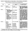
Part 1
Part 2


Bibliography
(On the development of the skeleton of the extremities in man. A few recent articles on variations and congenital deformities are included.)
Adrian, C: Uber kongenitale Humerusund Femurdefekte. Beitrage zur klin. Chir. Bd. 30. 1901.
Alexander, Bela: Die Entwicklung des knochemen Handskelettes vom Beginn der ersten Knochenpunkte. Wiener klin-therap. Wochenschrift Nr. 27-28. 1905. Die Entwicklung des menschlichen Handskeletts. Arch, fiir physikal. Med. Bd. 1. 1906.
Alsberg, M. : Die statisch-mechanischen Prinzipien der Extremitatenbildung beim Menschen und bei den Festlandtieren. Polit. Anthropol. Revue. Bd. 5, S. 605-6n. 1907.
Anthony, R.: L'^volution du pied humain. Bull, et Mem. Soc. d'Anthrop. Ser. 5, T. 3, p. 818-835. Paris 1903. Rev. Scientif. Ser. 4, T. 19, p. 129-139. 1903. Transl. in Annual Report of the Smithsonian Institution, p. 519-535. 1904.
Appraill^, G. : Malformations cong^nitales de I'extremite superieure du radius. Thhse. Paris 1901.
Bade, P.: Die Entwicklung der menschlichen Fussknochen nach Rontgogrammen. Sitz. Ber. Niederrhein. Ges. Bonn 1896. Die Entwicklung des menschlichen Skeletts bis zur Geburt. Arch. mikr. Anat. Bd. 55. 1899. Entwicklung des menschlichen Fussskeletts von der neunten Embryonalwoche bis zum 18. Jahre nach Rontgenbildem. Verb. d. Gesellsch. deutscher Naturf. und Arzte. S. 463-466. 1899-1900.
Barchielli, Alberto: Variazioni del margine superiore dello stemo umano e loro significato. Monit. Zool. Ital. Vol. 15. 1904.
Bardeen, Charles R. : Studies of the Development of the Human Skeleton. Amer. Joum. of Anat. Vol. 4, p. 265-305. 1905.
Bardeen, Charles R. and Lewis, W. H. : Development of the Limbs, Body-wall and Back in Man. Amer. Joum. of Anat. Vol. 1. 1901.
Bardelebex, v.: Das Intermedium tarsi beim Menschen. Sitzungsber. d. Jenaischen Gesellschaft fiir Med. und Naturw. 1883. Zur Entwicklung der Fusswurzel. Sitzungsber. d. Jenaischen Gesellschaft fur Med. und Naturw. Suppl. Bd. 19. 1885. Hand und Fuss. Verhandl. d. anat, Gesellsch. auf der achten Versammlung. 1894.
Bernats, a.: Die Entwicklungsgeschichte des Kniegelenkes des Menschen mit Bemerkungen uber die Gelenke im allgemeinen. Morphol. Jahrbuch. Bd. 4, p. 403. 1878.
Blencke: Uber kongenitalen Femurdefekt. Zeitschrift orthopad. Chirurgie. Bd. 9. 1901.
Blomme, G. : Considerations sur la Polydactylie. Th^se de doctorat en m6d. Paris 1901.
BoKAY, L. : Untersuchungen an der Handwurzel menschlicher und einiger Sauger embryonen. Magyar Orvosi Anthr. Bd. 5. 1904.
BoLK, L. : Beziehungen zwischen Skelett, Muskulatur und Nen^en der Extremitaten, dargelegt am Beckengiirtel. Morphol. Jahrbuch. Bd. 21. 1894. Die Segmentaldifferenzierung des menschlichen Rumpfes und seiner Extremitaten. Morphol. Jahrbuch. Bd. 26-28. 1898, 1899.
Bradley, 0. C. : A Contribution to the Development of the Interphalangeal Sesamoid Bone. Anat. Anz. Bd. 28, p. 528-536. 1906.
Braus : Die Entwicklung der Form der Extremitaten und des Extremitatenskeletts. Hertvvig's Handbuch der Entwicklungsgeschichte der Wirbeltiere. 1906 (appeared 1904).
Broom, R. : On the Arrangement of the Epiphyses of the Mammalian Metacarpals and Metatarsals. Anat. Anz. Vol. 28, p. 106-108. 1906.
Bruch, C. : Beitrage zur Entwicklungsgeschichte des Knochensystems. Neue Denkschriften der allg. Schweizerischen Gesellschaft fur die gesamten Naturwissenschaften. Bd. 12. Zurich 1852. Brunx, a. v.: Das Verhaltnis der Gelenkkapseln zu den Epiphysen der Extremitatenknochen. Leipzig 1881.
Brunner, K. : Uber Genese, kongenitalen Mangel und rudimentare Bildung der Patella. Virchow's Arch. Bd. 124. 1891.
Corson, E. R.: A Skiagraphic Study of the Normal Membral Epiphyses at the Thirteenth Year. Annals of Surgery. Vol. 32, p. 621-647. 1900.
Damany, p. le : Les torsions osseuses, leur role dans la transformation des membres. Joum. de lAnat. et de la Physiol. Annee 34, No. 2, p. 126-165, No. 3, p. 313 bis 337. No. 4, p. 426-450. No. 5, p. 534^545. 1903. Une nouvelle theorie pathogenique de la luxation congenitale de la hanehe. Rev. Chir. p. 175-179, 370-397. 1904. La cavity cotylo'ide. Evolution ontogenique compar6e de sa profondeur chez rhomme et les animaux. Joum. de FAnat. et de la Physiol. Ann^e 40, No. 1. 1904. L' Angle sacro-pelvien. Joum. de TAnat. 6t de la Physiol. Annee 42, p. 153. 1906.
Davbes, W. : Zur Frage der Fingermissbildungen. Dissert. Greifswald 1900.
Debierre: Contribution k Fetude de Tossification et de I'homotypie des pi^es du carpe et du tarse chez Phonune. Joum. de TAnat. et de la Physiol. T. 22. 1886.
Dixey, F. a.: On the Ossification of the Terminal Phalanges of the Digits. Proc. Roy. Soc. Vol. 31, p. 63-71. 1881. Drehmann: Uber kongenitalen Femurdefekt. Zeitsehrift orthop. Chir. Bd. 2, S. 220 bis 233. 1902.
DwiGHT, T. : Variations of the Bones of the Hand and Foot. 1907.
Eggeukg, H. : Uber den oberen Rand des menschlichen Brustbeinhandgriffes. Verhdl. Anat. Ges. (Heidelberg). 1903. Anat. Anz. Bd. 23. Erg.-Heft. 1903.
Engel: Uber kongenitale Ankylosen an den G«lenken der Hande und Fiisse. Inaug.rDiss. Berlin 1902.
Falk, E.: Zur Entwicklung des knochemen Beckens. Verb. Berl. med. Ges. Bd. 30, S. 290-292. 1899. Die Entwicklung und Form des fetalen Beckens. 6 Abbildungen und 5 Tafeln. Berlin 1908.
Farabeup, Lop.: Gaz. Hop. (Cited in Poirier et Charpy, Tlraite. Vol. 1, p. 702). 1895.
Fargeas, J. B. : Etude sur Fabsence congenitale de la rotule. Th^. Paris 1900.
Fawcett, E. : The Presence of Two Centres of Ossification in the Olecranon Process of the Ulna. Joum. of Anat. and Physiol. Vol. 38, p. xxvii. 1904.
Fehling, H. : Die Form des Beckens beim Fetus und Neugeborenen und ihre Beziehung zu der beim Erwachsenen. Archiv fiir G3niakologie. Bd. 10, p. 1-80. 1876. F6r6, 0. und Papin, Ed.: Fractures ou anomalies de developpement de la clavicle. Rev. Chir. Annee 23. 1903.
FiSCHEL, A.: Untersuchungen iiber die Wirbelsaule und den Brustkorb des Menschen. Anat. Hefte. Bd. 31. 1906.
Franz, K.: Zur Entwicklung des knoehernen Beckens nach der Geburt. Beitrag zur Geburtsh. imd Gynakol. Bd. 13, S. 12-29. 1908.
FURBRINGER, M. : Morphologische Streitfragen. Morpholog. Jahrbuch. Bd. 30 S. 85. 1902.
Gallois, E., et Cade, A.: Recherches anatomiques sur la date d'apparition et le developpem«it du ligament ou membrane interosseuse de Tavant-bras. Journ. de TAnat. et de la Physiol. Ahnee 39, p. 39^4. Paris 1903.
Gegenbaur, C. : Ein Fall von erblichem Mangel der Pars acromialis claviculae mit Bermerkungen iiber die Entwicklung der Clavicula. Jen. Zeitsehrift. Bd. 1, S. 1 bis 16. 1864. Vergleiehende Anatamie der Wirbeltiere mit Beriieksichtigung der Wirbellosen Bd. 1. Leipzig 1898.
Grafenberg, E. : Die Entwicklung der Knochen, Muskeln und Nerven der Hand und der f Ur die Bewegungen der Hand bestimmten Muskeln des Unterarms. Anat. Hefte. Bd. 30, S. 1-154. 1906.
Grashet, R. : Basale Epiphyse des Metacarpale I. und Pseudoepiphysen. Zeitsehrift fur Morphol. und Anthropol. Bd. 10, S. 94-97. 1906.
Gross, Alfr. : Uber angeborenen Mangel des Schliisselbeins. Miinch. med. Wochenschrift. Bd. 50, S. 1151-1153. 1903.
Gruber, W. : Vorlaufige Mitteilung iiber die sekundaren Fusswurzelknochen des Menschen. Archiv f iir Anat. und Physiol. 1884.
Grynteltt, E. : Sur les premiers stades de la formation de la cavity articulate du genou chez I'homme. Bull. Soc. Anat. Paris, Vol. 79, s^r. 7. 1904. Note sur le developpement de Particidation du genou chez Thomme. Montpellier md. No. 25, p. 613-624 ; No. 26, p. 641-655 ; No. 27, p. 13-21. 1904.
Goldberg, G. A. : Uber die Kriimmung des Oberschenkels. Intemat. Monatssehrif t Anat. und Physiol. Bd. 21. 1904.
Hagen, W. : Die Bildung des Knorpelskelettes beim menschlichen Embryo. Arch. f iir Anat. und Physiol. 1900.
Hagmeister, Ed. : Uber angeborenen Mangel der Fibula. Liaug.-Diss. Kiel 1902.
Hagopoff : De Forigine et du mode de developpement embryonnaire de Particulation de la hanche. Compt. Rend. Soc. Biol. Paris, Vol. 5, p. 51-54. 1898.
Hahn, Herm. : Rontgographische Untersuchungen iiber das Verhalten der Epiphysen der Unterschenkelknochen wahrend des Waehstums. Festschrift zum 70. Geburstag v. C. von Kuppfer. Bd. 3, S. 731. Jena 1899.
Hasselwander : Untersuchungen iiber die Ossifikation des menschlichen Fuss* skeletts. Zeitsehrift fiir Morphologie und Anthropologic. Bd. 5, S. 438508. 1903. Uber die Ossifikation des Fussskeletts. Anat. Anz. Bd. 32, S. 608. 1908.
Hassenstein, W. : Zur Reifebestimmung des Fetus aus dem Knochenkern der Oberschenkelepiphyse. Zeitsehrift fiir Medizinalbeamte. 1892.
Heimann, Alfr. und Potpeschnigg, K. : Uber die Ossifikation der kindlichen Hand. Jahrbuch fur Kinderheilkunde. Bd. 65, S. 437-456. 1907.
Heine, Otto : Uber den angeborenen Mangel der Kniescheibe. Berl. klin. Wochenschrift. Bd. 41. 1904.
Henke und Reyher : Studien iiber die Entwicklung der Extremitaten des Menschen insbes. der G^lenkflachen. Sitzungsb. d. K. Akad. der Wiss., Wien. Math.-naturw. Klasse. Bd. 70, 3. T., S. 217. Wien 1874. Hennig, C: Das kindliche Becken. Arch, fiir Anat. und Physiol. Anat. Abt. 1880.
Hepburn, D.: The Development of Diarthrodal Joints in Birds and Mammals. Journ. of Anat. and Physiol. Vol. 23, p. 507. 1889. Hissbach, Friedr.: Uber Polydactylie, deren Wesen und Bedeutung. Med. Diss. Leipzig 1902.
Hoffmann, C. K.: Zur Morphologie des Schultergiirtels und des Brustbeines bei Beptilien, Vogeln, Saugetieren und beim Menschen. Niederland. Archiv f iir Zoolo^e. Bd. 5. 1879.
HoLL, M. : Uber die Entwicklung der Stellung der Gliedmassen des Menschen. Sitzimgsb. d. K. Akad. d. Wiss. Wien Math.-naturw. Klasse. Bd. 100, 3 T., S. 12. Wien 1891.
HuETER, C: VirchoVs Arch. Bd. 25, 26, 28, 36. See also Klinik der Gelenkkrankheiten. S. 9. 1870.
HuLKRANTz, J. W. : Das Ellenbogengelenk und seine Mechanik. Jena 1897.
Humphrey: The Angle of the Neck with the Shaft of the Femur at Different Periods of Life and under Different Circumstances. Joum. of Anat. and Physiol. Vol. 23, p. 273, 387. 1889.
Jenkins : The Morphology of the Hip Joint. Brit. Med. Joum. No. 2393, p. 1702. 1906.
JoACHiMSTHAL, G. : Uber Brachydactylie und Hyperphalangie. Virchow's Arch. Bd. 151. 1898. Die angeborenen Verbildungen der oberen Extremitaten. Rontgenbilder. Fortschritte auf dem Gebiet der Rontgenstrahlen. Erganzungsh. 1900. Die angeborenen Verbildungen der unteren Extremitaten. Rontgenbilder. Fortschritte auf dem Gebiet der Rontgenstrahlen. Erganzungsh. Bd. 8. 1902. Uber angeborene Defektbildungen am Oberschenkel. Beitrag zur Gynakol. und Geburtshilfe. Festschrift f. Leopold Landau. Berlin 1902. Verschiedene Formen angeborener Fussdeformitaten. Verb. d. Deutsch. GeseUschaft fur Chir. Bd. 1, S. 66-67. 1906.
Johnston, H. M. : Epilunar and Hypolunar Ossicles, Division of the Scaphoid and Other Abnormalities in the Carpal Region. Joum. of Anat. and Physiol. Vol. 41, p. 59-65. 1906.
JuLiEN, A. : Loi de ^apparition du premier point epiphysaire des os longs. Compt. Rend. Acad. Sc. T. 114, p. 926-929. Paris 1892. Kastschenko, N. : Uber die Entwicklung der Finger beim menschlichen Embryo. Charkow 1884. (Russian.)
EIazzander, G. : Beitrag zur Lehre iiber die Entwicklungsgeschichte der Patella. Med. Jahrbuch. Bd. 1, S. 59-78. Wien 1886. Sullo svilluppo deir articolazione del ginocchio. Monitore Zool. Ital. Vol. 5, p. 220. 1894. Uber die Entwicklung des Kniegelenkes. Archiv f . Anat. und Physiol. Anat. Abt. S. 161. 1894.
Keskineff, G. : Contribution a Fetude des hypertrophies congenitales des membres. Th^se. Nancy 3900. KiNDL, J.: Fiinf Falle von angeborenen Defektbildungen an den Extremitaten. Zeitscht. f iir Heilkunde. Bd. 38, S. 110-138. 1907.
KiRCHNER, A. : Die Epiphyse am proximalen Ende des os metatarsale V. nebst Bemerkungen zur Calcaneus-Epiphyse. Anat. Hefte. Bd. 33, p. 513-551. 1907.
Klaatsch, H. : Die wichtigsten Variationen am Skelett der freien unteren Extremitat des Menschen und ihre Bedeutung fiir das Abstammungsproblem. Ergebnisse der Anatomic und Entwicklungsg. Bd. 10, S. 599-719. 1900.
Klaussner, Ferd. : Uber Missbildungen der menschl. Gliedmassen und ihre Entstehungsweise. Wiesbaden 1900.
KoLLMANN, J.: Handskelett und Hyperdactylie. Verhdl. Anat. Ges. (Wiirzburg). 1888. (Diskussion zu diesem Vortrag: A. Froriep, M. Fiirbringer.) Anat. Anz. Bd. 3. 1888.
KoNiKOW, M. : Zur Lehre von der Entwicklung des Beckens und seiner geschlechtlichen Differenzierung. Arch, fiir Gynakologie. Bd. 45, S. 19. 1894.
Krause, W.: Os acetabuli. Internationale Monatsschrift fiir Anatomie und Histologie. S. 150. 1885.
Lambertz : Die Entwicklung des mensehlichen Knochengeriistes wahrend des f etalen Lebens, dargestellt an Rontgenbildern. Fortschr. auf d. Gebiet d. Rontgenstrahlen. 1900.
Lazarus: Zur Morphologic des Fussskeletts. MorphoL Jahrbuch. Bd. 24. ' 1896. Leboucq, H. : Le developpement du premier metatarsien et de son articulation tarsienne chez I'homme. Archives de Biologie. T. 3, p. 335. 1882. Uber die Entwicklung der Fingerphalangen. Verhdl. Anat. Ges. (Tubingen) 1899. Anat. Anz. Bd. 16. Erghft. 1899. Recherches sur le developpement des phalanges terminales des doigts chez Fhomme et les mammifferes. Ann. soc. med. Gand. T. 89. 1904.
Legke^ W. : Das Vorkommen und die morphologisehe Bedeutung des Pfannen knochens (os acetabuli). Intern. Monatschr. f. Anat. u. Physiol. Bd. 1, p. 363. 1884. Lewis, W. H. : Development of the Arm in Man. Amer. Joum. of Anat. Vol. 2, page 145. 1902. Lubsen, J.: Zur Morphologic des Ilium bei Saugem. Petrus Camper, Dl. II. 3 Aufl. 1903.
LuciEN, M. : Developpement de Particulation du genou et formation du ligament adipeux. Bibl. Anat. Suppl. T. 13. 1904. Ludlopp: Uber Wachstum und Architektur der unteren Femurepiphyse und oberen Tibiaepiphyse. Ein Beitrag zur Rontgendiagnostik. Beitrage klin. Chir. Bd. 38, S. 64-75. 1903. Die Entwicklung der unteren Femurepiphyse und oberen Tibiaspitze im Rontgenbild. Ver. wissensch. Heilk. Konigsberg 1903.
Mall, F. P. : On Ossification Centers in Human Embryos Less than One Hundred Days Old. Amer. Joum. of Anat. Vol. 5, p. 433. 1906. Matet: Developpement de Fextremite du premier metatarsien. Bull, de la Societe Anat. de Paris. Annee 70, p. 384-388. 1895.
Mehnert, E. : Untersuchungen iiber die Entwicklung des Beckengiirtels bei einigen Saugetieren. Morphol. Jahrbuch. Bd. 16; S. 97-112. 1889.
Merkel: Beckenwachstum. Anat. Hefte. Bd. 1, S. 121-150. 1902.
MoLLiER : Die paarigen Extremitaten der Wirbeltiere. Anat. Hefte. Bd. 8, 16 und 24. 1893-97. Moser: Uber das ligamentum teres des Huftgelenkes. Schwalbe's Morphol. Arbeiten. Bd. 2, S. 36. 1893. Neuhauser, H. : Die Beckendrehung. Zeitschrif t fiir Morphol. u. Anthropol. Bd. 3. Stuttgart 1901.
NoLTE, Ad.: Ein Fall von kongenitalem totalem Tibiadefekt. Diss. Leipzig 1903. Pagenstecher, E.: Beitrage zu den Extremitatenmissbildungen. Deutsche Zeitschrift fur Chir. Bd. 60. 1901.
Parker and Shattuck: Pathology and Etiology of Club-Foot. Trans. Path. Soc. of London. Vol. 35. 1884.
Paterson, a. M. : Development of the Sternum and Shoulder Girdle in Mammals. Brit. Med. Joum. 1902.
Perna, G. : L'os trigonum ed il suo omologo nel carpo. Arch. Ital. Anat. e Embriol. Vol. 2, p. 237-254. 1903.
Petersen: Untersuchungen zur Entwicklung des mensehlichen Beckens. Arch. fiir Anat. und Physiol. Anat. Ab. S. 67-96. 1893.
Pfitzner, W. : Beitrage zur Kenntnis der Missbildungen und Variationen des mensehlichen Extremitatenskeletts. Morphol. Arb. Bd. 1-8. 1892-1898. Uber Brachyphalangie und Verwandtes. Verb. Anat. Ges. Anat. Anz. Bd. 14, Erghf. 1898. Die morphol. Elemente des menschlichen Handskeletts. Zeitschrift fiir Morphol. und Anthropol. Bd. 2. 1900. Bd. 4. 1901. PiCQu6: Formule de Fossification des phalanges, des metaearpiens de la clavieule et des cotes. Compt. rend. soe. biol. Paris. Vol. 4, p. 247-248. 1892.
Preleitner, K. : Zwei Falle von angeborenem partiellem Claviculardef ekt. Wiener klin. Wochenschrift. Bd. 16. 1903.
Pbyor, J. W. : The X-ray in the Study of Congenital Malformations. Medical Record. Nov. 1906. Ossification of the Epiphyses of the Hand. X-ray Method. Bulletin of the State College of Kentucky. Series 3, No. 4. 1906. The Chronology and Order of Ossification of the Bones of the Human Carpus. Bulletin of the State University, Lexington, Ky. April 1908.
Pye, W. : On the Growth Rate of the Bones of the Lower Extremities with Especial Reference to Rickety Curvatures. Joum. of Anat. and Physiol. Vol. 23, p. 116. 1889.
Rabl: Gedanken und Studien iiber den Ursprung der Extremitaten. 2Jeitschrift fiir wiss. Zool. Bd. 70, S. 474-558. 1901.
Rabl : Tiber einige Probleme der Morphologie. Verb. anat. Ges. Anat. Anz. Bd. 23. Erganz.-Heft. 1903.
Ranke, W. : Dia Ossification der Hand unter Rontgenbeleuchtung. MUnch. med. Wochenschr. S. 1365. 1898.
Reiner, M. : Uber die kongenitalen Femurdefekte. Zeitschrift fiir orthopad. Chirurgie. Bd. 9. 1901.
Rieder, H. : Uber gleichzeitige? Vorkommen von Brachyund Hyperphalangie an der Hand. Deutsch. Arch. klin. Med. Bd. 66, S. 330-348. 1899.
Retterer: De Tossification du pisiforme de Phonmie, du chien et du lapin. C. R. Soc. Biol. Paris. T. 5. 1898. Ebauche squelettog^ne des membres et developpement des articulations. Journal de I'Anat et de la Physiol. Annee 38, p. 473-509, 580-623. 1902. Rbtzius, G. : Uber die Auf richtung des fetal retrovertierten Kopf es der Tibia bei Menschen. Zeitschrift Morphol. und Anthrop. Bd. 2. 1900. Robertson, W. G.: A Case of Supernumerary and Webbed Fingers. Edinburgh Medical Journal. Vol. 14, p. 535-6. 1900.
RoMiTi, G. : Sui caratteri sessuali nel bacino del neonato. Atti della Societli Toscana di Science Natural!. Vol. 8. Pisa 1892.
Rosenberg, E. : Uber die Entwicklung der Wirbelsaule und das Centrale Carpi des Menschen. Morphol. Jahrbuch. Bd. 1. Leipzig 1876.
RiiCKERT: Ossifikation des menschlichen Fussskeletts. Sitzungsb. d. Konig. Bay. Akad. Math.-nat. Kl. S. 65-72. Munchen 1901. RuGE, Ernst: Die Entwicklung des Skeletts der vordenen Extremitat von Spinaxniger. Morphol. Jahrbuch. Bd. 30, S. 1-27. 1902. Sachs, Adalb. : Uberangeborene Def ekte der Schliisselbeine. Diss. Leipzig 1902.
ScHiCKELE, G. : Beitrag zur Lehre des normalen und gespaltenen Beckens. Beitrage Geburtsh. und Gynakol. Bd. 4, S. 243-272. 1901.
ScHOMBURG, H. : Untersuchungen der Entwicklung der Muskeln und Knochen des menschlichen Fusses. Dissertation. GWttingen 1900.
ScHRENK V. NoTziNG, Alb. Frh. : Beitrage zur Kenntnis der angeborenen Luxationen im Sprunggelenk. Diss. Leipzig 1902.
SOHUUN, K. : Uber die Entwicklung und weitere Ausbildung der Gelenke des menschlichen Korpers. Arch. f. Anat. u. Physiol. Anat. Abt. S. 240-274. 1879.
Schwegel: Die Entwicklungsgeschichte der Knochen des Stammes und der Extremitaten. Sitz. Ber. Akad. d. Wiss. V/ien. Bd. 30, S. 337-388. 1858. Sewell, R. B. S.: Lamella in Fcetal Astragalus. Joum. of Anat. and Physiol. Vol. 41, p. 45-48. London 1906.
Sick, C: Entwicklungsgeschichte der Ejiochen der unteren Extremitaten. Mit 58 Rontgenbildern. 1902. Also in Aixjh. und Atlas der normalen und patholog. Anatomie in typischen Rontgenbildern. Fortschritte aus dem Gebiete der Rontgenstrahlen. Erganzungsband. Bd. 9. 1902.
Spitzy, H. : Uber Bau und Entwicklung des kindlichen Fusses. Jahrbuch fiir Kinderheilk. Bd. 57, S. 731-762. 1903. Uber das Fussgewolbe. Wiener klin. Wochenschrift. Bd. 16, S. 890. 1903.
Stieda, Alpr. : Zur Kenntnis der Sesambeine der Finger und Zehen. Beitr. klin. Chir. Bd.42. 1894.
SteinhauS) F.: Kongenitaler Tibiadefekt. Virchow's Arch. Bd. 163. 1902.
Thilenius, G. : Die uberzahligen Carpuselemente menschlicher Embryonen. Anat. Anz. Bd. 9, S. 665. 1894. Die metacarpo-phalangealen Sesambeine menschlicher Embryonen. Anat. Anz. Bd. 9. 1894. Zur Entwicklungsgeschichte der Sesambeine der menschlichen Hand. Morphol. Arbeiten. Bd. 5. Jena 1896. Das OS intermedium antebrachii des Menschen. Ebenda. Bd. 5. 1896. Untersuchungen iiber die morphologische Bedeutung accessorischer Elemente am menschlichen Carpus. Ebenda. Bd. 5. 1896. accessorische und echte Skelettstiicke. Anat. Anz. Bd. 13. 1897.
Thomson, A. : The Sexual Differences of the Fetal Pelvis. Journal of Anat. and Physiol. Vol. 33, p. 3 and 359-380. 1899.
ToRXiER, G. : Das Entstehen der Gelenkformen. Arch, fiir Entw.-Mech. Bd. 1. Leipzig 1895.
Veit : Die Entstehung der Form des Beckens. Zeitschrif t fiir Geburtsh. und Gynakologie. Bd. 9, S. 347. 1889.
ViANNAY : Dcstot et Gallois, Note sur I'ossification des epiphyses du coude. Bull. Soc. Anat. Paris 1904.
VuiLiiAUME, G.: Contribution k P^tude Tabsence congenitale du tibia. Th^e de doctorat en m^d. Lyon 1898.
Walkhopp, E. : Die Architektur des menschlichen Beckens im Lichte der Entwicklungsmechanik. S.-B. phys. med. Ges. Wurzburg 1904.
Welcker: Die Einwanderung des Bicepssehne in das Schultergelenk. Arch, fiir Anat und Physiol. Anat. Abt. S. 20. 1878.
Whitely and Pearson: Data for the Problem of Evolution in Man. 1. A First Study of the Variability and Correlation of the Hand. Proc. R. Soc. Vol.65. 1899.
WiEDERSHEiM, R. : Das Gliedmassenskelett der Wirbeltiere. Jena 1892.
WiLGRESS, J. H. F. : A Note on the Development of the External Malleolus. Joum. of Anat. and Physiol. Vol. 34. 1900.
Wilms: Die Entwicklung der Knochen oberen Extremitaten, dargestellt in Rontgenbildern. Arch, und Atlas der normalen und patholog. Anatomie in typischen Rontgenbildern, Fortschritte auf dem Gebiete der Rontgenstrahlen. Erganzungsband. Bd. 9. 1902.
WiTTFELD, E. : Uber angeborenen Hochstand der scapula. Diss. Bonn 1901.
Wright, William: Case of accessory Patellae in the Human Subject, with Remarks on Emargination of the Patella. Joum. of Anat. and Physiol. Vol. 38. 1903.
WuTH, E. A.: Uber angeborenen Mangel sowie Herkunft und Zweck der Kniescheibe. Arch. klin. Chir. Bd. 58. 1899.
- Skeleton and Connective Tissues: Connective Tissue Histogenesis | Skeletal Morphogenesis | Chorda Dorsalis | Vertebral Column and Thorax | Limb Skeleton | Skull Hyoid Bone Larynx
| Historic Disclaimer - information about historic embryology pages |
|---|
|
Glossary Links
- Glossary: A | B | C | D | E | F | G | H | I | J | K | L | M | N | O | P | Q | R | S | T | U | V | W | X | Y | Z | Numbers | Symbols | Term Link
Cite this page: Hill, M.A. (2026, July 26) Embryology Book - Manual of Human Embryology 11D. Retrieved from https://embryology.med.unsw.edu.au/embryology/index.php/Book_-_Manual_of_Human_Embryology_11D
- © Dr Mark Hill 2026, UNSW Embryology ISBN: 978 0 7334 2609 4 - UNSW CRICOS Provider Code No. 00098G
- ↑ 1.0 1.1 Keibel F. and Mall FP. Manual of Human Embryology I. (1910) J. B. Lippincott Company, Philadelphia.