Book - An Atlas of the Medulla and Midbrain - Figures
| Embryology - 27 Apr 2024 |
|---|
| Google Translate - select your language from the list shown below (this will open a new external page) |
|
العربية | català | 中文 | 中國傳統的 | français | Deutsche | עִברִית | हिंदी | bahasa Indonesia | italiano | 日本語 | 한국어 | မြန်မာ | Pilipino | Polskie | português | ਪੰਜਾਬੀ ਦੇ | Română | русский | Español | Swahili | Svensk | ไทย | Türkçe | اردو | ייִדיש | Tiếng Việt These external translations are automated and may not be accurate. (More? About Translations) |
Sabin FR. and Knower H. An atlas of the medulla and midbrain, a laboratory manual (1901) Baltimore: Friedenwald.
| Online Editor | ||||||||||||||||||||||||||
|---|---|---|---|---|---|---|---|---|---|---|---|---|---|---|---|---|---|---|---|---|---|---|---|---|---|---|
| This 1901 book by Florence Rena Sabin (1871 - 1953) and her collaborator presents one of the very earliest atlases of the human central nervous system, describing the midbrain and brainstem. This atlas was extremely useful for later researchers attempting to both understand the development and mapping of the midbrain and medulla. Florence Sabin later work was as a key historic researcher in early 1900's establishing our early understanding of both vascular and lymphatic development in the embryo.
| ||||||||||||||||||||||||||
| Historic Disclaimer - information about historic embryology pages |
|---|
| Pages where the terms "Historic" (textbooks, papers, people, recommendations) appear on this site, and sections within pages where this disclaimer appears, indicate that the content and scientific understanding are specific to the time of publication. This means that while some scientific descriptions are still accurate, the terminology and interpretation of the developmental mechanisms reflect the understanding at the time of original publication and those of the preceding periods, these terms, interpretations and recommendations may not reflect our current scientific understanding. (More? Embryology History | Historic Embryology Papers) |
Description of Figures and Plates
Figs. 3-24. Series of horizontal sections passing through the medulla, pons and midbrain of a new-born babe. The series is traced from the dorsal to the ventral surface. The following sections, Figs. 6, 7, 9, 12, 13, 16 and 19 are after Barker, L. F.: The Nervous System and its Constituent Neurones. D. Appleton & Co., 1899. (Preparations by Dr. John Hewetson.)
Figs. 25-51. Series of transverse sections passing through the medulla, pons and midbrain of a new-born babe. The series is traced from the spinal cord toward the cerebrum. The following sections, Figs. 25, 28, 31, 33, 35, 36, 39, 41, 42, 46 and 49 are after Barker, L. F.: Op. cit. (Preparations by Dr. John Hewetson.)
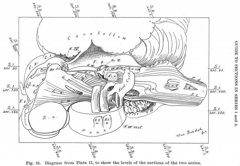
Fig. 52. Key to Planes of Sections
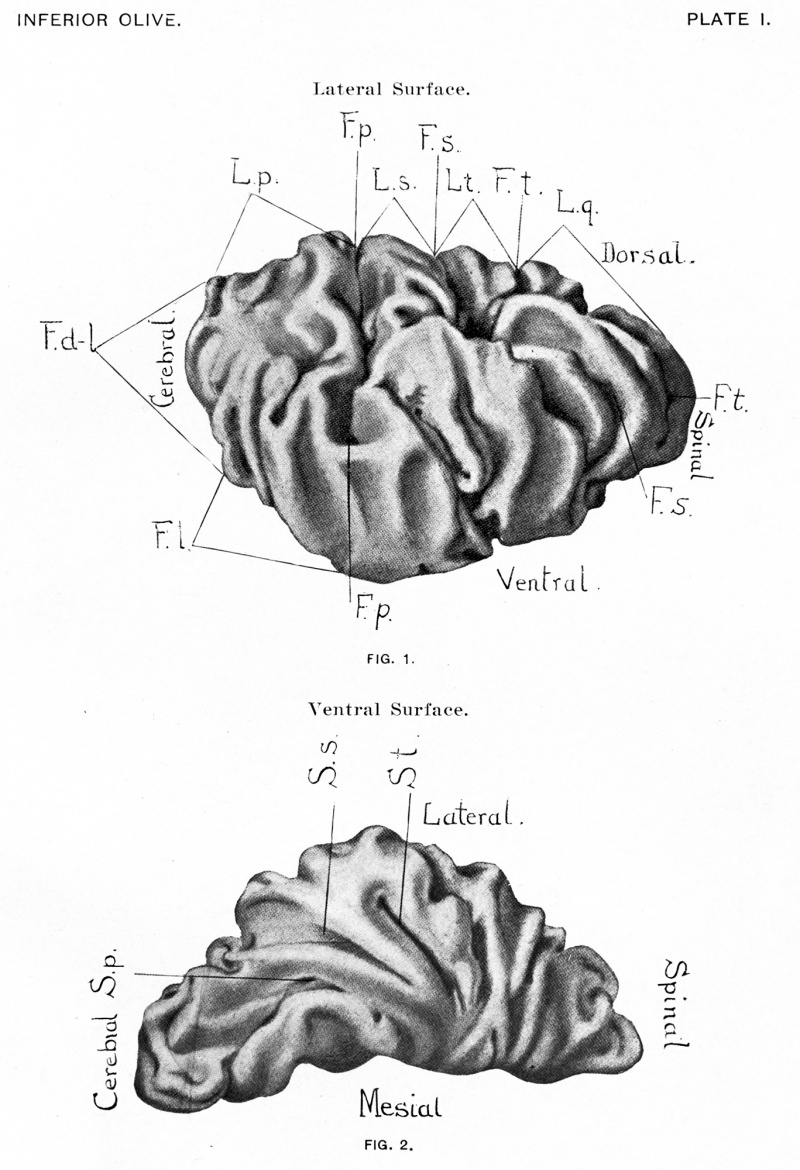
PLATE I
Fig. 1. View of the dorsolateral and lateral surfaces of the nucleus olivaris inferior
- F. dl. Facies dorsolateralis.
- F. 1. Facies lateralis.
- F. p. Fissura prima.
- F. s. Fissura secunda.
- F. t. Fissura tertia.
- F. q. Fissura quarta.
- L. p. Lobus primus.
- L. s. Lobus secundus.
- L. t. Lobus tertius.
- L. q. Lobus quartus.
Fig. 2. View of the ventral surface of the nucleus olivaris superior
- S. p. Sulcus primus.
- S. s. Sulcus secundus.
- S. t. Sulcus tertius.
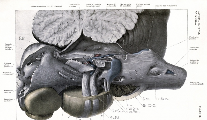
PLATE II
View of the model from the lateral surface. This view is designed to relate the model to the cord, the cerebellum and the cerebrum. The cut edge of the cord shows on the extreme right. The following points will make the position of the model clear: the dorsal and lateral funiculi and the dorsal horn of the spinal cord, the cerebellum, the fourth ventricle, the inferior and superior colliculi and the third ventricle.
The color system is as follows: all fibres are in white and black, all nuclei in colors. Red represents the nuclei of the motor cerebral nerves, blue the nuclei of the sensory cerebral nerves and yellow all other nuclei.
Nu. et Radix N. vestibuli: The nucleus is distinguishable from the root by its color. The ascending and descending parts of the root are to be determined by their relation to the entering root-bundle of the nerve. The part of the vestibular nucleus distal to the nucleus N. abducentis is the nucleus N. vestibuli medialis; the part proximal, is the nucleus N. vestibuli superior. The nucleus N. vestibuli lateralis (Deiters'), (pars lateralis) lies in the vestibular tract just dorsal to the corpus restiforme.
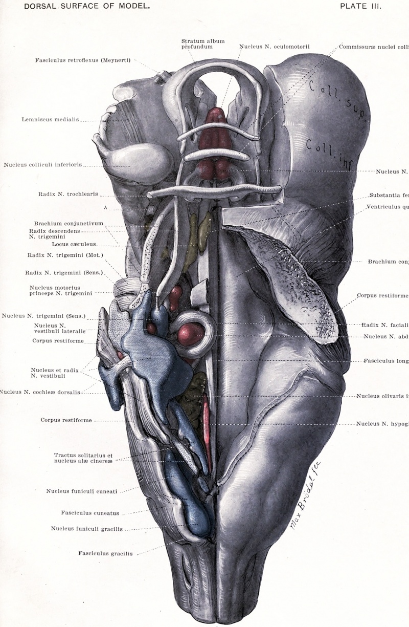
PLATE III
View of the model from the dorsal surface. On the right side is shown the floor of the fourth ventricle; on the left, the structures beneath are exposed. The position of these structures can be related to the dorsal funiculi of the spinal cord, the fourth ventricle, and the inferior and superior colliculi.
Nu. et Radix N. vestibuli: To be distinguished by the colors. The ascending root is marked by the most proximal of the three lines on the figure; the descending by the most distal line, while the nucleus N. vestibuli medialis is indicated by the middle of the three lines. The nucleus N. vestibuli superior is continuous with the medial nucleus and lies opposite the ascending root. The nucleus 1ST. vestibuli lateralis consists of two parts, one between the corpus restiforme and the ascending root, the other in the notch between the medial and superior nuclei.
Nucleus N. cochlew dor sails: The more proximal of the two lines points to the striae acusticae.
Traotus solitarius et Nu. alas cinerce: The former is in black and white, the latter in blue.
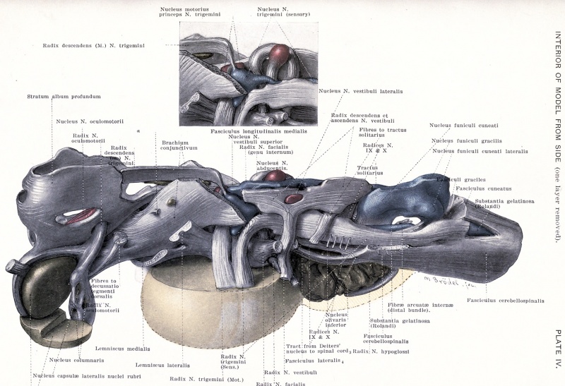
PLATE IV
View of the model from the lateral aspect. After removing from Plate i, the following structures: the corpus restiforme, the substantia nigra and the medial, lateral and superior lemnisci. The view is designed to show (1) the sensory nerves and their nuclei, and (2) the midbrain. The nuclei of the dorsal funiculi represent a way-station for the sensory fibres from the spinal cord; the sensory cerebral nerves are represented by the nuclei nervi glossopharyngei, vagi, vestibuli et trigemini. These include all of the sensory nerves of the region of the model except the N. cochleae, which was removed with the corpus restiforme.
Radix N. trigemini (Sens.) : The proximal line runs to the root bundle, the distal to the tractus spinalis N. trigemini.
Tract from Betters' nucleus to F. i. (3), and Fasciculus lateraMs (4): The numbers are explained in the text.
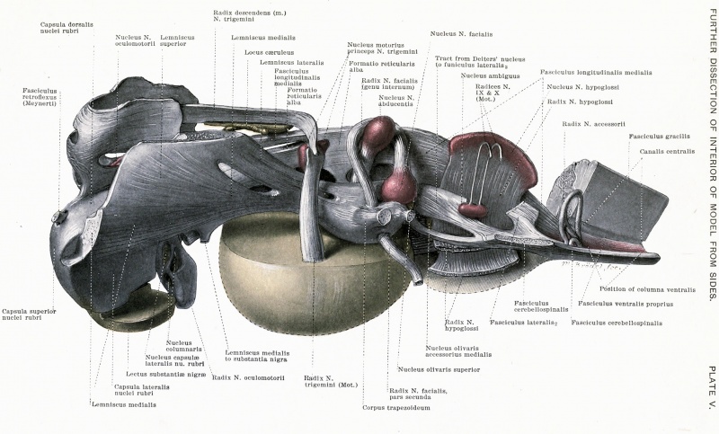
PLATE V
View of the model from the lateral aspect. The sensory nerves of Plate IV have been removed and all of the motor cerebral nerves except the N. trochlearis are now shown.
Fasciculus lateralis (2), and Fasciculus lateralis (3): The numbers are explained in the text.
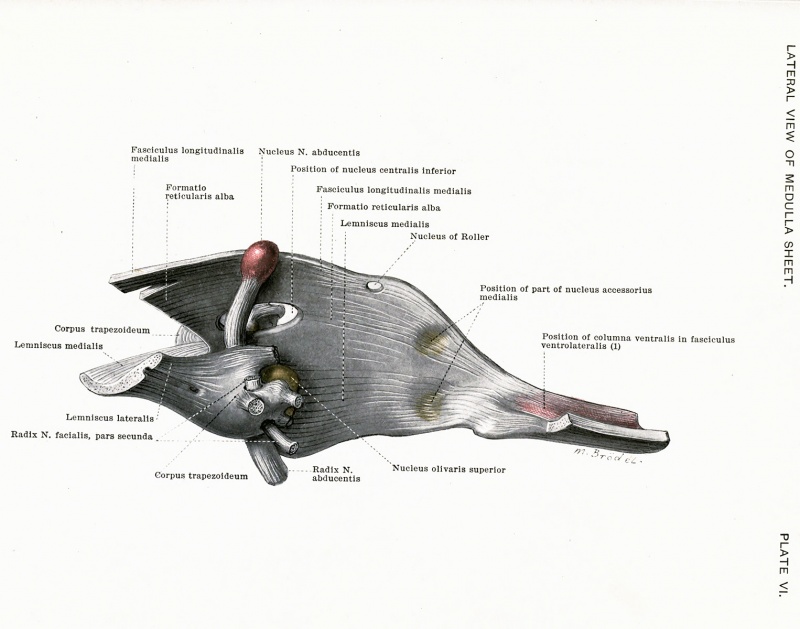
PLATE VI
View of the lateral surface of the medulla sheet. The view can be related to Plates n, iv and v, by the position of the nucleus N. abducentis. Fasciculus ventrolateralis (1): The number is explained in the text.
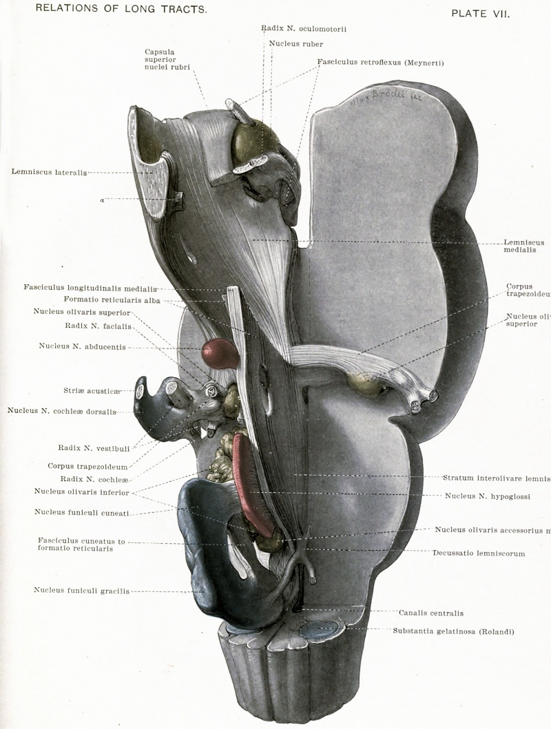
PLATE VII
View of the model from a dorsomedian aspect. This view is designed to show the central fibre mass, that is, the medulla, pontal and midbrain sheets, together with the corpus trapezoideum.
Fibres running from Lemniscus lateralis to the brachium conjunctivum.
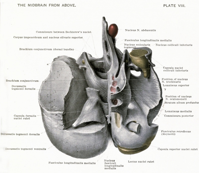
PLATE VIII
View of the midbrain from the superior or cerebral aspect. This view can be understood by comparing 1 it with Plates n, iv and v, which show the stratum profundum album, the lemniscus superior and the capsula nuclei rubri from the lateral aspect.
7 is a space in the model, in the stratum profundum album where fibres of the formatio reticularis alba are related to the substantia centralis grisea.
Fasciculus ventrolateralis (1) : The number is explained in the text.
HORIZONTAL (Frontal) SECTIONS 38, 56 and 62.
HORIZONTAL (Frontal) SECTIONS 66, 72 and 74.
HORIZONTAL (Frontal) SECTIONS 80, 86 and 94.
HORIZONTAL (Frontal) SECTIONS 100, 108, 114 and 116.
HORIZONTAL (Frontal) SECTIONS 122 and 126.
HOKIZONTAL (Frontal) SECTIONS 128 and 136.
HORIZONTAL (Frontal) SECTIONS 146 and 162.
HORIZONTAL (Frontal) SECTIONS 170, 180 and 202.
CROSS-SECTIONS 20-84.
CKOSS-SECTIONS 94-146.
CROSS-SECTION 158.
CROSS-SECTION 170.
CROSS-SECTION 182.
CROSS-SECTION 190.
CROSS-SECTIONS 200 and 212.
Fig-. 38, Series II, Section No. 200.
Fig. 39, Series II, Section No. 212.
CROSS-SECTIONS 254 and 268.
Fig. 40, Series II, Section No. 254.
Fig-. 41, Series II, Section No. 268.
CROSS-SECTIONS 290 and 304.
Fig-. 42, Series II, Section No. 290.
Fig. 43, Series II, Section No. 304.
CROSS-SECTIONS 316 and 330.
Fig-. 44, Series II, Section No. 316.
Fig. 45, Series II, Section No. 330.
CEOSS-SECT1ONS 338 and 354.
Fig-. 46, Series II, Section No. 338.
Fig-. 47, Series II, Section No. 354.
CROSS-SECT1ONS 372 and 384.
Fig. 48, Series II, Section No. 372.
Fig. 49, Series II, Section No. 384.
CROSS-SECTIONS 396 and 420.
Fig-. 50, Series II, Section No. 396.
Fig-. 51, Series II, Section No. 420.
GUIDE TO SECTIONS IN SERIES 1 and 2.
List of Illustrations
FIGURES : PAGE
1. Transverse Section of the Spinal Cord. Outline 36
2. Diagram of Medial Accessory Olive 91
3-24. Series of Horizontal (frontal) Sections, including Medulla and Midbrain 125-132
25-51. Series of Transverse Sections from the Cord to the Midbrain. .133-145
52. Diagram of the Model giving Levels of Sections here Figured 146
PLATES following page 146
I. The Inferior Olive. II. View of the Lateral Surface of the Reconstruction.
III. View of the Dorsal Surface of the Reconstruction.
IV. First Dissection of the Reconstruction. Lateral view, showing
Fibre Tracts, &c., and the Sensory Nuclei of Cerebral Nerves. V. Second Dissection of the Reconstruction. Lateral view, showing
Fibre Tracts, &c., and Motor Nuclei of Cerebral Nerves. VI. Third Dissection of the Reconstruction. Lateral view, showing
the Long Tracts of the Medulla.
VII. Fourth Dissection of the Reconstruction. Dorsomedian view, showing the Long Fibre Tracts as Related to Nuclei of Cerebral Nerves and to other Structures.
VIII. View of the Midbraiu from Above, showing Relations of Fibre Tracts.
| Historic Disclaimer - information about historic embryology pages |
|---|
| Pages where the terms "Historic" (textbooks, papers, people, recommendations) appear on this site, and sections within pages where this disclaimer appears, indicate that the content and scientific understanding are specific to the time of publication. This means that while some scientific descriptions are still accurate, the terminology and interpretation of the developmental mechanisms reflect the understanding at the time of original publication and those of the preceding periods, these terms, interpretations and recommendations may not reflect our current scientific understanding. (More? Embryology History | Historic Embryology Papers) |
An Atlas of the Medulla and Midbrain (1901): Chapter I. Introductory | Chapter II. The Long Tracts | Chapter III. The Columns Of The Spinal Cord | Chapter IV. Cerebellar Peduncles | Chapter V. The Cerebral Nerves And Their Nuclei | Chapter VI. The Cerebral Nerves And Their Nuclei (Continued). Lateral Group | Chapter VII. The Inferior And Accessory Olives | Chapter VIII. The Midbrain | Chapter IX. The Formatio Reticularis Alba And Grisea | General Summary of what Is shown In Reconstruction | References To Literature | Abbreviations | Description of Figures and Plates
Cite this page: Hill, M.A. (2024, April 27) Embryology Book - An Atlas of the Medulla and Midbrain - Figures. Retrieved from https://embryology.med.unsw.edu.au/embryology/index.php/Book_-_An_Atlas_of_the_Medulla_and_Midbrain_-_Figures
- © Dr Mark Hill 2024, UNSW Embryology ISBN: 978 0 7334 2609 4 - UNSW CRICOS Provider Code No. 00098G


