Abnormal Development - Anencephaly
| Embryology - 22 Jul 2026 |
|---|
| Google Translate - select your language from the list shown below (this will open a new external page) |
|
العربية | català | 中文 | 中國傳統的 | français | Deutsche | עִברִית | हिंदी | bahasa Indonesia | italiano | 日本語 | 한국어 | မြန်မာ | Pilipino | Polskie | português | ਪੰਜਾਬੀ ਦੇ | Română | русский | Español | Swahili | Svensk | ไทย | Türkçe | اردو | ייִדיש | Tiếng Việt These external translations are automated and may not be accurate. (More? About Translations) |
LA00 Anencephaly or similar anomalies
The above ICD-11 International Classification of Diseases code is from the next version update due for release in 2018. ICD-10 is the 10th revision, Australian Modification (ICD-10-AM) is currently in use in Australian hospitals for admitted patients.
ICD-10 Q00 Anencephaly and similar malformations
- Q00.0 Anencephaly, Acephaly, Acrania, Amyelencephaly, Hemianencephaly, Hemicephaly
- Q00.1 Craniorachischisis
- Q00.2 Iniencephaly
| About International Classification of Diseases |
|---|
|
The International Classification of Diseases (ICD) World Health Organization's classification used worldwide as the standard diagnostic tool for epidemiology, health management and clinical purposes. This includes the analysis of the general health situation of population groups and is used to monitor the incidence and prevalence of diseases and other health problems. Within this classification "congenital malformations, deformations and chromosomal abnormalities" are (Q00-Q99) but excludes "inborn errors of metabolism" (E70-E90). |
Introduction

A neural tube defect, anencephaly is a failure of the neural tube to close cranially. Also called exencephaly or craniorachischisis. The fortification of foods with folate has led to an overall decrease in the rate of this abnormality, showing that, at least in some cases, there is an association with maternal diet during pregnancy. Neural tube defects were historically more associated with Female infants, though a 2015 USA study showed no sex association in their population.[2] In contrast, a recent 2018 study of Latin American countries, has demonstrated a Female association and a substantial decrease in this ratio following folate fortification.[3]
- Links: Low Folic Acid and NTDs
Some Recent Findings
|
| More recent papers |
|---|
 This table allows an automated computer search of the external PubMed database using the listed "Search term" text link.
More? References | Discussion Page | Journal Searches | 2019 References | 2020 References Search term: Anencephaly <pubmed limit=5>Anencephaly</pubmed> |
| Fetal Anencephaly | |
|---|---|

|

|
| ventral view | lateral view |
Sex Ratios
Neural tube defects were historically more associated with Female infants, though a 2015 USA study showed no sex association in their population.[2] In contrast, a recent 2018 study of Latin American countries, has demonstrated a Female association and a substantial decrease in this ratio following folate fortification.[3]

Sex ratio changes for NTD cases and total births in Chile, Argentina, and Venezuela (1990–2013).[3]
NTD: neural tube defect; FAF: folic acid fortification; M/F: male/female; Sex ratio (male/female) for neural tube defect cases (full blue line), sex ratio for total births (dashed red line). Sex ratios estimated by multivariate regression models adjusted by hospital.
Ultrasound

Anencephaly in a fetus (GA week 18) from a diabetic mother. Ultrasound images (coronal) show a complete absence of the cranial vault and brain and enlarged orbits.[4]
- Links: maternal diabetes
Historic Anencephaly
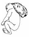
These drawings are from a 1921 study of a single embryonic anencephaly.[5]
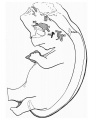
Fig. 1. Reconstruction of anencephalic embryo.
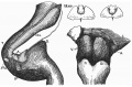
Fig. 2. Hind-brain 28 mm embryo (left view).


These drawings are from a 1925 study of 57 cases of anencephaly.[6]

anencephalic acranius

anencephalic craniorhachischisis
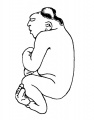
microcephalic acrauius
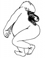
microcephalic craniorhachischisis
exocephalic acranius


Dodds GS. and Deangelis E. An anencephalic human embryo 16.5 mm long. (1937) Anat. Rec. 67(4): 499-505.
References
- ↑ Mathews TJ. Trends in spina bifida and anencephalus in the United States, 1991-2005, National Vital Statistics System.
- ↑ 2.0 2.1 Michalski AM, Richardson SD, Browne ML, Carmichael SL, Canfield MA, VanZutphen AR, Anderka MT, Marshall EG & Druschel CM. (2015). Sex ratios among infants with birth defects, National Birth Defects Prevention Study, 1997-2009. Am. J. Med. Genet. A , 167A, 1071-81. PMID: 25711982 DOI.
- ↑ 3.0 3.1 3.2 3.3 Poletta FA, Rittler M, Saleme C, Campaña H, Gili JA, Pawluk MS, Gimenez LG, Cosentino VR, Castilla EE & López-Camelo JS. (2018). Neural tube defects: Sex ratio changes after fortification with folic acid. PLoS ONE , 13, e0193127. PMID: 29538416 DOI.
- ↑ Alorainy IA, Barlas NB & Al-Boukai AA. (2010). Pictorial Essay: Infants of diabetic mothers. Indian J Radiol Imaging , 20, 174-81. PMID: 21042439 DOI.
- ↑ Frazer JE. Report on an anencephalic embryo. (1921) J Anat. 56(1): 12-9. PMID 17103933
- ↑ Nañagas JC. A comparison of the growth of the body dimensions of anencephalic human fetuses with normal fetal growth as determined by graphic analysis and empirical formulae. (1925) American J. Anatomy. 455-494.
Journals
Journal of Pediatric Neurosciences - is official publication of the Indian Society for Pediatric Neurosurgery.
Search PubMed
Search term: Neural Development Abnormalities | Anencephaly | Hydrocephalus | Encephalocele | Holoprosencephaly | Autism
Glossary Links
- Glossary: A | B | C | D | E | F | G | H | I | J | K | L | M | N | O | P | Q | R | S | T | U | V | W | X | Y | Z | Numbers | Symbols | Term Link
Cite this page: Hill, M.A. (2026, July 22) Embryology Abnormal Development - Anencephaly. Retrieved from https://embryology.med.unsw.edu.au/embryology/index.php/Abnormal_Development_-_Anencephaly
- © Dr Mark Hill 2026, UNSW Embryology ISBN: 978 0 7334 2609 4 - UNSW CRICOS Provider Code No. 00098G