ANAT2341 Lab 1 - Fertilization
| ANAT2341 Lab 1: Introduction | Gametogenesis | Oogenesis | Spermatogenesis | Fertilization | Sex Determination | Online Assessment | ANAT2341 Lab 1 - Quiz |
Introduction
This page covers the process of mammalian fertilization. A complex interaction between the two haploid gametes (oocyte and spermatozoa) resulting in a single diploid cell (zygote). Following entry of the spermatazoa into the oocyte a series of changes occur within the oocyte and zona pellucida that block further fertilization by additional bound spermatozoa (polyspermy).
Due to both scientific and medical research on this process, this can now occur outside the body (in vitro fertilization) as well as fertility therapies to aid normal (in vivo fertilization). In addition, our understanding of fertilization has led to the development of a number of alternative fertility control methods.
Fertilization Dynamics
|
|
|
|
| |||||||||||||||||||||||
|
|
|
|
|
|
|
Sperm Events
- Capacitation - removal of glycoprotein coat and seminal proteins, alteration of sperm mitochondria
- Binding - zona pellucida protein ZP3 acts as receptor for sperm
- Acrosome reaction - exyocytosis of acrosome contents (Calcium ion mediated), enzymes to digest the zona pellucida, exposes sperm surface proteins to bind ZP2
- Membrane fusion - between sperm and egg, allows sperm nuclei passage into egg cytoplasm
Egg Events
- Sperm membrane fusion - causes depolarization of egg membrane, primary block to polyspermy
- Cortical reaction - IP3 pathway elevates intracellular Calcium, exocytosis of cortical granules, enzyme alters ZP3 so it will no longer bind sperm plasma membrane (cortical reaction)
- Second meiotic division - completion of 2nd meiotic division, forms second polar body
Sperm Penetration
| <html5media height="430" width="600">File:Human fertilization 02.mp4</html5media> |
| Approximate Timing of Early Human Events (in vitro) | ||||
|---|---|---|---|---|

|

|

| ||
| 20 min - components | 28 min - spermatozoa penetrates zone pellucida | 31 min - spermatozoa penetrates oocyte - fertilization | ||
| See also clock in lower righthand corner for the approximate timing of events. | ||||
| Reference: PMID 22695746 J Assist Reprod Genet. | ||||
The animation shows:
|
Pronuclear Fusion
|
The animation shows:
|

|

|
| In the mouse zygote, separation of chromatin according to parental origin is preserved up to the four-cell embryo stage and then gradually disappears. |
Human Zygote Size
Actual Size ![]()
Enlarged Size 
Fertilization Overview

Model of mouse gamete recognition by zona pellucida[1] |
<html5media height="480" width="380">File:Mouse_zygote_division_02.mp4</html5media> |
Capacitation
- The mammalian spermatozoa once released, must remain for a time in the female genital tract before having the capacity to fertilize the oocyte. This process involves modifying the spermatozoa.
Acrosome Reaction
- Penetration of egg by spermatozoa is initiated by the acrosome reaction which takes different forms in different species.
- Mammalian acrosomal lysins contain proteinases which lyse the glycoproteins of the zona pellucida.
- The central part of the acrosome elongates into a tube which extends form the head of the spermatozoon. On contact with the egg the acrosomal membrane fuses with the sperm plasma membrane thus opening the acrosomal vesicle and liberating the granules containing acrosomal lysins.
- The inner portion of the acrosomal membrane everts and lengthens to form the acrosomal tubule through which the sperm nucleus enters the egg.
Sperm Contact
The act of fertilization changes the egg from a stage of slow structural and metabolic decline to one of renewed activation. Morphologically egg activation is a series of surface changes immediately following sperm contact.
- Mammals - No phenomenon comparable to the raising of the fertilization membrane is displayed. Mammalian eggs are surrounded by the zona pellucida which undergoes a structural change known as the zonal reaction after sperm penetration. On sperm contact with the egg plasma membrane, cortical granules break down as in above forms, substances liberated into the perivitelline space rapidly modify the zona pellucida resulting in a block to further sperm penetration.
Sperm Activation of Egg
- During fertilization sperm activates the egg by induction of a calcium ion (Ca2+) oscillation within the egg's cytoplasm.
- Induction occurs by a sperm protein factor (unidentified) which can stimulate only once calcium ion oscillations in metaphase eggs.
- Another sperm derived factor is then responsible for the inactivation of this oscillation.
- The activation of the egg by this calcium ion oscillation is essential for entry of the egg into the first mitotic cycle.
Zygote - Sperm Contribution
What the fertilizing sperm contributes in addition to the genetic material to the zygote differes between species.
- Centriole - most mammalian species, sperm contribute a centriole to reconstitute the zygotic centrosome. In rodents, only a maternal centrosomal inheritance occurs.
- Sperm Mitochondria - may enter the zygote, but are eliminated by a ubiquitin-dependent mechanism.
Perinuclear Theca - located in the sperm head perinuclear region. Contains a cytoskeletal element to maintain the shape of the sperm head and functional molecules leading to oocyte activation during fertilization.
Note - ART intracytoplasmic sperm injection techniques may introduce sperm components normally lost during in vivo fertilization.
Zona Pellucida
The specialized extracellular matrix layer lying directly around the oocyte underneath follicular cells. The structure consists of glcosaminoglycans and three main glycoproteins (ZP1, ZP2, ZP3).
After fertilization, the zona pellucida:
- blocks polyspermic fertilization
- physically protects the preimplantation embryo during early embryonic development (aided by an initial "hardening" after fertilization)
- aids uterine tube transport
- impacts upon blastocyst development
Zygote Contributions
Maternal - mitochondria, nucleolus, oocyte contributes one centriole during fertilisation.
Paternal - spermatozoon contributes one centriole during fertilisation.
Both Parents - chromosomes (inherent epigenetic differences between the paternal and maternal pronuclei), plasma membranes spermatozoon/oocyte mingle to form a mosaic plasma membrane.
Additional Information
| Additional Information - Content shown under this heading is not part of the material covered in this class. It is provided for those students who would like to know about some concepts or current research in topics related to the current class page. |

- Mouse Spermatozoa Mitochondria Movie - What happens to the spermatozoa mitochondria?
- Textbook overviews - Dev Biol - Fertilization | MBoC - Fertilization
- Assisted Reproductive Technology - More about modern reproductive technologies.
- ↑ <pubmed>24934154</pubmed>
Terms
- adplantation - Initial adhesion of blastocyst (released from zona pellucida) to uterine wall. Adplantation is followed by implantation.
- ampulla - longest segment (approximately 2/3 of overall length) of uterine tube (oviduct or Fallopian tube). Medial segment forming the remainder of the tube is called the isthmus.
- antrum- (L. a cave), cavity; a nearly-closed cavity or bulge. In the ovary this refers to the follicular fluid-filled space within the follicle.
- blastocyst - the developmental stage following morula, as this stage matures, the zona pellucia is lost allowing the coceptus to adplant and then implant into the uterine wall.
- capacitation - the process of activation of sperm, requires removal of surface glycoproteins and increased motility. The sperm now become capable of fertilizing an egg.
- cavitates- to form a space within a solid object.
- conceptus - the entire structure generated from the zygote.
- fertilization - (fertilisation) The process of penetration of the oocyte (egg) by the spermatozoa and the combining of their genetic material that initiates development of the embryo. The union of two haploid gametes to form the first diploid cell, the zygote. (More? Fertilization | Spermatozoa Development | Testis Development | Ovary Development | Lecture - Fertilization)
- fimbriae- ( L. = a fringe) fingerlike projections at the ovarian end of uterine tube.
- follicular fluid- (or follicular fluid) the fluid found in the antrum of a secondary follicle. Secreted by cells in the wall of the follicle. This fluid is released along with the oocyte at ovulation.
- infundibulum- funnel-shaped initial segment of uterine tube (oviduct or Fallopian tube) opening into peritoneal cavity and connected to the ampulla. The peritoneal opening sitting over the ovary.
- morula -(L. morus = mulberry) early stage of development (12-15 cells) when conceptus is a solid ball of cells, further cell division forms the blasocyst.
- oocyte - (egg or ovum) female germ cell.
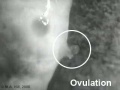
- ovulation- release of the oocyte from the mature follicle.
- uterine tube- (also called oviduct or Fallopian tube) the laterally paired tubes that connect the ovary to the uterus. Is the site for oocyte fertilization and initial development of the conceptus.
- uterine wall - the site of normal blastocyst implantation.
- zona pellucida- glycoprotein shell that surrounds the oocyte through to blastula stage of development
- uterus- site of embryo implantation and development. Uterine wall has 3 layers; endometrium, myometrium, and perimetrium.
- zona pellucida- extracellular layer lying directly around the oocyte underneath follicular cells. Consists of glcosaminoglycans and glycoproteins (ZP1, ZP2, ZP3).
- ZP1, ZP2, ZP3 - acronyms for the zona pellucida glycoproteins.
- zygote - The first diploid cell that forms following fertilization by fusion of haploid oocyte (egg or ovum) and [S#spermatozoa|spermatozoa]] (sperm), resulting in the combination of their separate genomes. This single cell will divide by mitosis to form all cells of the embryo, fetal membranes and the embryonic component of the placenta. The term conceptus is used to describe all these cells derived from the fertilization event forming the zygote.
| ANAT2341 Lab 1: Introduction | Gametogenesis | Oogenesis | Spermatogenesis | Fertilization | Sex Determination | Online Assessment | ANAT2341 Lab 1 - Quiz |
Glossary Links
- Glossary: A | B | C | D | E | F | G | H | I | J | K | L | M | N | O | P | Q | R | S | T | U | V | W | X | Y | Z | Numbers | Symbols | Term Link
Cite this page: Hill, M.A. (2026, July 6) Embryology ANAT2341 Lab 1 - Fertilization. Retrieved from https://embryology.med.unsw.edu.au/embryology/index.php/ANAT2341_Lab_1_-_Fertilization
- © Dr Mark Hill 2026, UNSW Embryology ISBN: 978 0 7334 2609 4 - UNSW CRICOS Provider Code No. 00098G