Paper - The vascular drainage of the endolymphatic sac and its topographical relation to the transverse sinus in the human: Difference between revisions
(Created page with "{{Header}} {{Ref-Streeter1916}} {| class="wikitable mw-collapsible mw-collapsed" ! Online Editor |- | 90px|left This 1916 paper by Streeter is on...") |
mNo edit summary |
||
| Line 4: | Line 4: | ||
! Online Editor | ! Online Editor | ||
|- | |- | ||
| [[File:Mark_Hill.jpg|90px|left]] This 1916 paper by Streeter is on the embryonic the endolymphatic sac and its relationship to the transverse sinus. In the adult, the | | [[File:Mark_Hill.jpg|90px|left]] This 1916 paper by [[Embryology History - George Streeter|George Streeter (1873-1948)]] is on the embryonic the endolymphatic sac and its relationship to the transverse sinus. These human embryos are [[Carnegie Embryos]] and fetuses from the [[Carnegie Collection]]. In the adult, the endolymphatic sac regulates the inner ear volume and pressure of endolymph, immune responses and the elimination of endolymphatic waste products by phagocytosis. The transverse sinuses (left and right lateral) allow blood to drain from the back of the head. | ||
endolymphatic sac regulates the inner ear volume and pressure of endolymph, immune responses and the elimination of endolymphatic waste products by phagocytosis. The transverse sinuses (left and right lateral) allow blood to drain from the back of the head. | |||
<br> | <br> | ||
Historically, see also by Streeter: {{Ref-Streeter1915}} | |||
{{Ref-Streeter1917innerear1}} | |||
:Links: [[ | {{Ref-Streeter1917innerear2}} | ||
<br> | |||
Below are shown links to modern resources on inner ear and vascular development. | |||
:Links: [[Hearing_-_Inner_Ear_Development|Inner Ear]] | [[Neural - Meninges Development]] | [[Neural - Vascular Development]] | |||
{{Hearing Links}} | {{Hearing Links}} | ||
| Line 18: | Line 22: | ||
|} | |} | ||
{{Historic Disclaimer}} | {{Historic Disclaimer}} | ||
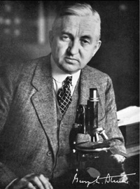
[[File:George_L._Streeter.jpg|thumb|200px|alt=Embryology History George Streeter|link=Embryology History - George Streeter|George Linius Streeter (1873-1948)]] | |||
=The Vascular Drainage of the Endolymphatic Sac and its Topographical Relation to the Transverse Sinus in the Human= | |||
[[Embryology History - George Streeter|George L. Streeter]] | |||
Department of Embryology, Carnegie Institution of Washington, Johns Hopkins Medical School, Baltimore, Maryland | |||
{{Footer}} | |||
[[Category:Human]][[Category:Hearing]][[Category:Inner Ear]] | |||
[[Category:George Streeter]][[Category:1910's]][[Category:Draft]] | |||
Revision as of 10:07, 1 March 2017
| Embryology - 3 May 2024 |
|---|
| Google Translate - select your language from the list shown below (this will open a new external page) |
|
العربية | català | 中文 | 中國傳統的 | français | Deutsche | עִברִית | हिंदी | bahasa Indonesia | italiano | 日本語 | 한국어 | မြန်မာ | Pilipino | Polskie | português | ਪੰਜਾਬੀ ਦੇ | Română | русский | Español | Swahili | Svensk | ไทย | Türkçe | اردو | ייִדיש | Tiếng Việt These external translations are automated and may not be accurate. (More? About Translations) |
Streeter GL. The vascular drainage of the endolymphatic sac and its topographical relation to the transverse sinus in the human. (1916) Amer. J Anat. 19(1): 67-89.

| Historic Disclaimer - information about historic embryology pages |
|---|
 |
The Vascular Drainage of the Endolymphatic Sac and its Topographical Relation to the Transverse Sinus in the Human
Department of Embryology, Carnegie Institution of Washington, Johns Hopkins Medical School, Baltimore, Maryland
Cite this page: Hill, M.A. (2024, May 3) Embryology Paper - The vascular drainage of the endolymphatic sac and its topographical relation to the transverse sinus in the human. Retrieved from https://embryology.med.unsw.edu.au/embryology/index.php/Paper_-_The_vascular_drainage_of_the_endolymphatic_sac_and_its_topographical_relation_to_the_transverse_sinus_in_the_human
- © Dr Mark Hill 2024, UNSW Embryology ISBN: 978 0 7334 2609 4 - UNSW CRICOS Provider Code No. 00098G