Paper - The development of the limbs, body-wall and back: Difference between revisions
m (→Second Week) |
mNo edit summary |
||
| (40 intermediate revisions by the same user not shown) | |||
| Line 2: | Line 2: | ||
{{Ref-BardeenLewis1901}} | {{Ref-BardeenLewis1901}} | ||
{| class="wikitable mw-collapsible mw-collapsed" | {| class="wikitable mw-collapsible mw-collapsed" | ||
! Online Editor | ! Online Editor | ||
|- | |- | ||
| [[file:Mark_Hill.jpg|90px|left]] | | [[file:Mark_Hill.jpg|90px|left]] This historic 1901 paper by [[Embryology History - Charles Bardeen|CharlesBardeen]] and Lewis described the development of the limbs, body-wall and back using human embryos from the [[Carnegie Collection]]: {{CE2}}, {{CE12}}, {{CE22}}, {{CE43}}, {{CE76}}, {{CE106}} {{CE109}}, {{CE144}}, {{CE144}}, {{CE148}}, {{CE163}}, {{CE167}}, {{CE175}}. | ||
<br> | |||
[[ | See also: {{Ref-Bardeen1905}} | ||
<br> | |||
{{Ref-Bardeen1905a}} | |||
<br> | |||
[[Embryology History - Charles Bardeen|Charles Bardeen]] | |||
<br> | |||
<br> | |||
'''Modern Notes:'''[[Musculoskeletal System - Limb Development|Limb Development]] | |||
<br> | |||
{{Musculoskeletal Links}} | |||
|} | |} | ||
{{Historic Disclaimer}} | {{Historic Disclaimer}} | ||
=The Development of the Limbs, Body-wall and Back= | =The Development of the Limbs, Body-wall and Back= | ||
{| | |||
| width=215px|[[File:Charles Russell Bardeen.jpg|210px|alt=Charles Bardeen|link=Embryology History - Charles Bardeen|Charles Bardeen (1871 – 1935)]] | |||
| [[File:Warren H Lewis.jpg|210px|alt=Warren H Lewis|link=Embryology History - Warren Lewis|Warren H Lewis (I870 - 1964)]] | |||
|- | |||
| [[Embryology History - Charles Bardeen|Charles Russell Bardeen]], M.D. | |||
| and [[Embryology History - Warren Lewis|Warren Harmon Lewis]], M.D. | |||
|} | |||
From the Anatomical Laboratory of the Johns Hopkins University , Baltimore Md. | |||
With 9 Plates and 27 Text Figures. | With 9 Plates and 27 Text Figures. | ||
The purpose of the following paper is a description of various typical | ==Introduction== | ||
stages in the development of the back, the limbs, and the body-wall | The purpose of the following paper is a description of various typical stages in the development of the back, the limbs, and the body-wall in man. The work is based primarily upon reconstructions, according to the method of Born,<ref>See Bardeen : Wax plate reconstruction according to the method of Born as utilized in the Anatomical Laboratory of the Johns Hopkins University. The Johns Hopkins Bulletin, April-May-June, 1901.</ref> of parts of five human embryos; it has been extended and controlled by a study of the external form and of serial sections of several other human embryos. Dr. Lewis has devoted special study to the formation of the arm, Dr. Bardeen to that of the leg, the body-wall and the back. | ||
in man. The work is based primarily upon reconstructions, according | |||
to the method of Born, | |||
extended and controlled by a study of the external form and of serial | |||
sections of several other human embryos. Dr. Lewis has devoted special | |||
study to the formation of the arm, Dr. Bardeen to that of the leg, the | |||
body-wall and the back. | |||
In the accompanying table a list is given of the embryos utilised those marked with an asterisk have been reconstructed. | In the accompanying table a list is given of the embryos utilised those marked with an asterisk have been reconstructed. | ||
| Line 33: | Line 42: | ||
==I. External Form== | ==I. External Form== | ||
The external form of the embryos we have used has been compared | The external form of the embryos we have used has been compared with that of embryos of a corresponding stage of development pictured in the His Atlas.<ref>Anatomic menschlicher Embryonen, Leipzig, 188.5. </ref> Figs. 1-15, on pages 3 to 9 represent a series of embryos belonging some to the Mall collection and some to the His collection. The general relation of the limbs and body-wall in embryos between two and seven weeks of age,<ref>The ages given are for the most part only roughly approximate.</ref> and between 2.1 and 20 mm. in length, are here represented by simple outline diagrams, based in part upon published drawings and in part upon photographs and upon sketches of the embryos indicated. On Plate I the photographs utilized are reproduced. On Plates II to IX are represented several typical stages in the general development of the body-wall and limbs. Figs. A and B, Plate II, are drawn from wax-plate reconstructions. Figs. C to E, Plates III to Y, are based, in the main, upon a reconstruction of the regions of the arm, abdomen and leg of embryos CLXIII and CIX, and upon excellent photographs. Figs. F to I, Plates VI to IX, are based upon wax-plate reconstructions of Embryo XXII. | ||
with that of embryos of a corresponding stage of development pictured | |||
in the His Atlas. | |||
embryos belonging some to the Mall collection and some to the His | |||
collection. The general relation of the limbs and body-wall in embryos | |||
between two and seven weeks of age, | |||
length, are here represented by simple outline diagrams, based in part | |||
upon published drawings and in part upon photographs and upon | |||
===Table 1=== | ===Table 1=== | ||
{{BardeenLewis1901 table1}} | |||
The Roman numerals refer to embryos in the collection of human embryos belonging to Prof. Mall, in the Anatomical Laboratory of the Johns Hopkins University. To Dr. Mall we are greatly indebted for the use of these embryos. | |||
of the | |||
Reference to the embryos given in Table I will be found in the following articles by [[Embryology History - Franklin Mall|Dr. Franklin P. Mall]] | |||
* No. II, A Human Embryo Twenty-six Days Old, Jour, of Morph., Vol. V; | |||
* Nos. II, XII, XXII and XLIII, Development of the Human Coelom, Jour, of Morph., Vol. XII; Nos. II, XII and XXII, Ueber die Bntwickelung des Menschlichen Darmes, Arch, fiir Anat. und Phys., Special Bd., 1897; | |||
* Nos. II, XLIII and LXXVI, Development of the Internal Mammary and Deep Epigastric Arteries in Man, Johns Hopkins Hospital Bulletin, 1898; Nos. II, XH, XXII, XLIII, LXXVI and CIX, Development of the Ventral Abdominal Walls in Man, Jour, of Morph., Vol. XIV, 1898; | |||
* Nos. II, XII, XXII. LXXX CVI. CIX and CXLVIII, A Contribution to the Study of Pathology of Early Human Embryos, Johns Hopkins Hospital Reports, Vol. IX, 1901. | |||
the | |||
Fig. 1. X about 10 d. | Fig. 1. X about 10 d. | ||
Fig. 2. | Fig. 2. | ||
Fig. 3. | |||
Fig. 4. X about .5 d. | |||
Fig | Fig. 5. Embryo XII. | ||
The development of the neuro-muscular apparatus begins in the | The development of the neuro-muscular apparatus begins in the human embryo in the cervical region. In Fig. 1 is represented Embryo XII, 2.1 mm. in length and about two weeks of age. The axis of the embryo is curved in a semicircle about the heart and the umbilical vesicle. The axis contains neural tube, notochord, myotomes, dorsal-aortas, and mesenchyme (see Fig. 16). There are fourteen myotomes on each side. Mall considers three of them occipital, eight cervical, and three thoracic. The first cervical and the first thoracic myotomes are numbered " 1 " in Fig. 1. Caudal to the fourteenth myotome, an unsegmented band of tissue extends along each side of the spinal cord. The neural tube is open dorsally anterior to the fourth and posterior to the fourteenth myotome. Opposite the twelfth myotome a solid band of cells, the "' neurenteric canal," unites spinal-cord and entoderm. The notochord extends from a point opposite the cephalic margin of the heart to the region of the neurenteric canal. The dorsal aortae run a course parallel with the notochord, but extend further than the myotomes caudally. A considerable amount of mesenchyme is formed at the cephalic extremity of the axis of the body, in the region of the heart, but toward the caudal extremity little exists. The heart and the pericardial and pleural cavities are developed in the cephalic region of the wall of the umbilical vesicle. Between the region of the heart and the neural tube the pharynx extends forwards. Trom it project the hrst and second branchial pockets, Seessel's pocket, and the thyroid diverticulum. Into the caudal end of the embryo the hind-gut extends. The umbilical vesicle projects forwards from the region opposite the 1-6 cervical myotomes (see Fig. 1). At this period the amnion arises on each side along the length of the axis of the embryo as far forward as the region of the heart (Fig. 1). Externally and internally the amnion is covered by a layer of epithelial cells. From epithelium lining the coelom, several layers of cells have arisen (Fig. 16). There is, however, as yet, no true body-wall caudal to the region of the heart. There are no external visible signs of limb buds. | ||
human embryo in the cervical region. In Fig. 1 is represented Embryo | |||
XII, 2.1 mm. in length and about two weeks of age. The axis of the | |||
embryo is curved in a semicircle about the heart and the umbilical vesicle. | |||
The axis contains neural tube, notochord, myotomes, dorsal-aortas, and | |||
mesenchyme (see Fig. 16). There are fourteen myotomes on each side. | |||
Mall considers three of them occipital, eight cervical, and three thoracic. | |||
The first cervical and the first thoracic myotomes are numbered " 1 " in | |||
Fig. 1. Caudal to the fourteenth myotome, an unsegmented band of | |||
tissue extends along each side of the spinal cord. The neural tube is | |||
open dorsally anterior to the fourth and posterior to the fourteenth | |||
myotome. Opposite the twelfth myotome a solid band of cells, the | |||
"' neurenteric canal," unites spinal-cord and entoderm. The notochord | |||
extends from a point opposite the cephalic margin of the heart to the | |||
region of the neurenteric canal. The dorsal aortae run a course parallel | |||
with the notochord, but extend further than the myotomes caudally. | |||
A considerable amount of mesenchyme is formed at the cephalic extremity of the axis of the body, in the region of the heart, but toward | |||
the caudal extremity little exists. The heart and the pericardial and | |||
pleural cavities are developed in the cephalic | |||
umbilical vesicle. Between the region of the heart and the neural tube the pharynx extends forwards. Trom it project the hrst and | |||
second branchial pockets, Seessel's pocket, and the thyroid diverticulum. | |||
Into the caudal end of the embryo the hind-gut extends. The umbilical | |||
vesicle projects forwards from the region opposite the 1-6 cervical | |||
myotomes (see Fig. 1). At this period the amnion arises on each side | |||
along the length of the axis of the embryo as far forward as the region | |||
of the heart (Fig. 1). Externally and internally the amnion is covered | |||
by a layer of epithelial cells. From epithelium lining the coelom, several layers of cells have arisen (Fig. 16). There is, however, as yet, no | |||
true body-wall caudal to the region of the heart. There are no external | |||
visible signs of limb buds. | |||
Embryo Lr | ===Embryo Lr=== | ||
In Fig. 2 is represented the His embryo Lr; length, neck-breach, | In Fig. 2 is represented the His embryo Lr; length, neck-breach, 4.2 mm.; age, about three weeks. The back of this embryo presents a slight concavity opposite the ninth (first thoracic) myotome. It is probable that this is an artefact, due to the removal of the embryo from the ovum, and that in the natural condition the back curved about the viscera as it does in the embryos represented in Figs. 1, 3, 4 and 5. "Lr" shows externally thirty-one myotomes (8c, 12t, 51, 5s, Ic). The ninth (first thoracic) and twenty-first (first lumbar) myotomes are designated by the numeral " 1.'' Lateral to the region of the myotomes lies the Wolffian ridge, a band of tissue which represents the anlage of the limbs and body-wall. The arm is represented by a slight swelling opposite the 5th to 8th cervical and 1st thoracic myotomes. The leg is represented by a slight swelling opposite the 1st to 5th lumbar and 1st sacral myotomes. The amnion was probably attached, in this embryo, to the umbilical cord. Between the Wolffian ridge and the umbilical cord the menibrana reuniens extends, at this period, so as to cover over the thoracic and abdominal viscera. It is represented as torn along the heavy irregular line. | ||
4.2 mm.; age, about three weeks. The back of this embryo presents a | |||
slight concavity opposite the ninth (first thoracic) myotome. It is | |||
probable that this is an | |||
the ovum, and that in the natural condition the back curved about the | |||
viscera as it does in the embryos represented in Figs. 1, 3, 4 and 5." | |||
" | |||
ninth (first thoracic) and twenty-first (first lumbar) myotomes are designated by the numeral " 1.'' Lateral to the region of the myotomes | |||
lies the Wolffian ridge, a band of tissue which represents the anlage of | |||
the limbs and body-wall. The arm is represented by a slight swelling | |||
opposite the 5th to 8th cervical and 1st thoracic myotomes. The leg is | |||
represented by a slight swelling opposite the 1st to 5th lumbar and 1st | |||
sacral myotomes. The amnion was probably attached, in this embryo, | |||
to the umbilical cord. Between the Wolffian ridge and the umbilical | |||
cord the menibrana reuniens extends, at this period, so as to cover over | |||
the thoracic and abdominal viscera. It is represented as torn along | |||
the heavy irregular line. | |||
Embryo CXLVIII | ===Embryo CXLVIII=== | ||
In Fig. 3 is represented Embryo CXLVIII; length, neck-breach, 4.3 | In Fig. 3 is represented Embryo CXLVIII; length, neck-breach, 4.3 mm.; age, about three weeks. A photograph of the embryo is given on Plate I. Though more advanced in development than Lr, but twenty-seven myotomes are present (2o, 8c, lOt, 51, 2s). This has been determined by careful counting of the myotomes in serial sections of the embryo. The base of the arm-bud appears to lie opposite the seventh to the eleventh myotomes. It is, therefore, probable that two occipital myotomes are present. But nine myotomes lie in the area between the arm-bud and the leg-bud. The base of the latter lies opposite 31st to the 25th or 86th myotomes. If two myotomes be considered occipital myotomes, the leg, in this instance, lies two segments nearer the head than usual. It is therefore probable that this embryo has an unusually short body-wall. | ||
mm.; age, about three weeks. A photograph of the embryo is given | |||
on Plate I. Though more advanced in development than Lr, but | |||
twenty-seven myotomes are present (2o, 8c, lOt, 51, 2s). This has been | |||
determined by careful counting of the myotomes in serial sections of | |||
the embryo. The base of the arm-bud appears to lie opposite the seventh to the eleventh myotomes. It is, therefore, probable that two | |||
occipital myotomes are present. But nine myotomes lie in the area | |||
between the arm-bud and the leg-bud. The base of the latter lies | |||
opposite 31st to the 25th or 86th myotomes. If two myotomes be | |||
considered occipital myotomes, the leg, in this instance, lies two segments nearer the head than usual. It is therefore probable that this | |||
embryo has an unusually short body-wall. | |||
4 See Mall, Human Coelom, op. cit., p. 421. | 4 See Mall, Human Coelom, op. cit., p. 421. | ||
Embryo LXXVI (length, 4.5 mm.; age, about three weeks) is of essentially the same stage of development as CXLVTII. It has thirty five myotomes (3o, 8c, 13t, 51, 5s, 2c). The base of the arm lies opposite the eighth (fifth cervical) to the twelfth (first thoracic) myotomes. The base of the leg lies opposite the twenty-fourth (first lumbar) to the twenty-ninth (first sacral) myotomes. Eleven myotomes lie between the regions of the arm and leg-buds. In CXLVIII the limb buds protrude more than in LXXVI and the body-wall extends further ventrally. | |||
Embryo LXXX (length, 5 mm.; age, about three weeks), a photograph of which is given in Plate I, is similar, though slightly more advanced in development than Embryo CXLVIII. | |||
Embryo | ===Embryo a=== | ||
In Fig. 4 is represented the His embryo a ; length, neck-breach, 4 mm.; age, about 23 days. The back of this embryo is very greatly flexed. Thirty-five myotomes are present (8c, 12t, 51, 5s, 5c). The armbud lies opposite the 5th to 8th cervical and 1st thoracic myotomes; the leg-bud opposite the 1st to 5th lumbar and 1st sacral myotomes. Both protrude further than in CXLVIII. The arm-bud projects in a caudal direction. The leg is represented, not as in the original His drawing, but instead in the more normal position shown in the His drawing of the right side of the same embryo. | |||
===Embryo R=== | |||
In Fig. 5 is represented the His embryo E; length, 5 mm.; age, about 3^ weeks. Thirty-five myotomes are pictured (8c, 12t, 51, 5s, 5c). The arm-bud lies opposite the 5th to 8th cervical and 1st thoracic myotomes; the leg-bud opposite the 1st to 5th lumbar and 1st sacral. Both point somewhat caudally. | |||
===Embryo II=== | |||
Embryo II. | In Fig. G is represented Embryo II; length, neck-breach, 7 mm.; vertex-breach, 6 mm.; age, about 4 weeks. Thirty-eight myotomes are present (3o, 8c, 12t, 51, 5s, 5-6c). The extensions of the myotomes within the body Avail are pictured. The base of the arm-bud lies opposite the 5th to 8th cervical and 1st thoracic myotomes; that of the legbud opposite the 1st to 5th lumbar and 1st sacral myotomes. The armbud projects caudally, the leg-bud outwards and slightly caudally. | ||
===Embryo A=== | |||
In Fig. 7 is represented the His embryo A; length, 7.5 mm.; age, about 4 weeks. Thirty-five myotomes are pictured (8c, 12t, 51, 5s, 5c). The arm-bud lies opposite the 5th to 8th cervical and 1st thoracic; the leg-bud lies opposite the 1st to 5th lumbar and 1st sacral myotomes.^ Both project caudally. Both show a slight division into segments. This, however, is much more marked in the following embryo. | |||
Fig. 7. X about 5 d. | |||
===Embryo CLXIII=== | |||
In Fig. 8 is represented Embryo CLXIII; length, 9 mm.; age, about 4.5 weeks. Two photographs of this embryo are shown on Plate I. Thirty-three myotomes are present (8c, 12t, 51, 5s, 3c). The base of the arm lies opposite the 4th to 8th cervical and 1st thoracic, and that of the leg opposite the 1st to 5th lumbar and 1st to 2nd sacral myotomes. The arm projects nearly caudally. A constriction on the cephalic and caudal borders separates the rounded upper arm from the flattened lower arm and hand. The constriction on the caudal border is close to where the arm joins the body-wall, while that on the cephalic border is at a point some distance from the body-wall. This difference on the two borders is to be correlated with the caudal projection of the arm. | |||
In Fig. 8 is represented Embryo CLXIII; length, 9 mm.; age, about | |||
4 | |||
Thirty-three myotomes are present (8c, 12t, 51, 5s, 3c). The base of | |||
the arm lies opposite the 4th to 8th cervical and 1st thoracic, and that of | |||
the leg opposite the 1st to 5th lumbar and 1st to 2nd sacral myotomes. | |||
The arm projects nearly caudally. A constriction on the cephalic and | |||
caudal borders separates the rounded upper arm from the flattened | |||
lower arm and hand. The constriction on the caudal border is close | |||
to where the arm joins the body-wall, while that on the cephalic border | |||
is at a point some distance from the body-wall. This difference on the | |||
two borders is to be correlated with the caudal projection of the arm. | |||
5 This statement is based on the drawing given in Fig. 3, Plate I* of the Atlas. | 5 This statement is based on the drawing given in Fig. 3, Plate I* of the Atlas. | ||
| Line 231: | Line 112: | ||
The medio-lateral flattening of the distal portion of the arm-bud is | The medio-lateral flattening of the distal portion of the arm-bud is especially well marked. Proximal to this flattened portion swellings on both medial and lateral surfaces indicate -where pre-muscle tissue is developed. A constriction may likewise be seen dividing the leg-bud into two distinct divisions. Owing to a slight torsion the lower portion of the leg-bud presents to view the anterior margin instead of the flattened lateral surface. | ||
especially well marked. Proximal to this flattened portion swellings on | |||
both medial and lateral surfaces indicate -where pre-muscle tissue is | |||
developed. A constriction may likewise be seen dividing the leg-bud | |||
into two distinct divisions. Owing to a slight torsion the lower portion | |||
Fig. 9. X about 5 d. | |||
Fig. 10. | Fig. 10. | ||
===Embryo Br=== | |||
In Fig. 9 is represented the His embryo Br,; length, 11 mm.; age, about 4.5 weeks. Thirty-five myotomes are pictured (8c, 12t, 51, 5s' 5c).The base of the arm lies opposite the 4th to 8th cervical and 1st thoracic spinal ganglia. The division of the arm into its main segments is advanced beyond that pictured in Embryo CLXIII. The upper arm still projects caudally. The lower arm, owing to flexion at the elbow projects caudo-ventrally. The hand is flattened and can be distinguished from the forearm. Swellings of the digits are visible. The first indications of the shoulder are present. The posterior limb shows a differentiation of foot, leg and thigh regions. | |||
In Fig. 9 is represented the His embryo Br,; length, 11 mm.; age, | |||
about 4 | |||
The base of the arm lies opposite the 4th to 8th cervical and | |||
spinal ganglia. The division of the arm into its main segments is | |||
advanced beyond that pictured in Embryo CLXIII. The upper arm | |||
still projects caudally. The lower arm, owing to flexion at the elbow | |||
projects caudo-ventrally. The hand is flattened and can be distinguished | |||
from the forearm. Swellings of the digits are visible. The | |||
* Externally visible segmentation at this stage is due to the spinal ganglia not to myotomes. The latter have lost their identity. | |||
ganglia. The | |||
===Embryo CIX=== | |||
Fig. 10 represents Embryo CIX; length, 11 mm.; age, about 5 weeks Two photographs of this embryo are reproduced in Plate I. The base of the arm lies opposite the 3d to 8th cervical and 1st thoracic spinal ganglia. The upper arm still projects caudally. The forearm is more flexed and projects ventrally; it is now quite well marked off from the upper arm and hand. The digital swellings have increased and are now visible on the margin as well as on the flattened surface of the hand. The shoulder is more marked. The base of the arm is larger and extends higher in the cervical region than in the younger stages. The posterior limb shows a distinct differentiation of the foot. The kneebend may be distinguished. The hip region is not clearly marked externally. | |||
| Line 293: | Line 142: | ||
Fig. 13. | Fig. 13. | ||
Embryo CLXXV | ===Embryo CLXXV=== | ||
Fig. 11 represents Embryo CLXXV; length, 13 mm.; age, about 5-| weeks. Two photographs of this embryo are given in Plate I. The various regions of the arm and the swellings of the digits are well marked. The forearm has a more caudal projection than in Fig. 10, In the posterior limb the various regions are more or less distinctly indicated. In the foot digitation has begun. The body wall has advanced half-way across the surface of the liver. | |||
Fig. | |||
===Embryo CVI=== | |||
Plate I. | Fig. 12 represents Embryo CVI; length, vertex-breach, 17 mm., neckbreach, 15.5 mm.; age, about 5^ weeks. A photograph is reproduced on Plate I. The limbs and body-wall are similar in development to those of Embryo CLXXV, but the flexion at the elbow and knee is more marked, and the body-wall lias advanced further across the abdomen. | ||
the | |||
===Embryo CLXVII=== | |||
Fig. 13 represents Embryo CLXVII; length vertex-breach 14.5 mm., neck-breach 13.5 mm.; age, 5-|- weeks. A photograph is shown on | |||
Plate I. While the embryo is similar in general differentiation to Embryos CLXXV (Fig. 11) and CVI (Fig. 13), the development of the digits of the hands and feet is further advanced. The flexion of the forearm is also more marked. | |||
Fig:. 14. X about 3^ d. | |||
Fig. 15. | Fig. 15. | ||
===Embryo XLIII=== | |||
Fig. | Fig. 14 represents Embryo XLIII; length, vertex-breach 16 mm., neck-breach 14 mm.; age, about 6 weeks. The forearm and leg have grown considerably beyond the stage shown in Figs. 11-13, but without marked alteration in external form. | ||
Embryo | ===Embryo XXII=== | ||
Fig. 15 represents Embryo XXII; length, vertex-breach 20 mm., neck-breach 18 mm.; age, about 7 weeks. A photograph is reproduced on Plate I. The limbs have begun to resemble those of the adult. In the hand the clefts between the digits are well marked. The forearm and hand are somewhat pronated. The tips of the digits now reach nearly to the ventral mid-line. In the hind-limb the foot still lies in the same plane with the leg. The twist at the ankle which brings the foot into adult position has not begun. The toes are fairly distinct. | |||
neck-breach | |||
* A number of embryos have been pictured which correspond essentially in external form to those above described. The following list gives reference to the articles in which several of these have been described and pictured. To the His embryo R. Fig. 5, corresponds : | |||
* A number of embryos have been pictured | |||
form to those above described. The following list gives reference to the articles in | |||
which several of these have been described and pictured. | |||
To the His embryo R. Fig. 5, corresponds : | |||
Foil's 5.6 mm. embryo, Fig. 217, p. 386, Minot's Embryology. To No. II, Fig. 6, corresponds : | Foil's 5.6 mm. embryo, Fig. 217, p. 386, Minot's Embryology. To No. II, Fig. 6, corresponds : | ||
| Line 367: | Line 181: | ||
==Relations of the Limbs and Body-Wall to the Spinal Segments== | ==Relations of the Limbs and Body-Wall to the Spinal Segments== | ||
In noting the main features of development in early human embryos, | In noting the main features of development in early human embryos, the most convenient landmarks for describing relative positions of structures are the myotomes. By the end of the second week of embryonic development about fourteen myotomes have been distinctly differentiated in the human embryo (see Fig. 1). The formation of myotomes continues until by the end of the fourth week about thirty eight have been differentiated (see Embryo II, Fig. 6). Anterior to the eight cervical myotomes, three occipital myotomes seem usually to be developed; posterior to the cervical region, twelve thoracic, five lumbar, five sacral, and five to six coccygeal myotomes are formed. The division of the myotomes into occipital, cervical, thoracic, luinbar, sacral and coccygeal groups depends upon the nerves and skeletal structures related to the body segments in which the myotomes lie. | ||
the most convenient landmarks for describing relative positions of | |||
structures are the myotomes. By the end of the second week of embryonic development about fourteen myotomes have been distinctly | |||
differentiated in the human embryo (see Fig. 1). The formation of | |||
myotomes continues until by the end of the fourth week about | |||
eight cervical myotomes, three occipital myotomes seem usually to be | |||
developed; posterior to the cervical region, twelve thoracic, five lumbar, | |||
five sacral, and five to six coccygeal myotomes are formed. The division of the myotomes into occipital, cervical, thoracic, luinbar, sacral | |||
and coccygeal groups depends upon the nerves and skeletal structures | |||
related to the body segments in which the myotomes lie. | |||
The occipital myotomes are transient structures developed in conjunction with roots of the Twelfth Cranial Nerve. Before the spinal nerves have appeared they cannot with certainty be distinguished. No account of occipital myotomes is given in the description of the embryos pictured in the His Atlas. In the Mall embryos, LXXVI. CXLVIII and II, the occipital myotomes have been determined by a careful study of serial sections. | |||
His's embryo Pr, length 10 mm., Atlas, Taf. XIII, Fig. 4, and Taf. X, Fig. 10. | To the [[Embryology_History_-_Wilhelm_His|His]] embryo A, Fig. 7, correspond : His's embryo Pr, length 10 mm., Atlas, Taf. XIII, Fig. 4, and Taf. X, Fig. 10. His's embryo B, length 7 mm.. Atlas, Taf. I, Fig. 1. | ||
To No. CLXIII, Fig. 8, correspond: Kollmann's 10.3 mm. embryo, Arch, of Anat. and Phys., 1891, Taf. Ill, Fig. 1. Ruge's 9.1 mm. embryo, His's Atlas, Taf. XIII, Fig. 5, and Taf. X, Fig. 12. | |||
His's embryo | To the His embryo Br, Fig. 9, corresponds : Keibel's embryo, H. S. Bui., length 11. .5 mm.. Arch, fiir Anat. und Phys., 1891, Taf. XIX, Fig. 13a. | ||
To No. CIX, Fig. 10, corresponds : His's embryo S, length 13..5 mm.. Atlas, Taf. XIII, Fig. 7, and Taf. X, Fig. 16. | |||
To No. CLXXV, Fig. 11, corresponds: His's embryo Schj, length 13.8 mm., Atlas, Taf. XIV, Fig. 3, and Taf. X, Fig. 18. | |||
To No. CVI, Fig. 13, correspond : His's embryo Br, length 13.3 mm.. Atlas, Taf. XIV, Fig. 1. Ruge's 13.6 mm., embryo, His Atlas, Taf. XIV, Fig. 4, and Taf. X, Fig. 19. | |||
Taf. | |||
His's embryo | To No. CLXVII, Fig. 13, corresponds : His's embryo Pr, length 14..5 mm.. Atlas, Taf. XIV, Fig. .5, and Taf. X, Fig. 30. | ||
His's | To No. XLIII, Fig. 14 corresponds : His's embyro 8^, length 1.5..5 mm., Atlas, Taf. X, Fig 31. | ||
To No. XXII, Fig. 1.5 correspond : His's embryo XCI, length 16 mm., Atlas, Taf. X, Fig. 33. His's embryo Ltj, length 17..5 mm., Atlas, Taf. X, Fig. 33. His's embryo Z. W., Atlas, Taf. X, Fig. 34. | |||
The cervical myotomes are those developed in conjuction with the cervical spinal nerves. In the vast majority of instances these are eight in number. The occasional absence in the adult of a cervical vertebra indicates that in the embryo less than the normal number of cervical spinal segments sometimes develop. In all of the embryos we have studied, the arm-bud begins its development opposite the fifth to the eighth cervical segments and opposite the following spinal segment. The most caudal myotome lying opposite the arm-bud may, therefore, be taken to represent the first thoracic segment. | |||
His | Between the base of the arm-bud and that of the leg-bud, as a rule, eleven myotomes intervene. A marked exception to this is found in Embryo CXLVIII, where but nine myotomes seem to lie in this region. In other instances, twelve myotomes have been pictured. Such is apparently the case in the His embryo B (Atlas, Taf. I, Fig. 1) and in the His embryo Pr (Atlas, Taf. XIII, Fig. 4). It is difficult to determine this with certainty from the figures. Variation in the number of myotomes intervening between, the regions of the arm and leg corresponds with well-known variations in the length of the spinal axis in the adult.<ref>Bardeen, Costo-vertebral Variation in Man, Anat. Anz., 1900.</ref> | ||
and | In the His wax-models of young human embryos, Embryo Lr (No. 6) shows nine myotomes between the regions of the swellings which indicate the arm- and leg-buds. Embryo A shows fifteen myotomes between the arm and leg areas. The number of the myotomes in the thoraco-abdominal region in each of the models is probably incorrect. Fig. .5, Plate XX, of the His Atlas seems to show the usual number of thoraco-abdominal myotomes in Lr. Fig. 2, Plate I*, shows twelve instead of fifteen thoraco-abdominal myotomes in A. The model of A presents, in case of the thoracoabdominal myotomes, conditions characteristic of the pig. Out of twelve young pig embryos of various sizes, in eleven instances we have found sixteen myotomes intervening on each side between the arm and leg regions, and in one instance fifteen. In the pig, therefore, five more body segments than in man lie between the arm and leg areas. This is also to be seen in the adult. In the pig there are two more thoracic segments than in man, and, as indicated by the nerve distribution to the abdominal wall, three more abdominal segments. The third lumbar nerve in the pig corresponds in distribution somewhat to the first lumbar in man, but it has a more extensive abdominal distribution. While therefore in man the first lumbar segment is counted as belonging to the leg area, in the pig the third lumbar segment may be considered to belong to the thoraco-abdominal region. There are six lumbar vertebrie in the pig. The furcal nerves are the fifth and sixth lumbar usually, but sometimes the fifth, probably sometimes also the sixth, may be the sole furcal nerve. | ||
In the Keibel " Normentafeln " of the pig the number of myotomes pictured between the arm and leg areas varies from fifteen to eighteen in different embryos. It | In the Keibel " Normentafeln " of the pig the number of myotomes pictured between the arm and leg areas varies from fifteen to eighteen in different embryos. It | ||
The leg-bud arises opposite six myotomes. These usually are the | The leg-bud arises opposite six myotomes. These usually are the twenty-fourth to the twenty-ninth, corresponding to the five lumbar and 1st sacral myotomes. Caudal to this region arise the remaining sacral and the coccygeal myotomes. | ||
twenty-fourth to the twenty-ninth, corresponding to the five lumbar | |||
and 1st sacral myotomes. Caudal to this region arise the remaining | |||
sacral and the coccygeal myotomes. | |||
The myotomes give rise to the dorsal musculature, to the thoracoabdominal musculature, and to the musculature of the neck, tongue (?) | The myotomes give rise to the dorsal musculature, to the thoracoabdominal musculature, and to the musculature of the neck, tongue (?) and caudal region. When differentiation of body musculature takes place the distinction between the myotomes becomes lost, and they can no longer be used as landmarks. This occurs by the time the embryo has reached a length of 11 mm. and an age of five weeks. Hereafter segmental skeletal structures and spinal ganglia may be used as landmarks. The former present the more stable relative conditions. | ||
and caudal region. When differentiation of body musculature takes | |||
place the distinction between the myotomes becomes lost, and they | |||
can no longer be used as landmarks. This occurs by the time the | |||
embryo has reached a length of 11 mm. and an age of five weeks. Hereafter segmental skeletal structures and spinal ganglia may be used as | |||
landmarks. The former present the more stable relative conditions. | |||
==General Summary of the Most External Features noted in the Early Development== | ==General Summary of the Most External Features noted in the Early Development== | ||
Development of the spinal axis begins in the cervical region. At the | Development of the spinal axis begins in the cervical region. At the end of the second week fourteen myotomes are present (3o, 8c, 3t); at the end of the fourth week, thirty-eight (3o, 8c, 12t, 51, 5s and 5c). During the fifth week the myotomes, owing to fusion of their dorsal surfaces, cease to be externally visible. The spinal ganglia, however, give rise to a segmentation externally visible for a somewhat longer period. | ||
end of the second week fourteen myotomes are present (3o, 8c, 3t); at | |||
the end of the fourth week, thirty-eight (3o, 8c, 12t, 51, 5s and 5c). During the fifth week the myotomes, owing to fusion of their dorsal surfaces, | |||
cease to be externally visible. The spinal ganglia, however, give rise | |||
to a segmentation externally visible for a somewhat longer period. | |||
The limbs and body-wall arise in the region where the amnion joins | The limbs and body-wall arise in the region where the amnion joins the axis of the embryo. By the end of the second week, at the stage represented in Embryo XIT, the amnion is attached directly to the axis along a line extending from a region anterior to the heart to the caudal extremity (Fig. 1). The amniotic cavity rapidly enlarges. That part of the amnion near the axis of the embryo is carried ventrally so that it closes in the viscera which have previously protruded free into the general coelomic cavity. Finally the amnion reaches the allantoic stalk (or umbilical cord) and becomes attached to this. That part of the amnion extending from the umbilical cord to the axis of the embryo is now known as the menibrana reuniens. It forms the chief covering of the pericardial, pleural and peritoneal cavities. Fig. 2 probably, and Fig. 3 certainly, represent stages in which the viscera are completely inclosed by the menibrana reuniens. | ||
the axis of the embryo. By the end of the second week, at the stage | |||
represented in Embryo XIT, the amnion is attached directly to the axis | |||
along a line extending from a region anterior to the heart to the caudal | |||
extremity (Fig. 1). The amniotic cavity rapidly enlarges. That part | |||
of the amnion near the axis of the embryo is carried ventrally so that | |||
it closes in the viscera which have previously protruded free into the | |||
general coelomic cavity. Finally the amnion reaches the allantoic stalk | |||
(or umbilical cord) and becomes attached to this. That part of the | |||
amnion extending from the umbilical cord to the axis of the embryo is | |||
now known as the menibrana reuniens. It forms the chief covering of | |||
the pericardial, pleural and peritoneal cavities. Fig. 2 probably, and | |||
Fig. 3 certainly, represent stages in which the viscera are completely | |||
inclosed by the menibrana reuniens. | |||
is possible tliat no great care was taken in determining accurately the number of | is possible tliat no great care was taken in determining accurately the number of myotomes in tbis region. In the " Normentafeln " of the chick the number of myotomes pictured in this area varies from seven to ten. Apparently seven in the normal number. | ||
myotomes in tbis region. In the " Normentafeln " of the chick the number of | |||
myotomes pictured in this area varies from seven to ten. Apparently seven in | |||
the normal number. | |||
During the second half of the third week the membrana reuniens | During the second half of the third week the membrana reuniens becomes markedly thickened along its line of attachment between, usually, the fourth and twenty-sixth spinal segments. This thickening is known as the WoMan ridge. In Figs. 2-7 the ventral margin of this ridge is emphasized by a heavy line. Opposite the fifth to the ninth and the twenty-first to the twenty-sixth spinal myotomes this thickening is especially marked. The two latter areas represent the inception of the limb-buds; the intervening area represents the rudiment of the lateral body-wall. These three areas first become well marked toward the end of the third week (Figs. 2-3). | ||
becomes markedly thickened along its line of attachment between, | |||
usually, the fourth and twenty-sixth spinal segments. This thickening | |||
is known as the WoMan ridge. In Figs. 2-7 the ventral margin of this | |||
ridge is emphasized by a heavy line. Opposite the fifth to the ninth and | |||
the twenty-first to the twenty-sixth spinal myotomes this thickening is | |||
especially marked. The two latter areas represent the inception of the | |||
limb-buds; the intervening area represents the rudiment of the lateral | |||
body-wall. These three areas first become well marked toward the end | |||
of the third week (Figs. 2-3). | |||
The limb-buds increase very rapidly in size. At first the limb-buds | The limb-buds increase very rapidly in size. At first the limb-buds extend directly laterally from the Wolffian ridge (Figs. 2 and 3), but as development proceeds they project more and more in a caudal direction (Figs. 4, 5 and 6). Meanwhile two distinct divisions appear in each limbbud. The part which arises directly from the Wolffian ridge we may call the basal division. This portion continues to grow in the ventrolateral direction taken by the limb-bud originally. Beyond this basal part the limb-bud bends in a ventro-median direction. The part of the limb-bud beyond the bend we may call the " distal part " of the limbbud. For some time the basal portion of the limb-bud continues to grow in a ventro-lateral direction, while the distal part grows in a ventro-median direction. The limb as a whole meanwhile points distinctly in a caudal direction. | ||
extend directly laterally from the Wolffian ridge (Figs. 2 and 3), but as | |||
development proceeds they project more and more in a caudal direction | |||
(Figs. 4, 5 and 6). Meanwhile two distinct divisions appear in each limbbud. The part which arises directly from the Wolffian ridge we may | |||
call the basal division. This portion continues to grow in the ventrolateral direction taken by the limb-bud originally. Beyond this basal | |||
part the limb-bud bends in a ventro-median direction. The part of the | |||
limb-bud beyond the bend we may call the " distal part " of the limbbud. For some time the basal portion of the limb-bud continues to | |||
grow in a ventro-lateral direction, while the distal part grows in a | |||
ventro-median direction. The limb as a whole meanwhile points distinctly in a caudal direction. | |||
Dorsally the base of the limb becomes continuous with the dorsal | Dorsally the base of the limb becomes continuous with the dorsal margin of the Wolffian ridge, ventrally with its ventral margin. The dorso-lateral surface of the base of the limb-bud is therefore extensive; the ventro-median surface is small in area. Owing to the caudal direction assumed by the limb, the anterior (cephalic) surface of the base is | ||
margin of the Wolffian ridge, ventrally with its ventral margin. The | |||
dorso-lateral surface of the base of the limb-bud is therefore extensive; | |||
the ventro-median surface is small in area. Owing to the caudal direction assumed by the limb, the anterior (cephalic) surface of the base is | |||
extensive, while the posterior (caudal) surface is limited in extent. | extensive, while the posterior (caudal) surface is limited in extent. | ||
The distal part of each limb-bud is flattened so that it presents median | The distal part of each limb-bud is flattened so that it presents median and lateral surfaces and anterior (cephalic), ventral and posterior (caudal) margins. A constriction may be seen immediately beyond the region where the distal flattened portion of the limb joins the thicker rounded base. In Fig. 8 the constriction is emphasized by heavy lines. In the arm it is seen as one looks at the limb directly from the side. In the leg, which is here slightly twisted, one may see the constriction as it appears when the limb is viewed on the anterior margin. The constriction is just beginning to appear in the leg-bud shown in Fig. B, | ||
and lateral surfaces and anterior (cephalic), ventral and posterior | |||
(caudal) margins. A constriction may be seen immediately beyond the | |||
region where the distal flattened portion of the limb joins the thicker | |||
rounded base. In Fig. 8 the constriction is emphasized by heavy lines. | |||
In the arm it is seen as one looks at the limb directly from the side. In | |||
the leg, which is here slightly twisted, one may see the constriction as it | |||
appears when the limb is viewed on the anterior margin. The constriction is just beginning to appear in the leg-bud shown in Fig. B, | |||
Plate II, and in Fig. C, Plate III. | Plate II, and in Fig. C, Plate III. | ||
From the basal portion of each limb-bud are developed the limbgirdle (shoulder and pelvic-girdles), and the upper limb (upper arm and thigh), from the distal portion are developed forelimb (forearm and leg) | From the basal portion of each limb-bud are developed the limbgirdle (shoulder and pelvic-girdles), and the upper limb (upper arm and thigh), from the distal portion are developed forelimb (forearm and leg) and extremity (hand and foot). The place where the distal part of the limb joins the base represents the future elbow or knee. These two joints are formed in a flexed position (see especially Figs. F, G, H and I, Plates VI-IX). | ||
and extremity (hand and foot). The place where the distal part of the | |||
limb joins the base represents the future elbow or knee. These two | |||
joints are formed in a flexed position (see especially Figs. F, G, H and I, | |||
Plates VI-IX). | |||
The constriction mentioned above serves to designate the differentiation of the distal part of the limb-bud into the forelimb and extremity. | The constriction mentioned above serves to designate the differentiation of the distal part of the limb-bud into the forelimb and extremity. Opposite the constriction the forelimb is formed; immediately distal to it the extremity (Figs. 8, 9, 10, 11, 12, 13, 14, 15). | ||
Opposite the constriction the forelimb is formed; immediately distal to | |||
it the extremity (Figs. 8, 9, 10, 11, 12, 13, 14, 15). | |||
The hand and foot are at first flattened, disk-shaped bodies. Digitation is first marked on the lateral surface (see the hand in Fig. 9), and | The hand and foot are at first flattened, disk-shaped bodies. Digitation is first marked on the lateral surface (see the hand in Fig. 9), and then in the free margin also (Fig. 10). | ||
then in the free margin also (Fig. 10). | |||
Differentiation of the base of the limb-bud is somewhat less easy to | Differentiation of the base of the limb-bud is somewhat less easy to follow than that of the distal part. This is due mainly to the fact that many distinguishing structures are at first deep-seated. As the musculature, however, becomes distinctly differentiated, the various main groups of muscles give rise to distinctive external characteristics. | ||
follow than that of the distal part. This is due mainly to the fact that | |||
many distinguishing structures are at first deep-seated. As the musculature, however, becomes distinctly differentiated, the various main | |||
groups of muscles give rise to distinctive external characteristics. | |||
The limbs at the stage shown in Fig. 15, p. 9, and in Figs. F, Gr, | The limbs at the stage shown in Fig. 15, p. 9, and in Figs. F, Gr, H and I, Plates VI-IX, exhibit rudiments of most of the structures characteristic of the adult limbs. Much growth and shifting of parts, however, is necessary before adult conditions are reached. | ||
H and I, Plates VI-IX, exhibit rudiments of most of the structures | |||
characteristic of the adult limbs. Much growth and shifting of parts, | |||
however, is necessary before adult conditions are reached. | |||
The great curvature of the axis of the body opposite the limbs | The great curvature of the axis of the body opposite the limbs during their formation has been noticed by His.<ref>Zur Geschichte des Gehirns etc., Bd. XIV, der Abhandl. der Mathematisch-pbysiclien Classe der Konigl. Sachs. Gesellschaft der Wissenschaften, p. 381. </ref> | ||
during their formation has been noticed by His. | |||
The arm-bud at first lies opposite the 5th to 8th cervical and 1st | The arm-bud at first lies opposite the 5th to 8th cervical and 1st thoracic myotomes. As it grows in size the area of attachment to the body enlarges and extends on its cephalic side to the level of the 3rd cervical neural process (see Embryo CIX, Fig. 10). We find also that the shoulder comes to lie closer and closer to the precervical sinus until the embryo reaches a length of about 20 mm. As development still further proceeds the shoulder gradually migrates caudally and the distance between the shoulder and precervical sinus increases imtil the adult position is attained. Simultaneous with this caudal migration of the arm, the lower portion of the ventral wall of the neck appears. | ||
thoracic myotomes. As it grows in size the area of attachment to the | |||
body enlarges and extends on its cephalic side to the level of the 3rd | |||
cervical neural process (see Embryo CIX, Fig. 10). We find also that the | |||
shoulder comes to lie closer and closer to the precervical sinus until the | |||
embryo reaches a length of about 20 mm. As development still further | |||
proceeds the shoulder gradually migrates caudally and the distance between the shoulder and precervical sinus increases imtil the adult position | |||
is attained. Simultaneous with this caudal migration of the arm, the | |||
lower portion of the ventral wall of the neck appears. | |||
The posterior limb arises slightly later than the anterior. Throughout its development the leg lingers somewhat behind the arm. The | The posterior limb arises slightly later than the anterior. Throughout its development the leg lingers somewhat behind the arm. The base of the leg-bud is at first opposite the 1st to 5th lumbar and the 1st sacral myotomes. Gradually the base extends so as to include the region opposite the second and third sacral myotomes and the upper extremity of the base ceases to extend over the region of the first lumbar segment. As adult condition is approached, the leg assumes a much more caudal position. | ||
base of the leg-bud is at first opposite the 1st to 5th lumbar and the 1st | |||
sacral myotomes. Gradually the base extends so as to include the | |||
region opposite the second and third sacral myotomes and the upper extremity of the base ceases to extend over the region of the first lumbar | |||
segment. As adult condition is approached, the leg assumes a much | |||
more caudal position. | |||
During the growth of the limbs the body-wall grows forwards at the expense of the membrana reuniens. In the- various figures the ventral line of the body-wall is indicated. The abdominal wall has closed in against the umbilical cord by the time the embryo has reached a length of 6 cm. and an age of about 3 months. | |||
Having thus considered the more general external features presented in the early development of the limbs and body-wall, let us turn to a consideration of the formation of the main structures within these areas. | |||
Fig. 16. Cross section through the fourth cervical segment of Embryo XII. X 55 d. | |||
==II. Internal Structural Differentiation== | |||
===Second Week=== | |||
Fig. 17. Cross section through the fifth thoracic segment of embryo LXXVI. x 55 d. The right side of the section passes through the middle, the left side through the posterior third of the segment. | |||
Fig. 16 | In an embryo of two weeks, No. XII, Fig. 1, the viscera are attached to the ventral surface of the spinal axis of the embryo, and the amnion is attached to its lateral margin. Fig. 16 shows the conditions which are seen in a transverse section through the fourth cervical segment. The spinal cord is, in this region, a closed tube, the lateral walls of which are composed of five or six layers of epithelial cells. On each side lie the myotomes. These are oval in cross-section, square with rounded corners when viewed from the lateral surface. Each has a well-marked myocoel, surrounded on all sides by four or five layers of epithelial cells. On the left in Fig. 16 the section passes through the centre, at the right it passes through the anterior margin of a myotome. Ventral to the spinal-cord lies the notochord; on each side of this a dorsal aorta. Between notochord, aortse, spinal-cord and myotomes lies a certain amount of mesenchyme, which arises apparently from the ventro-median surface of the myotomes. | ||
In the region between the dorsal margin of the myotomes, the spinal cord and the ectoderm lie spinal ganglia cells. | |||
Ventral to the aorta on each side the splanchnopleure is attached to the axis of the embryo; ventral to the myotomes on each side, the somatopleure. The latter is continuous with the amnion. The coelom is surrounded by epithelium, and this is surmounted by several layers of mesenchyme cells which apparently have arisen from the epithelium lining the coelom. | |||
== | ===Third Week=== | ||
During the third week of embryonic development marked changes take place in the spinal axis of the embryo and in the adjoining somatopleure. A section through the fifth thoracic segment of an embryo of the third week. No. LXXVI, is shown in Fig. 17. The spinal ganglia are definite, well-developed groups of cells on each side of the spinal-cord. In the spinal-cord the ventral-root zone is well marked, and the first ventral-root fibres have appeared. The myotomes have become flattened and elongated. The median surface of each of these has become converted into muscle cells, shown in cross-section in the figure. There is a single dorsal aorta, on each side of which a Wolfl&an body has developed. Dorsal to the Wolffian body lies the cardinal vein. The axial mesenchyme, which arises, at first at least, apparently from the myotomes, and the mesenchyme which springs from the coelomic wall have increased very greatly in amount, and have fused so as to form a common mass of tissue which surrounds- the other structures. None, however, intervenes between the dorso-lateral surface of the myotomes and the ectoderm. | |||
A vascular plexus is developing in the mesenchyme. The axial mesenchyme of the caudal third of each segment has become dense. This is shown at the left in Fig. 17. It represents the anlage of the intervertebral disc and of the vertebral arch and costal processes. The somatopleure is considerably thicker than at the stage shown in Fig. 16. This increased thickness is due to an increase in the amount of the mesenchyme. This mesenchyme extends for a short distance dorsally between the ventral tip of the myotome and the ectoderm. The myotomes have not yet entered the body-wall. | |||
In the region of the posterior limb the increase of mesenchyme between coelom and ectoderm precedes the formation of muscle on the median layer of the myotomes, the formation of the spinal ganglia, and the appearance of the ventral nerve roots. This is shown in Fig. 18, taken through the leg region of Embryo LXXVI. | |||
Fig. 18. Cross section tbrougli tlie lumbar region of Embryo LXXVI. x 55 d. | |||
Fig. 19, taken through the leg region of Embryo CXLVIII, shows an older stage in which the limb-bud is considerably more advanced in development. The spinal ganglia are distinct. Formation of muscle cells has begun on the median surface of the myotomes. | |||
Fig. 19. Cross section tbrough the lumbar region of Embryo CXLVIII. x 55 d. | |||
The Wolffian ridge and limb-buds, which appear during the third week, as shown in Figs. 2, 3 and 4, consist, therefore, at the end of this period, merely of a mass of mesenchyme which intervenes between the coelom and the ectoderm lateral to the axis of the body. This mesenchyme contains a vascular plexus. Along the free edge of the limbbuds the ectoderm consists of several layers of epithelial cells (Fig. 19). | |||
the third week | |||
a | |||
and the | |||
===Fourth Week=== | |||
During the fourth week certain structures extend from the spinal axis into the Wolffian ridge and the limb-buds. Let us consider first the relation of these axial structures to the body-wall and then their relation to the limb-buds. | |||
the | |||
Fig. 20. Cross section through the fifth thoracic segment of Embryo II. x .55 d. The right half of the section passes through the middle, the left half through the posterior third of the segment. | |||
Fig. | Fig. 21. Diagrammatic cross section through the mid-thoracic region of Embryo II. About 2.5 d. In looking at the section the si)ectator is supposed to be facing towards the head of the embryo. In the background one sees the fourth thoracic scleromere with arch and costal process, and at the right the intersegmental artery and the distal edge of the fourth myotome. In the foreground the spinal cord and spinal ganglia in section, and the spinal nerves of the fifth thoracic segment are shown. At the left the fifth thoracic myotome is shown in section. | ||
Fig. 20 shows the general appearance of a typical thoracic segment (the fifth) in an embryo at the end of the fourth week. In Fig. 21 similar structures are shown somewhat more diagrammatically. Fig. A, Plate II, shows a reconstruction of the axis of the body and a part of the right lateral wall and the leg in the same embryo. | |||
The skeletal portion of each axial segment consists of a condensation of the mesenchyme at the caudal third of the segment as shown at the left in Fig. 20. To this skeletal tissue the term scleromere may be applied. It represents the intervertebral disc, the arch or neural process and the costal process of a segment of the future spinal column. The general form of the scleromeres at this stage may be readily seen in Fig. A. The scleromeres do not as yet extend into the body-wall. | |||
The general form of the myotomes may be seen in Fig. A. At the right the 11th and 13th thoracic and the 1st and 3d lumbar myotomes are viewed from the side and slightly in front. At the left a portion of the median surface of several myotomes may be seen. In Fig. 20 two myotomes are shown in cross-section. The myocoel has disappeared. The median surface of each myotome has been entirely converted into musculature. The lateral surface and ventral and dorsal tips are covered by epithelium. A certain portion of the dorsal surface of the thoracic myotomes is, however, converted into musculature. This is shown in the myotomes at the right in Fig. A. The thoracic myotomes extend for a short distance into the body-wall. In Fig. 20 this is shown in a cross-section. In Fig. A the projecting tips of the myotomes may be seen through tlie membrane lining the coelom. | |||
The thoracic spinal nerves project for a shorter distance into the bodywall than do the myotomes. Each is divided at the dorsal margin of the coelom into two portions, a median which becomes the sympathetic ramus, and a lateral which extends outwards between the median surface of the myotome and the lining membrane of the coelom and becomes the ventral trunk of the spinal nerve (see Figs. A, 20 and 21). | |||
From the aorta intersegmental arteries arise and send branches dorsally toward the spinal ganglia, laterally between the myotomes and ventrally into the body-wall (see Fig. 21). A considerable vascular plexus is developed in the mesenchyme. The general relations of the latter are shown in Fig. 20. The venous blood is collected in the cardinal veins and in branches of the umbilical vein. | |||
The general relation of the formed structures of the axis of the body to the leg-bud at the end of the fourth week are shown in Fig. A, Plate II. The limb-bud lies opposite the five lumbar and the first sacral segments. The coelom extends to a point opposite the first sacral segment, but in the region of the limb it does not extend as far dorsally as in the thoracic region. From the model represented in Fig. A, several of the myotomes of the left side, the axial mesenchyme, the aorta, the left cardinal vein, the intestines and urino-genital organs have been removed. A ^^ortion of the right cardinal vein and a portion of the right umbilical artery are shown, reduced in size for the sake of clearness. The umbilical artery curves about the distal extremity of the coelom. From the umbilical artery a branch passes into the limb-bud. Veins pass from the limb-bud into the cardinal vein. The blood-vessels of the limb exist at this time in the form of an irregular sinusoidal plexus. | |||
The second, third and fourth lumbar nerves may be seen sending spreading bundles of nerve fibres into the dense tissue of the limb, dorsal to the cardinal vein. They extend, however, for no considerable distance into the limb-bud. The myotomes do not extend into the limbbud. | |||
By following the successive spinal segments from the fourth sacral to the first lumbar in Fig. A, an idea may be gained of the development of segmental structures in the axial region. The scleromeres are represented at the caudal third of each segment. On the left side of the body the myotomes are omitted from the 3d sacral to the 3d lumbar segments. The spinal nerves first appear in the first and second sacral segments. | |||
Fig. 33. Tangential section tlirougli the leg region of Embryo II. 35 d. | |||
Fig. 22 shows the general conditions existing in the limb region of Embryo II when seen in section. At the left the leg-bud is shown cut through an area near the distal extremity of the coelom. At the right the cut is more dorsal and extends through the tips of the lumbar spinal nerves. | |||
In the arm region of Embryo II, the cervico-brachial plexus is formed and limb nerves extend into the limb-bud. The conditions are essentially similar to the conditions found in the leg region of Embryo CLXIII and described below. | |||
similar | |||
===Fifth Week=== | |||
During the fifth week of development a considerable amount of organization occurs in the spinal axis, the body-wall and the limbs. The nature of the processes taking place are indicated in Embryo CLXIII (length, 9 mm.; probable age, 4^ weeks). | |||
of the | |||
The structure of the back, the limbs and body-wall in this embryo is shown in Fig. B, Plate II, and Fig. C, Plate III. The areas mentioned are drawn from reconstructions. The remaining parts of Pig. C are drawn from an excellent photograph. | |||
Fig. 28. Diagrammatic cross section through the 5th-6th thoracic segments of Embryo CLXIII. x 25 d. The general arrangement of the structures represented is like that described for Fig. 21. | |||
Fig. | Fig. 23 shows diagrammatically the general nature of the structures in a typical thoracic segment (the 6th) of Embryo CLXIII. The changes taking place in the thoracic region during the first half of the 5th week may be readily followed by comparing Fig. A with Fig. B, and Fig. 21 with Fig. 23. | ||
The skeletal portion of the segment consists in Embryo CLXIII, as in Embryo II, of a condensed mesenchyme at the distal third of the segment, but the scleromere is far more definitely outlined. Xeural and costal processes are well developed. The latter present something of the general form of ribs in the thoracic region (see Fig. 23 and Fig. B). A sheet of condensed mesenchyme connects the neural and costal process of the scleromeres of neighboring segments. That connecting the neural processes is shown in Fig. B at the right. That portion of each scleromere representing an intervertebral disc is likewise united ventrally and dorsally with its neighbors by a dense sheet of tissue at the periphery of the disc. The ventral portions of these sheets of tissue are represented in Fig. B. Within the space lying between the scleromeres is developed the chondrogenous tissue which gives rise to the vertebral bodies. Marked alterations have taken place in the myotomes. In the thoracic region the two layers of the myotome have, with the exception of the dorsal and ventral tips in the more distal segments, become converted into musculature (see Fig. 23 and Fig. B). As shown at the left in Fig. 23, the myogenous tissue which has arisen from the myotomes is being divided into three portions — a dorsal, a lateral and a ventral — by the ingrowth of a vascular mesenchyme. The finer changes taking place during this period are essentially similar to those previously described as taking place in the body-wall of the pig.<ref>Bardeen, Development of the musculature of the body wall in the pig, including its histogenesis, and its relations to the myotomes and to the skeletal, and nervous apparatus. Vol. IX, Johns Hopkins Hospital Reports, 1900.</ref> | |||
The tissue of the superficial lateral layers of the myotomes has formed into a continuous layer (see Fig. C, Plate III), Segmentation, however, is still visible. | |||
Both the costal processes of the scleromeres and the myotomes have extended well into the body-wall (Fig. 23). | |||
The thoracic spinal nerves likewise have kept pace in growth with the myotomes. The ventral extremities of the spinal nerves are caught between the tips of the myotomes (see Fig. B, Plate II). From the spinal nerves, dorsal and lateral branches have arisen as well as the sympathetic. A sympathetic cord has arisen from the extremities of the sympathetic rami. | |||
The segmental arteries (see Fig. 23) are similar in nature to those of the stage shown in Fig. 21, but the branching is more extensive. The mesenchyme is much more developed and now surrounds all formed structures in each spinal segment. It has begun to invade the myotomes. | |||
In the region of the posterior limb bundles of fibres from the five lumbar and first two sacral nerves have become anastomosed into a plexus, from which in turn four nerves have sprung. These represent the femoral, obturator, tibial and peroneal nerves (Fig. B). Within the legbud the central mesenchyme, near the axis of the embryo, has become condensed. This condensed mesenchyme represents the femur and hip bone of the adult limb. In the drawing the outline of this sclerogenous mass is made dia grammatically sharp. The femoral portion of the skeletal mass fades gradually into the undifferentiated mesenchyme of the distal portion of the limb. It. is this skeletal mass which seems to divide the bundles of nerve fibres of the plexus into the four main divisions which form the origin of the four chief nerves of the limb. The main artery and vein of the limb are represented, but smaller in proportion to the other structures than in nature. The border vein at this period is well developed. The conditions just described are well shown in I'ig. C, Plate III, which represents the conditions seen in the limb from the lateral side after removing the ectoderm and the undifferentiated mesenchyme. It is to be noted that the myotomes do not extend into the limb-bud. | |||
The arm is somewhat more advanced in development than the leg. The following description applies to the conditions represented in Fig. C, Plate III. A detailed account of the structure of the arm at this stage is reserved for a later paper. | |||
Lateral to the myotome system in the arm region is an ill-defined mass of mesenchyme extending from the upper cervical to the 7th thoracic myotomes. At the level of the 1st and 2d ribs it divides into several masses. The first passes ventral to the arm and brachial plexus. The main portion of it joins the arm pre-muscle sheath. From this mass the pectoral muscles develop, hence we may designate it the pectoral pre-muscle mass. It is continuous ventrally with an in-egular mass of condensed tissue, the ventral neck pre-muscle mass. The second division of the lateral pre-muscle mass passes dorsal to the arm and brachial plexus and joins the arm pre-muscle sheath. It constitutes the latissimus dorsi pre-muscle mass. The third division, parallel to the ventral portion of the cervical myotome column, represents the levator scapulge and serratus anterior pre-muscle mass. Lateral to it is an ill-defined mass of pre-muscle tissue. The fourth and most dorsal division is thinner and less clearly marked than the others. We may call it the rhomboid pre-muscle mass. The caudal limit of the trapezius pre-muscle mass appears at the upper end of the arm region. It is at the level of the 4th cervical neural processes, and from here the muscle mass extends to the occipital region. Most of the arm premuscle sheath which surrounds the skeletal core has been dissected away. Toward the distal end of the arm the skeletal and pre-muscle tissues blend (Fig. C). Beyond this point differentiation is less advanced. The proximal portion of the arm sheath is continuous with that around the scapula. | |||
in | |||
and Fig. | |||
The tissue of the | Part of the skeletal core is seen projecting caudally beyond the cut edge of the pre-muscle sheath. The lowej end of the humerus, ulna and radius are represented. The ulna and radius are continuous with the hand plate, which is composed of less differentiated tissue. There is a slight bend at the elbow. The upper end of the humerus is continuous with the scapula. The scapula is a flattened oval mass embedded in the scapular pre-muscle tissue. No coracoid or arromiom processes are present. The clavicula is not present. The skeletal core is composed of dense mesenchyme. It shades oft", however, into the surrounding pre-muscle sheath. | ||
The thoracic spinal nerves | The brachial plexus is formed by the 5th to 8th cervical and 1st thoracic nerves. The spinal nerves, as well as the plexus they form, have scarcely any caudal inclination, but pass ventrolaterally into the arm. The plexus is fairly well formed. The main nerves arising from it are present. The presence of the condensed skeletal core separates these into two groups. The musculo-spiral and circumflex on the dorsal and the ulnar, median and musculo-cutaneous on the ventral side. In Fig. C only the tips of the musculo-spiral and median nerves are shown. At this stage they have scarcely grown to the elbow. | ||
the | |||
===Sixth Week=== | |||
During the sixth week the limbs and the body-wall and the back begin to approach the structural conditions characteristic of the adult. | |||
The conditions present at the end of the fifth week are shown in Embryo CIX; length, 11 mm.; age, about 5 weeks. This embryo is pictured in Figs. D and E, Plates IV and V. These figures, with the exception of the head in Fig. D, are drawn from wax-plate reconstructions. Fig. 24 represents a cross-section through the 6th to 7th thoracic segment of a slightly older embryo, CVI (length, 15.5 mm.; age, about 5-| weeks). | |||
The | The typical thoracic segment during the first half of the sixth week exhibits the following conditions: | ||
The skeletal structures have begun to assume adult form. Between the intervertebral discs the bodies of the vertebrae are now formed of chondrogenous tissue. This may be seen in the dotted area of the scleromere in Fig. 24 and in the darker areas of the spinal column in Fig. E. The chondrogenous tissue extends into the neural and transverse processes of the scleromere (see the left side of Fig. 24). Each costal process extends considerably further ventrally than at the stage shown in Fig. 23. Within the dense tissue of the costal process chondrogenous tissue is formed similar to, but without direct connection with, that of the vertebral body and transverse process. A lateral view of the neural and transverse processes of the vertebrae is shown in the cervical region in Fig. D. The extent of development of the ribs is shown in Fig. E. | |||
the | |||
is | |||
Fig. 24. Diagrammatic cross section through the 6th-7th thoracic segments of Embryo CVI. x 35 d. This figure is made to correspond with Figs. 31 and 23. See legend for Fig. 31. | |||
The musculature has undergone most marked changes. The dorsal musculature is slightly separated from the ventro-lateral by vascular mesenchyme. The dorsal musculature is further subdivided into three muscle columns. These correspond to the ileo-costal, longissimus dorsi and spinalis groups of dorsal muscles in the adult. The ventral musculature is likewise subdivided into two main portions, the rectus muscle and the lateral musculature. The last is further subdivided into external oblique, the intercostals, the internal oblique, and the transversalis muscles. In the intercostal muscles alone is the segmentation characteristic of the myotomes fully maintained. | |||
In Fig. D the separation of dorsal from ventro-lateral musculature is clearly shown. The dorsal musculature is shown divided into three columns in the upper thoracic region; caudally differentiation is not so extensive. The musculature of the external oblique may be seen, in Fig. D, covering the external intercostal muscle, the ribs and in part the rectus musculature. In Fig. E the rectus musculature, internal intercostal and internal oblique may be seen. Differentiation, however, is not so far advanced at the stage shown in Figs. D and E as it is at the stage shown in Fig. 34. The ventro-lateral abdominal musculature of Embryo CTX is connected by an irregular dense band of tissue Avith the pubic process of the anlage of the pelvic bone. This band of tissue is represented with diagrammatic distinctness in the draAvings. | |||
The neural apparatus has undergone rapid development. Fig. 21 shows clearly the main branches arising from the typal thoracic nerve. Muscle twigs are arising. The general appearance of the spinal nerves at this stage is shown in Fig. E. | |||
The | The mesenchyme is extensive in amount. The blood-vessels are similar in general distribution to those described in Embryo CLXIII, but the vascular plexus is more extensively developed. | ||
In the posterior limb the central skeletal mass has assumed somewhat definite outlines. Fairly good views of it are presented in Figs. D and E. The pelvic portion of the skeleton of the limb consists of a central region continuous with the head of the femur. From this central acetabular portion spring iliac, ischial and pubic processes. The femur is short and thick. It is indistinctly shown in Fig. D. The tibia and fibula are of fairly definite form (Figs. D and E). The skeleton of the foot has the form shown in Figs. D and E. It is composed of condensed mesenchyme. No accurate division into parts can be distinguished. | |||
The main nerve trunks have continued their growth into the limb. From them many of the principal muscular and cutaneous branches have sprung. The general distribution of the lateral nerves of the limb, the femoral and peroneal, and their branches, may be seen in Fig. D. That of the median nerves of the limb, the obturator and tibial, in Fig. E, In both figures the anterior border nerves (the ilio-hypogastric and genito-crural) and the posterior border nerves (the pudic and posteriorcutaneous) are shown. | |||
Fig. D | |||
About the main branches of the nerves of the limb a differentiation of musculature has begun. This is indicated in Figs. D and E. In Fig. 25 are shown the appearances in cross-section presented by the early developing musculature of the leg. The blood-vessels of the limb are shown in Figs. D and E. The sciatic artery is still the chief source of supply, but the femoral and obturator arteries also have appeared. The blood is carried into the cardinal (iliac) vein partly by the femoral vein and partly by the sciatic. The formed structures of the limb are surrounded by a vascular mesenchyme. | |||
Fig-. 25. Section through Embryo CIX. x 25 d. | |||
The | The conditions existing in the arm are considerably in advance of those in the leg. The following description of the conditions in the arm region can be followed from Figs. D and E, Plates IV and V. | ||
the | |||
E, | |||
The lateral pre-muscle mass has become completely divided into several groups. The tissue of these groups is now fibrillated. The first division, the one which passes ventral to the brachial plexus, is seen in Plate V. It constitutes the pectoral muscle mass, representing both pectoralis major and minor muscles. This mass extends from the level of the third rib to the humerus and clavicle. There is no attachment to the ribs. The intercostal muscles have been dissected away in Plate V to show its costal end. The second division of the lateral pre-muscle mass has developed into the latissimus dorsi and teres major muscle mass. It extends, from the level of the 4th rib to the humerus, where it blends with the scapulo-humeral mass. Its development and differentiation have not proceeded so far as the pectoral mass. The third division of the lateral pre-muscle mass has developed into a long muscle extending from the 1st cervical vertebra to the 9th rib, Digitations extend to the transverse processes of the cervical vertebra and to the cephalic 9 ribs. The muscle lies in a more median plane than the scapula and has as yet no attachment to it. It represents the levator scapula? and serratus anterior muscles. The trapezius mass has extended to a lower level than found in CLXIII. There is no scapular attachment. Only the ventral half of the mass pictured in Plate IV consists of muscle fibres, the remaining connective tissue portion connects with the dorsal ends of the neural processes. | |||
Considerable differentiation in the pre-muscle sheath has taken place. The scapulo-humeral mass is with difficulty separated into the various muscles. These are more blended into a single mass than would appear from Plates IV and V. Here portions of the deltoid and trapezius have been dissected away. | |||
Fig | The extensor mass of the forearm can be separated into three groups which are more or less blended however. The larger superficial mass has been partially dissected away. It represents the extensor digitorum communis, extensor carpi ulnaris, and extensor digiti quinti proprius muscles. The second group arises beneath the first group, taking a course at nearly right angles to it. It represents the deep extensor muscle of the forearm. These two groups fuse distally with the general condensed mesenchyme of the digits, where all traces between premuscle and pre-cartilage are lost. The third group represents the brachio-radialis and the extensor carpi radiates longus et brevis. A • portion of the brachialis can be seen in Plate IV. The flexor surface of the arm is shown in Fig. E, Plate V. The biceps and coracobrachialis are obscured by the pectoral mass. The flexor mass of the forearm has split into two layers. It shows less differentiation than the extensor mass. | ||
The | The arm skeleton shows considerable advance. The shape of the scapula has changed, both coracoid process and acromion are present and of large size. The clavicle has begun to develop and consists of an ill-defined mass of condensed tissue projecting about one-half the distance from the acromion to the tips of the first rib. The humerus is fairly well defined and is continuous with the scapula as well as the ulna and radius. The elbow bend is well marked. The ulna and radius are farther advanced than in Embryo CLXIII. They end distally in the carpal plate. In the carpal plate indications of formation of the carpal elements are seen. The digits consist of undifferentiated tissue in which are blended skeletal muscle and tendinous elements. The humerus, ulna and radius have a core of hyaline cartilage. The rest of the skeletal tissue consists of condensed mesenchyme and pre-cartilage. | ||
the | |||
In Plate IV are shown the circumflex, radial and musculo-cutaneous nerves. The brachial plexus with portions of the nerves arising from it are seen in Plate V. The plexus is well formed. It has only a slight caudal inclination. The spinal accessory nerve is seen on the median surface of the trapezius. Branches from the 3d and 4th cervical nerves join it. | |||
===Seventh Week=== | |||
By the end of the seventh week most of the structures characteristic of the adult back, body-wall and limbs have appeared. Subsequent development depends in the main upon growth and upon relative shifting of parts. | |||
The structures of the abdominal wall in a seven-week embryo (XXII, length 20 mm.) are shown in the Figs. F, G, H and I, Plates VI, VII, VIII and IX. The vertebra are composed of embryonic hyaline cartilage. Each presents a neural and a transverse process on each side. The cartilaginous portions of the vertebrae are shown in the upper thoracic region of Fig. H. The. ribs are likewise composed of embryonic cartilage, shown in the same portion of the figure. The cartilage of the ribs is at no time continuous with that of the vertebrae. The ribs and vertebras are surrounded by a dense mesenchyme. This is continuous from the ribs to the transverse process of the vertebra. Between the bodies of the vertebrae it forms the intervertebral discs (Fig. I). It is continuous over the surface of the spinal column. From it are developed the ligaments characteristic of the thorax and spinal column together with the perichondrium and periosteum. No thirteenth rib is present in this embryo or in any of the other young embryos we have studied. Fig. 26 shows at the right the portion of the skeleton composed of embryonic cartilage, at the left the covering of dense mesenchyme. | |||
The | The musculature of the back and abdominal walls has a general resemblance to that of the adult. Fig. I shows the musculature of the abdomen and thorax as seen from within, Figs. F and G that of the more superficial layers of the abdomen and thorax, and Fig. H that of the deeper layers of the abdomen and thorax. The dorsal muscles are not clearly shown in any of the figures, but they are divisible into the three distinct groups, the ileo-costal, longissimus dorsi, and spinalis muscles, characteristic of the adult. | ||
of the | |||
and | |||
Fig. 26. Skeleton of distal half of Embryo XXII. At the left side the covering of dense embryonic connective tissue is shown, at the right the parts composed of embryonic cartilage, x 10 d. | |||
The nerves, like the muscles, have a distribution essentially similar to that found in the adult. The figures indicate with sufficient clearness the distribution of the thoraco-abdominal and border nerves. | |||
The | The main blood-vessels are those characteristic of the adult. â– In the posterior limb the skeletal tissue has undergone extensive differentiation. The rudiments of all of the bones of the leg may be seen in the form of cartilage except that the terminal phalanges of the three onter toes have not yet appeared (see Fig. 26, right side). The cartilaginous skeleton of the limb, like that of the spine, is covered by a dense mesenchyme. Torsion has not yet begun at the ankle-joint. | ||
that | |||
the | |||
The musculature of the posterior limb is so far differentiated that all | The musculature of the posterior limb is so far differentiated that all of the individual muscles characteristic of the adult may be distinguished except the lumbricales. The muscles lie in distinct groups, as is shown | ||
of the individual muscles characteristic of the adult may be distinguished | |||
except the lumbricales. The muscles lie in distinct groups, as is shown | |||
in the various figures. | in the various figures. | ||
The femoral or extensor group of muscles is shown in Figs. F and H. | The femoral or extensor group of muscles is shown in Figs. F and H. The groups of muscles belonging to the peroneal nerve and its branches, the gluteal, peroneal, and pedal extensor muscles may be seen in Figs. F and H. The adductor or obturator group of muscles is best seen in Fig. G. The groups of muscles belonging to the tibial nerve may be seen in Fig. I. A detailed account of these muscles is reserved for a subsequent article. | ||
The groups of muscles belonging to the peroneal nerve and its branches, | |||
the gluteal, peroneal, and pedal extensor muscles may be seen in Figs. | |||
F and H. The adductor or obturator group of muscles is best seen in | |||
Fig. G. The groups of muscles belonging to the tibial nerve may be | |||
seen in Fig. I. A detailed account of these muscles is reserved for a | |||
The nerves of the posterior limb, like the muscles, are so well developed | The nerves of the posterior limb, like the muscles, are so well developed that most of them may be readily compared with those of the adult. To reach the adult position a considerable amount of shifting, however, must take place. | ||
that most of them may be readily compared with those of the adult. | |||
To reach the adult position a considerable amount of shifting, however, | |||
must take place. | |||
At the period under consideration the blood-vessels of the limb are | At the period under consideration the blood-vessels of the limb are those characteristic of the adult. The main artery and the chief vein are the femoral. | ||
those characteristic of the adult. The main artery and the chief vein | |||
are the femoral. | |||
An idea of the general condition of the tissues of the limb at this | An idea of the general condition of the tissues of the limb at this stage may be obtained from Fig. 27, which represents a photograph taken through a section passing through the limb region. | ||
stage may be obtained from Fig. 27, which represents a photograph | |||
taken through a section passing through the limb region. | |||
The anterior limb presents similar conditions of structure. | The anterior limb presents similar conditions of structure. | ||
The muscular system shows well-marked fibrillation. All of the | The muscular system shows well-marked fibrillation. All of the muscles of the adult arm are present and in about the relative adult positions. | ||
muscles of the adult arm are present and in about the relative adult | |||
positions. | |||
The bones of the arm are represented by hyaline cartilage except the | The bones of the arm are represented by hyaline cartilage except the distal row of phalanges of the 2d to 5th digits. This row is represented by masses of undifferentiated condensed tissue, into which the long extensor and flexor tendons merge. | ||
distal row of phalanges of the 2d to 5th digits. This row is represented | |||
by masses of undifferentiated condensed tissue, into which the long extensor and flexor tendons merge. | |||
The clavicle is well developed and extends to the 1st rib, where it | The clavicle is well developed and extends to the 1st rib, where it comes in contact with the sternal anlage. The sternal anlage is composed of condensed mesenchyme. The ribs, vertebrse and their neural processes are composed of cartilage. All of the cartilages of the arm are surrounded by a condensed mesenchymal sheath, the perichondrium. | ||
comes in contact with the sternal anlage. The sternal anlage is composed of condensed mesenchyme. The ribs, vertebrse and their neural | |||
processes are composed of cartilage. All of the cartilages of the arm are | |||
surrounded by a condensed mesenchymal sheath, the perichondrium. | |||
The nervous system presents nearly the adult conditions. It has not | The nervous system presents nearly the adult conditions. It has not been possible, however, to resolve the brachial plexus into its usual distinct cords. They appear from study of sections and the reconstruction to be fused into one mass. | ||
been possible, however, to resolve the brachial plexus into its usual | |||
distinct cords. They appear from study of sections and the reconstruction to be fused into one mass. | |||
| Line 1,177: | Line 480: | ||
In an embryo of two weeks, XII, Figs. 1 and IG, fourteen myotomes are present, three occipital and eleven spinal (8c, 3t). In an embryo of four weeks, II, Figs. 6, 20, 21 and A, thirty-eight myotomes have been | In an embryo of two weeks, XII, Figs. 1 and IG, fourteen myotomes are present, three occipital and eleven spinal (8c, 3t). In an embryo of four weeks, II, Figs. 6, 20, 21 and A, thirty-eight myotomes have been counted, three occipital and thirty-five spinal (8c, 12t, 51, 5s and 5c). At this stage the formation of myotomes ceases. Soon hereafter the occipital myotomes disappear. From the thoracic myotomes processes enter the hody wall. During the fifth week the myotonies give rise to a dorso-ventral muscle-mass in which the segmentation characteristic of the myotomes mainly disappears. This muscle-mass becomes divided longitudinally into two great divisions — a dorsal and a ventro-lateral. Into the composition of the dorsal division all of the spinal myotomes enter. From it is derived the dorsal musculature of the adult. The ventro-lateral muscle-mass is formed from the processes which extend from the thoracic myotomes into the body wall. From it are derived the intrinsic muscles of the thorax and abdomen. The differentiation of these muscles takes place during the fifth, sixth and seventh weeks. | ||
counted, three occipital and thirty-five spinal (8c, 12t, 51, 5s and 5c). | |||
At this stage the formation of myotomes ceases. Soon hereafter the occipital myotomes disappear. From the thoracic myotomes processes | |||
enter the hody wall. During the fifth week the myotonies give rise to | From the median surface of the myotomes near the ventral margin mesenchyme springs to surround the intrinsic structures of the spinal axis (Fig. 16). This mesenchyme is at first non-segmental in distribution. Gradually, however, it becomes denser at the posterior third of each spinal segment. This condensed tissue forms the scleromeres. From the scleromeres are developed the intervertebral discs, the arches and transverse processes of the vertebrae, and the ribs. Between the scleromeres the bodies of the vertebras are formed. The vertebral column at first surrounds only the ventral half of the spinal-cord. It is at a comparatively late period that the vertebral arches from each side meet dorsally to form the vertebral spines. | ||
a dorso-ventral muscle-mass in which the segmentation characteristic | |||
of the myotomes mainly disappears. This muscle-mass becomes divided | |||
longitudinally into two great divisions — a dorsal and a ventro-lateral. | |||
Into the composition of the dorsal division all of the spinal myotomes | |||
enter. From it is derived the dorsal musculature of the adult. The | |||
ventro-lateral muscle-mass is formed from the processes which extend | |||
from the thoracic myotomes into the body wall. From it are derived | |||
the intrinsic muscles of the thorax and abdomen. The differentiation | |||
of these muscles takes place during the fifth, sixth and seventh weeks. | |||
Owing to the accurate studies by His, the main stages in the development of the spinal-cord and early formation of the spinal nerves are too | Owing to the accurate studies by His, the main stages in the development of the spinal-cord and early formation of the spinal nerves are too well known to demand further description. We find, however, that the dorsal divisions of the spinal nerves are given off after the division of the spinal nerve into somatic and sympathetic branches, a period later than that described by His. When the dorsal musculature begins to be formed from the tissues derived from the myotomes, the dorsal divisions appear and pass into the differentiating musculature, where they give rise to the characteristic median and lateral trunks from which muscular and cutaneous branches spring. The ventral trunks of the spinal nerves in the cervical and lumbo-sacral regions unite to form plexuses from which in turn the nerves of the neck and limbs arise. In the thoracic region the ventral trunks pass inio the body wall as intercostal nerves. Sympathetic branches are given off at the end of the fourth week, at the period when the thoracic nerves reach the dorsal margin of the coelom. The lateral and ventral cutaneous branches are given off during the fifth, the main muscular branches during the sixth week. | ||
well known to demand further description. We find, however, that the | |||
dorsal divisions of the spinal nerves are given off after the division of | |||
the spinal nerve into somatic and sympathetic branches, a period later | |||
than that described by His. When the dorsal musculature begins to be | |||
formed from the tissues derived from the myotomes, the dorsal divisions | |||
appear and pass into the differentiating musculature, where they give | |||
rise to the characteristic median and lateral trunks from which muscular | |||
and cutaneous branches spring. The ventral trunks of the spinal | |||
nerves in the cervical and lumbo-sacral regions unite to form plexuses | |||
from which in turn the nerves of the neck and limbs arise. In the | |||
thoracic region the ventral trunks pass inio the body wall as intercostal nerves. Sympathetic branches are given off at the end of the | |||
fourth week, at the period when the thoracic nerves reach the dorsal | |||
margin of the coelom. The lateral and ventral cutaneous branches | |||
are given off during the fifth, the main muscular branches during the | |||
sixth week. | |||
Soon after the two dorsal aortas fnse into a single dorsal aorta intersegmental arteries are given off. From these, main branches pass between the myotomes, between the spinal ganglia and to the ventral surface of the spinal-cord. Longitudinal anastomosing branches are formed between these vessels and an extensive vascular plexus arises. The blood is collected again in the cardinal veins and into the abdominal branches of the umbilical vein. | |||
The | |||
The limbs and body-wall are developed in the Wolffian ridge. This first appears as a thickening of the membrana reuniens along its attachment to the axis of the body between the 4th and 26th spinal segments. The limbs are developed from special bud-like projections from the Wolffian ridge, the anterior extremity appearing opposite the 5th to 9th spinal segments, the posterior extremity opposite the 21st to 26th. At the end of the third week the Wolffian ridge and limb-buds are well marked, but are without special internal differentiation. | |||
the | |||
The body-wall is developed by an ingrowth into the Wolffian ridge of processes from the myotomes, scleromeres, nerves and blood-vessels belonging to the twelve thoracic spinal segments, and a gradual differentiation of adult structures from embryonic. Ingrowth begins during the fourth week, structures essentially similar to those characteristic of the adult are differentiated by the end of the sixth week. The body-wall does not complete its growth to the midline, however, until toward the end of the third month. | |||
Into the limb-buds blood-vessels and nerves extend from the axis of the embryo, but neither myotomes nor scleromeres send processes into the limbs. The skeletal and muscular structures of the limb are differentiated from the mesenchyme of the limb-bud. Ingrowth of bloodvessels precedes ingrowth of nerves. The brachial plexus is formed and nerves grow into the anterior limb during the latter half of the fourth week. The lumbo-sacral plexus is formed, and nerves grow into the posterior limb during the first half of the fifth week. Skeletal differentiation begins in the region of the shoulder or hip, and extends distally and proximally. This differentiation immediately precedes ingrowth of the nerves of the limbs. The skeletal structures serve in part to guide the nerves in their distribution. Muscle differentiation immediately follows the entrance of a motor nerve into a given region. From this, however, it must not be concluded that a causal connection exists between the nerves and differentiation of muscles.<ref>See: Leonowa. Ein Fall von Anencephalle combinirt mit totaler Amyelie Neurol. Centralbl. Leipsic. Bd. XII (1893), s. 218, 363.</ref> | |||
The structures of the upper arm and thigh are differentiated before | The structures of the upper arm and thigh are differentiated before those of the forearm and leg, and the latter before the hand and foot. Differentiation in the anterior limb precedes that in the posterior limb. Most of the main structures of the anterior limb may be distinguished at the end of the sixth week; most of those of the posterior limb at the end of the seventh week. | ||
those of the forearm and leg, and the latter before the hand and foot. | |||
Differentiation in the anterior limb precedes that in the posterior limb. | |||
Most of the main structures of the anterior limb may be distinguished | |||
at the end of the sixth week; most of those of the posterior limb at the | |||
end of the seventh week. | |||
During the first two months of embryonic life, therefore, are developed | During the first two months of embryonic life, therefore, are developed the rudiments of the muscles, nerves, blood-vessels, and skeletal structures characteristic of the back, the body-wall and the limbs. Adult conditions are reached by an increase in size and complexity of the various organs and by a relative shifting of parts. | ||
the rudiments of the muscles, nerves, blood-vessels, and skeletal structures characteristic of the back, the body-wall and the limbs. Adult | |||
conditions are reached by an increase in size and complexity of the | |||
various organs and by a relative shifting of parts. | |||
==Plates== | |||
===Plate I=== | ===Plate I=== | ||
A series of photographs of human embryos in the collection belonging to | A series of photographs of human embryos in the collection belonging to Dr. Mall in the Anatomical Laboratory of the Johns Hopkins University The Roman numerals refer to the numbers by which these embryos are designated. The Arabic numbers indicate the ratio between the size of the photographic image and the size of the embryo. | ||
Dr. Mall | |||
The Roman numerals refer to the numbers by which these embryos are | |||
designated. The Arabic numbers indicate the ratio between the size of | |||
the | |||
===Plate II=== | ===Plate II=== | ||
Figure A. Magnification | Figure A. Magnification about 20 d. | ||
Drawing from a reconstruction of the region of the posterior extremity | Drawing from a reconstruction of the region of the posterior extremity in Embryo II; length, 7 mm.; age, 26 days. The xvii (9th thoracic) to the xxix (4th sacral) spinal segments, and, at the left, the right leg and a portion of the body-wall are represented. From the region ventral to the spinal-cord unformed mesenchyme, the aorta and left cardinal vein, and the smaller blood-vessels, the intestines and urino-genital organs have been removed. From the spinal segments the myotomes of the left side have been removed with the exception of the last two thoracic, the first two lumbar and the fourth sacral. Half encircling the spinal-cord the scleromeres may be seen at the distal third of each spinal segment. The chorda dorsalis runs ventral to the midline of the spinal-cord. The 9th to the 12th right thoracic myotomes and nerves of the right side may be seen extending for a short distance behind the lateral surface of the coelom. The thoracic nerves give off sympathetic branches at the dorsal margin of the ccelom. The first four lumbar nerves give off spreading branches towards the limb bud. The 5th lumbar and 1st sacral nerves are but slightly developed. The umbilical artery curves about the posterior tip of the coelom and sends an arterial branch into the leg-bud. Below this the cardinal vein sends a branch into the limb. | ||
in Embryo II; length, 7 mm.; age, 26 days. The xvii (9th thoracic) to the | |||
xxix (4th sacral) spinal segments, and, at the left, the right leg and a | |||
portion of the body-wall are represented. From the region ventral to the | |||
spinal-cord unformed mesenchyme, the aorta and left cardinal vein, and | |||
the smaller blood-vessels, the intestines and urino- | |||
removed. From the spinal segments the myotomes of the left side have been | |||
removed with the exception of the last two thoracic, the first two lumbar | |||
and the fourth sacral. Half encircling the spinal-cord the scleromeres may | |||
be seen at the distal third of each spinal segment. The chorda dorsalis | |||
runs ventral to the midline of the spinal-cord. The 9th to the 12th right | |||
thoracic | |||
for a short distance behind the lateral surface of the coelom. The thoracic | |||
nerves give off sympathetic branches at the dorsal margin of the ccelom. | |||
The first four lumbar nerves give off spreading branches towards the | |||
umbilical artery curves about the posterior tip of the coelom and sends | |||
an arterial branch into the leg-bud. Below this the cardinal vein sends a | |||
branch into the limb. | |||
Figure B. Magnification about 20 d. | Figure B. Magnification about 20 d. | ||
Drawing from a reconstruction of the region of the posterior extremity | Drawing from a reconstruction of the region of the posterior extremity in Embryo CLXIII; length, 9 mm.; age, about 4% weeks. The xvi (8th thoracic) to the xxx (fifth sacral) spinal segments are represented. The right leg and a portion of the right body-wall are shown. From the region ventral to the spinal-cord unformed mesenchyme, the blood-vessels, the lining membrane of the coelom, the intestine, and the uro-genital organs have been removed. The 11th and 12th thoracic myotomes only are represented on the left side. The 9th to the 12th myotomes, costal processes and spinal nerves extend ventrally in the right body-wall. Lateral and dorsal branches have arisen from the spinal nerves. The sympathetic cord receives branches from the thoracic and the first two lumbar nerves. The five lumbar and the first two sacral nerves combine to form a plexus. From this the four main nerve trunks of the posterior limb are beginning to spring. The sciatic artery and vein are represented entering the limb. At the centre of the base of the limb-bud the femur and hip-bone are beginning to be differentiated by the formation of a dense mass in the mesenchyme. The mesenchyme lying median to the limb-bud has been removed so as to expose this skeletal mass and the lumbo-sacral plexus. | ||
in Embryo CLXIII; length, 9 mm.; age, about 4% weeks. The xvi (8th | |||
thoracic) to the xxx (fifth sacral) spinal segments are represented. The | |||
right leg and a portion of the right body-wall are shown. From the region | |||
ventral to the spinal-cord unformed mesenchyme, the blood-vessels, the | |||
lining membrane of the coelom, the intestine, and the | |||
have been removed. The 11th and 12th thoracic myotomes only are represented on the left side. The 9th to the 12th myotomes, costal processes and | |||
spinal nerves extend ventrally in the right body-wall. Lateral and dorsal | |||
branches have arisen from the spinal nerves. The sympathetic cord receives | |||
branches from the thoracic and the first two lumbar nerves. The five lumbar | |||
and the first two sacral nerves combine to form a plexus. From this the | |||
four main nerve trunks of the posterior limb are beginning to spring. The | |||
sciatic artery and vein are represented entering the limb. At the centre | |||
of the base of the limb-bud the femur and hip-bone are beginning to be | |||
differentiated by the formation of a dense mass in the mesenchyme. The | |||
mesenchyme lying median to the limb-bud has been removed so as to expose | |||
this skeletal mass and the lumbo-sacral plexus. | |||
===Plate III=== | ===Plate III=== | ||
Figure C. Magnification about 15 d. | Figure C. Magnification about 15 d. | ||
Lateral view of Embryo CLXIll; length, 9 mm.; age, 41/0 weeks. The | Lateral view of Embryo CLXIll; length, 9 mm.; age, 41/0 weeks. The areas from which the skin has been removed are drawn from reconstructions, the remaining portions are drawn from excellent photographs. The myotomes hide from view most of the deeper structures of the back and body-wall. The superficial tissue of the myotomes has to a certain extent fused, so that segmentation is becoming indistinct. In the region of the arm, certain dense masses of tissue are represented in which later the musculature of the arm is differentiated (see p. 23). In the region of the forearm and hand this " premuscle " tissue has been removed so as to disclose the dense mass of mesenchyme which at the centre of the limb-bud represents the forerunner of the skeleton. The skeletal tissue is represented with sharper outlines than in nature. Ulna, radius and hand plate are shown. The musculo-spiral and median nerves may be seen reaching about to the elbow. | ||
areas from which the skin has been removed are drawn from reconstructions, the remaining portions are drawn from excellent photographs. The | |||
myotomes hide from view most of the deeper structures of the back and | |||
body-wall. The superficial tissue of the myotomes has to a certain extent | |||
fused, so that segmentation is becoming indistinct. In the region of the | |||
arm, certain dense masses of tissue are represented in which later the musculature of the arm is differentiated (see p. 23). In the region of the | |||
forearm and hand this " premuscle " tissue has been removed so as to | |||
disclose the dense mass of mesenchyme which at the centre of the limb-bud | |||
represents the forerunner of the skeleton. The skeletal tissue is represented | |||
with sharper outlines than in nature. Ulna, radius and hand plate are | |||
shown. The musculo-spiral and median nerves may be seen reaching about | |||
to the elbow. | |||
In the region of the leg the superficial tissue has been moved so as to | In the region of the leg the superficial tissue has been moved so as to disclose the border vein, the sciatic artery, the skeletal rudiment of the femur and hip-bone and the lumbo-sacral plexus. Into the formation of the latter enter the five lumbar nerves and the first two sacral. | ||
disclose the border vein, the sciatic artery, the skeletal rudiment of the | |||
femur and hip-bone and the lumbo-sacral plexus. Into the formation of | |||
the latter enter the five lumbar nerves and the first two sacral. | |||
===Plate IV=== | ===Plate IV=== | ||
| Line 1,346: | Line 533: | ||
Figure D. Magnification about 12 d. | Figure D. Magnification about 12 d. | ||
Lateral view of Embryo CIX; length, 11 mm.; age, about 5 weeks. The | Lateral view of Embryo CIX; length, 11 mm.; age, about 5 weeks. The areas from which the skin has been removed are drawn from wax-plate reconstructions, the remaining portions are drawn partly from photographs, partly from an embryo of corresponding age. The arches and transverse processes of the 4th to 8th cervical vertebrae have been exposed by the removal of the dorsal musculature in that region. The embryonic cartilage of which these structures are formed has been exposed by the removal of the perichondrial mesenchyme. The dorsal musculature has likewise been removed from the 5th lumbar and first three sacral segments. In this region, however, is shown the dense mesenchyme which incloses the cartilaginous portions of the spinal column. | ||
areas from which the skin has been removed are drawn from wax-plate | |||
reconstructions, the remaining portions are drawn partly from photographs, | |||
partly from an embryo of corresponding age. The arches and transverse | |||
processes of the 4th to 8th cervical vertebrae have been exposed by the | |||
removal of the dorsal musculature in that region. The embryonic cartilage | |||
of which these structures are formed has been exposed by the removal of | |||
the perichondrial mesenchyme. The dorsal musculature has | |||
removed from the 5th lumbar and first three sacral segments. In this | |||
region, however, is shown the dense mesenchyme which incloses the cartilaginous portions of the spinal column. | |||
The heads of the first three ribs may be seen median to the transverse | The heads of the first three ribs may be seen median to the transverse processes of the first three thoracic vertebrae. The third to the eleventh ribs may be seen through the lateral musculature of the body-wall. | ||
processes of the first three thoracic vertebrae. The third to the eleventh | |||
ribs may be seen through the lateral musculature of the body-wall. | |||
The dorsal musculature is distinctly separated from the ventro-lateral. | The dorsal musculature is distinctly separated from the ventro-lateral. In the thoracic region little evidence remains of segmentation in the dorsal musculature. In the lumbar, sacral and coccygeal regions myomeric structure is still visible. The ventro-lateral musculature, which has developed from processes from the twelve thoracic myotomes, is beginning to assume a differentiation into the muscles characteristic of the thorax and abdomen. | ||
In the thoracic region little evidence remains of segmentation in the dorsal | |||
musculature. In the lumbar, sacral and coccygeal regions myomeric structure is still visible. The ventro-lateral musculature, which has developed | |||
from processes from the twelve thoracic myotomes, is beginning to assume | |||
a differentiation into the muscles characteristic of the thorax and abdomen. | |||
The dorsal divisions of the spinal nerves are shown in the regions where | The dorsal divisions of the spinal nerves are shown in the regions where the vertebrae are exposed. The lateral branches of the ventral divisions are shown in the thoracic region. | ||
the vertebrae are exposed. The lateral branches of the ventral divisions are | |||
shown in the thoracic region. | |||
In the region of the anterior limb superficial tissues have been removed | In the region of the anterior limb superficial tissues have been removed so as to expose the main structural features. Near the spinal column the trapezius and serratus anticus muscles are shown, the former being represented as semi-transparent. From the shoulder the greater portion of the deltoid muscle has been removed, from the upper arm the greater portion of the triceps, and from the forearm the greater portion of the extensor digitorum communis. | ||
so as to expose the main structural features. Near the spinal column the | |||
trapezius and serratus anticus muscles are shown, the former being represented as semi-transparent. From the shoulder the greater portion of the | |||
deltoid muscle has been removed, from the upper arm the greater portion | |||
of the triceps, and from the forearm the greater portion of the extensor | |||
digitorum communis. | |||
In the region of the leg the more superficial tissue has been removed so | In the region of the leg the more superficial tissue has been removed so as to expose the skeletal, muscular, nervous and vascular apparatus. | ||
as to expose the skeletal, muscular, nervous and vascular apparatus. | |||
The skeleton consists of hip-bone, femur, tibia and fibula, which are composed of embryonic pre-cartilage covered by a dense mesenchyme, and of | The skeleton consists of hip-bone, femur, tibia and fibula, which are composed of embryonic pre-cartilage covered by a dense mesenchyme, and of a dense mass of tissue which represents the anlage of the ankle and foot. | ||
a dense mass of tissue which represents the anlage of the ankle and foot. | |||
The five lumbar and the first three sacral nerves enter into the formation | The five lumbar and the first three sacral nerves enter into the formation of the lumbo-sacral plexus. From this arise the femoral nerve, which enters a mass of tissue that represents the extensor muscles of the thigh; the peroneal nerve, which gives off gluteal branches to the gluteal muscle mass, a posterior cutaneous branch, branches to the extensor musculature of the foot, and a peroneal branch, which extends a short distance along the fibula. The peroneal musculature has not yet become differentiated from the mesenchyme. At the posterior extremity of the plexus the pudic nerve may be seen. | ||
of the lumbo-sacral plexus. From this arise the femoral nerve, which | |||
enters a mass of tissue that represents the extensor muscles of the thigh; | |||
the peroneal nerve, which gives off gluteal branches to the gluteal muscle | |||
mass, a posterior cutaneous branch, branches to the extensor musculature | |||
of the foot, and a peroneal branch, which extends a short distance along | |||
the fibula. The peroneal musculature has not yet become differentiated | |||
from the mesenchyme. At the posterior extremity of the plexus the pudic | |||
nerve may be seen. | |||
The border vein empties into the femoral and sciatic veins. The sciatic | The border vein empties into the femoral and sciatic veins. The sciatic artery is shown terminating in the extensor musculature of the foot. | ||
artery is shown terminating in the extensor musculature of the foot. | |||
===Plate V=== | ===Plate V=== | ||
| Line 1,401: | Line 555: | ||
Figure E. Magnification about 15 d. | Figure E. Magnification about 15 d. | ||
Drawing from a reconstruction of the regions of the arm, leg and bodywall of Embryo CIX; length, 11 mm.; age, about 5 weeks. In the arm | Drawing from a reconstruction of the regions of the arm, leg and bodywall of Embryo CIX; length, 11 mm.; age, about 5 weeks. In the arm region the cervico-brachial plexus, and the nerves and muscles of the arm and hand are exposed (see text, page 27); in the region of the body-wall the ribs, the spinal nerves, and the thoracic musculature (see text, page 26); ' and in the region of the leg the lumbo-sacral plexus and the main nerves of the limb, the anlage of the limb musculature, the skeletal rudiment and the blood-vessels. | ||
region the cervico-brachial plexus, and the nerves and muscles of the arm | |||
and hand are exposed (see text, page 27); in the region of the body-wall | |||
the ribs, the spinal nerves, and the thoracic musculature (see text, page | |||
26); ' and in the region of the leg the lumbo-sacral plexus and the main | |||
nerves of the limb, the anlage of the limb musculature, the skeletal rudiment and the blood-vessels | |||
The five lumbar and the first three sacral nerves enter into the formation of the lumbo-sacral plexus. The twelfth thoracic nerve, however, sends a communicating branch to the ileo-hypogastric. From the first lumbar nerve arise the ileo-hypogastric nerve and a communicating branch to the lumbar plexus. From the lumbar plexus the femoral nerve may be seen passing behind the pubic process of the hip-bone into the extensor muscle mass. Just above the femoral nerve the genito-crural nerve arises from the plexus. Between the pubic and ischial processes of the hip-bone the obturator nerve passes forward into a mass of tissue which represents the adductor musculature. Below the ischial process the sciatic nerve passes into the limb, and from this the tibial nerve extends distally on the median surface of the skeleton of the leg and terminates in the flexor musculature of the foot. Along the course of the tibial nerve several muscle masses may be distinguished. These represent the perineal, obturator internus and quadratus femoris, ham-string and soleus-gastroenemus muscle masses. Posterior to the tibial nerve the pudic nerve arises. The sciatic artery passes in company with the tibial nerve, the obturator artery with the obturator nerve, and the femoral with the femoral nerve. The border, sciatic and femoral veins may be distinguished. | |||
A portion of the interosseal musculature has been removed near the tips of the first three ribs. | |||
===Plate VI=== | ===Plate VI=== | ||
Figure F. | Figure F. Magnification about 10 d. | ||
Drawing- from a reconstruction of Embryo XXII; length, 20 mm.; age, | Drawing- from a reconstruction of Embryo XXII; length, 20 mm.; age, about 7 weeks (see also Figs. G, H and I). From the arm, leg, body-wall, and the adjacent dorsal region the ectoderm and the superficial tissues have been removed. The muscles and nerves of the body-wall may be recognized readily from their likeness to adult structures. The shoulder muscles and the brachialis and triceps muscles of the upper arm are likewise plain. In the forearm the following muscles may be distinguished from above downwards: brachio-radialis, extensor carpi radialis longus et brevis, abductor pollicis longus, extensor pollicis brevis, extenso;r pollicis longus and extensor indicis proprius, extensor digitorura communis, extensor carpi ulnaris, and flexor carpi ulnaris; and in the hand the abductor minimi digiti. Branches from the circumflex and radial nerves may be seen. | ||
about 7 weeks (see also Figs. G, H and I). From the arm, leg, body-wall, | |||
and the adjacent dorsal region the ectoderm and the superficial tissues have | |||
been removed. The muscles and nerves of the body-wall may be recognized | |||
readily from their likeness to adult structures. The shoulder muscles and | |||
the brachialis and triceps muscles of the upper arm are likewise plain. In | |||
the forearm the following muscles may be distinguished from above downwards: brachio-radialis, extensor carpi radialis longus et brevis, abductor | |||
pollicis longus, extensor pollicis brevis, extenso;r pollicis longus and extensor indicis proprius, extensor digitorura communis, extensor carpi | |||
ulnaris, and flexor carpi ulnaris; and in the hand the abductor minimi | |||
digiti. Branches from the circumflex and radial nerves may be seen. | |||
In the posterior limb the sartorius and the extensor muscles (the vastus | In the posterior limb the sartorius and the extensor muscles (the vastus internus, rectus and vastus externus) may be seen above the femur. Between the femur and ilium the tensor vaginae femoris and the gluteus minimus, medius and maximus muscles may be seen. The biceps curves below the knee-joint. In the leg the tibialis anticus, extensor hallucis longus, extensor digitorum communis, and peroneus tertius muscles may be distinguished, and below the last the peroneal muscles. The middle and lateral cutaneous nerves lie over the thigh; the long saphenus, musculocutaneous and lateral saphenus lie exposed in the region of the leg and foot. | ||
internus, rectus and vastus externus) may be seen above the femur. Between the femur and ilium the tensor vaginae femoris and the gluteus | |||
minimus, medius and maximus muscles may be seen. The biceps curves | |||
below the knee-joint. In the leg the tibialis anticus, extensor hallucis | |||
longus, extensor digitorum communis, and peroneus tertius muscles may be | |||
distinguished, and below the last the peroneal muscles. The middle and | |||
lateral cutaneous nerves lie over the thigh; the long saphenus, musculocutaneous and lateral saphenus lie exposed in the region of the leg and foot. | |||
The perichondrium has been dissected away from the phalanges of the | The perichondrium has been dissected away from the phalanges of the hand, leaving the cartilaginous cores visible. In the leg and foot the condensed mesenchyme or perichondrium surrounding the cartilages has been left intact. | ||
hand, leaving the cartilaginous cores visible. In the leg and foot the condensed mesenchyme or perichondrium surrounding the cartilages has been | |||
left intact. | |||
| Line 1,462: | Line 574: | ||
Figure G. Magnification about 10 d. | Figure G. Magnification about 10 d. | ||
Drawing- from a reconstruction of Embryo XXII; length, 20 mm.; age, | Drawing- from a reconstruction of Embryo XXII; length, 20 mm.; age, about 7 weeks (see also Figs. F, H and I). The left arm was not reconstructed, but has been drawn in part from the reconstruction of the right arm, in part from a photograph. In the region of the right arm the pectoralis major, biceps, coraco-brachialis, brachialis, brachio-radialis, extensor carpi radialis longus et brevis, the extensor communis digitorum and extensor carpi ulnaris; and the interossenis muscles may be seen. In the abdominal region the external oblique and rectus muscles are exposed. In the region of the posterior limb the adductor and ham-string muscles, and the tibialis anticus, the extensor pollicis longus and extensor digitorum communis muscles may be distinguished. The ventral tips of the thoracic nerves and the long saphenus nerve, and the tip of the anterior tibial nerve are shown. | ||
about 7 weeks (see also Figs. F, H and I). The left arm was not reconstructed, but has been drawn in part from the reconstruction of the right | |||
arm, in part from a photograph. In the region of the right arm the | |||
pectoralis major, biceps, coraco-brachialis, brachialis, brachio-radialis, extensor carpi radialis longus et brevis, the extensor communis digitorum and | |||
extensor carpi ulnaris; and the interossenis muscles may be seen. In the | |||
abdominal region the external oblique and rectus muscles are exposed. In | |||
the region of the posterior limb the adductor and ham-string muscles, and | |||
the tibialis anticus, the extensor pollicis longus and extensor digitorum | |||
communis muscles may be distinguished. The ventral tips of the thoracic | |||
nerves and the long saphenus nerve, and the tip of the anterior tibial nerve | |||
are shown. | |||
The dense mesenchyme covering the cartilaginous parts of the skeleton | The dense mesenchyme covering the cartilaginous parts of the skeleton is pictured intact. | ||
is pictured intact. | |||
| Line 1,482: | Line 583: | ||
Figure H. Magnification aboiit 10 d. | Figure H. Magnification aboiit 10 d. | ||
Drawing from a reconstruction of Embryo XXII; length, 20 mm.; age, | Drawing from a reconstruction of Embryo XXII; length, 20 mm.; age, about 7 weeks (see also Figs. F, G and I). The dorsal musculature has been removed. From the thoracic region all intrinsic muscles lateral to the internal intercostal muscles have been removed down to the 8th rib. The serratus anterior, however, has been left in position. The attachments of the external oblique musculature are shown. The rest of the external oblique muscle and a considerable portion of the internal oblique muscles have been removed. The thoraco-abdominal nerves are exposed. The most anterior nerve shown is the 7th cervical. The 7th cervical vertebra bears a rib-like process. The cartilaginous portions of the vertebrae and ribs of the first eight thoracic segments are represented. The ribs and vertebrae distal to this point are shown covered with a dense embryonic connective tissue. | ||
about 7 weeks (see also Figs. F, G and I). The dorsal musculature has | |||
been removed. From the thoracic region all intrinsic muscles lateral to | |||
the internal intercostal muscles have been removed down to the 8th rib. | The skeletal portions of the arm are drawn without the condensed mesenchyme or perichondrium which surrounds all the cartilages. Portions of the cartilages of the scapula, clavicle, humerus, ulnacarpus, metacarpus and phalanges are shown. | ||
The serratus anterior, however, has been left in position. The attachments | |||
of the external oblique musculature are shown. The rest of the external | |||
oblique muscle and a considerable portion of the internal oblique muscles | |||
have been removed. The thoraco-abdominal nerves are exposed. The most | |||
anterior nerve shown is the 7th cervical. The 7th cervical vertebra bears a | |||
rib-like process. The cartilaginous portions of the vertebrae and ribs of the | |||
first eight thoracic segments are represented. The ribs and vertebrae | |||
distal to this point are shown covered with a dense embryonic connective | |||
Most of the superficial muscles of the arm have been partially dissected | Most of the superficial muscles of the arm have been partially dissected away. Most of the deltoid, the infraspinatus and teres minor, the teres major and latissimus dorsi, the triceps and anconeus, the extensor digitorum communis and the extensor carpi ulnaris muscles have been dissected away except for their attached ends which can be readily recognized. The senatus anterior, supraspinatus brachialis, brachioradialis, extensor carpi radialis longus et brevis, supinator brevis, abductor pollicis longus, extensor pollicis brevis, extensor pollicis longus and extensor indicis muscles are left intact. The intrinsic muscles of the hand as well as the insertions there of the muscles of the forearm have been entirely cut away. | ||
away. Most of the deltoid, the infraspinatus and teres minor, the teres | |||
major and latissimus dorsi, the triceps and anconeus, the extensor digitorum communis and the extensor carpi ulnaris muscles have been dissected | |||
away except for their attached ends which can be readily recognized. The | |||
senatus anterior, supraspinatus brachialis, brachioradialis, extensor carpi | |||
radialis longus et brevis, supinator brevis, abductor pollicis longus, extensor pollicis brevis, extensor pollicis longus and extensor indicis muscles | |||
are left intact. The intrinsic muscles of the hand as well as the insertions | |||
there of the muscles of the forearm have been entirely cut away. | |||
The suprascapular, circumflex and radial nerves with their main | The suprascapular, circumflex and radial nerves with their main branches are shown. | ||
branches are shown. | |||
In the posterior limb portions of the rectus and sartorius muscles have | In the posterior limb portions of the rectus and sartorius muscles have been removed so as to expose the chief branches of the femoral nerve; and portions of the tensor vaginae femoris, gluteus medius and gluteus maximus muscles, so as to expose the chief branches of the gluteal nerves. In the leg a portion of the extensor digitorum communis muscle has been removed so as to expose the extensor hallucis longus and the extensor digitorum brevis muscles, and the distribution of the anterior tibial nerve. Portions of the peroneal muscles have been removed so as to expose the distribution of nerves to these muscles. | ||
been removed so as to expose the chief branches of the femoral nerve; and | |||
portions of the tensor vaginae femoris, gluteus medius and gluteus maximus | |||
muscles, so as to expose the chief branches of the gluteal nerves. In the | |||
leg a portion of the extensor digitorum communis muscle has been removed | |||
so as to expose the extensor hallucis longus and the extensor digitorum | |||
brevis muscles, and the distribution of the anterior tibial nerve. Portions | |||
of the peroneal muscles have been removed so as to expose the distribution | |||
of nerves to these muscles. | |||
In the anterior limb the cartilaginous portions of the skeleton are represented except at the joints, where some condensed tissue is pictured. | In the anterior limb the cartilaginous portions of the skeleton are represented except at the joints, where some condensed tissue is pictured. In the posterior limb is shown the condensed tissue which incloses the cartilaginous portions of the skeleton. | ||
In the posterior limb is shown the condensed tissue which incloses the | |||
cartilaginous portions of the skeleton. | |||
===Plate IX=== | ===Plate IX=== | ||
Figure I. Magnification about 12 d. | Figure I. Magnification about 12 d. | ||
Drawing from a reconstruction of Embryo XXII; length, SO mm.; age, | Drawing from a reconstruction of Embryo XXII; length, SO mm.; age, about 7 weeks (see also Figs. F, G and H). The thoracic, abdominal and pelvic viscera have been removed. The attachment of the diaphragm to the body-wall is shown. The intrinsic muscles and nerves of the thorax, abdomen and pelvis are shown intact, and may be readily distinguished by their relative positions. | ||
about 7 weeks (see also Figs. F, G and H). The thoracic, abdominal and | The ventral ends of the upper four ribs and the median end of the clavicle are not shown. | ||
pelvic viscera have been removed. The attachment of the diaphragm to | |||
the body-wall is shown. The intrinsic muscles and nerves of the thorax, | |||
abdomen and pelvis are shown intact, and may be readily distinguished | |||
by their relative positions. | |||
In the region of the shoulder and upper arm the deltoid, biceps, brachialis, coracobrachialis and subscapular muscles are shown intact. The attached ends of the pectoral muscles may be seen. In the forearm the following muscles may be distinguished from above downwards: brachio-radialis, pronator teres, flexor carpi radialis, palmaris longus, flexor digitorum sublimis and flexor carpi ulnaris. The brachial plexus is shown together with the main nerves of the arm. Some of the interossei muscles are shown in the hand. | |||
In the region of the shoulder and upper arm the deltoid, biceps, brachialis, | |||
coracobrachialis and subscapular muscles are shown intact. The attached | |||
ends of the pectoral muscles may be seen. In the forearm the following | |||
muscles may be distinguished from above downwards: brachio-radialis, | |||
pronator teres, flexor carpi radialis, palmaris longus, flexor digitorum | |||
sublimis and flexor carpi ulnaris. The brachial plexus is shown together | |||
with the main nerves of the arm. Some of the interossei muscles are shown | |||
in the hand | |||
The brachial plexus arising- from the 5th to 8th cervical and 1st thoracic nerves is seen. The relatively large size of the nerves and plexus is at once noticed. The plexus itself forms a closely packed mass of fibers in which it is just possible to distinguish the position of the three main cords from which the principal nerves of the arm arise. The posterior cord is not visible in this figure. The suprascapular, small nerve to the subclavious, branch to the pectoral muscles, musculocutaneous, median, ulnar, and internal cutaneous nerves are seen arising from the plexus. | |||
In the region of the posterior limb the psoas muscle is shown cut away over the lumbar plexus. The sartorius and vastus medius muscles may be seen above the femur. In the region of distribution of the obturator nerve the belly of the gracilis muscle has been removed so as to expose the adductor muscles. The bellies of the semi-membranous and semi-tendinosus muscles have been removed so as to expose the sciatic nerve, below which The long head of the biceps may be seen. In the leg the gastroenemeus, soleus, popliteus muscles, and the flexors of the toes may be distinguished. The lumbo-sacral plexus arises from the 12th thoracic to the 3d sacral spinal nerves. The inguinal nerve arises from in front of the first lumbar nerve, the genito-crural from in front of the second, and the lateral cutaneous from a point just above the region where the obturator and femoral nerves are given off. From the lumbar plexus a large nerve bundle passes into the psoas muscle. After passing Pouparts ligament the main trunk of the femoral nerve may be seen below the sartorius muscle. The long saphenus nerve may be traced to the ankle. The main branches of the obturator nerve are shown. The pudic nerve may be seen passing out between the great sacro-iliac ligament and the levator ani muscle, the posterior cutaneous nerve is given ofE on the lateral side of sacro-iliac ligament. The main branches of the tibial nerve in the leg and foot may be readily followed. | |||
==Footnotes== | |||
<references/> | |||
{{Historic Disclaimer}} | |||
{{Footer}} | {{Footer}} | ||
[[Category:Draft]] | |||
[[Category:Limb]][[Category:Draft]][[Category:1900's]] | |||
[[Category:Charles Bardeen]] | |||
[[Category:Carnegie Embryo 12]] | |||
[[Category:Carnegie Embryo 22]] | |||
[[Category:Carnegie Embryo 43]] | |||
[[Category:Carnegie Embryo 176]] | |||
[[Category:Carnegie Embryo 106]] | |||
[[Category:Carnegie Embryo 109]] | |||
[[Category:Carnegie Embryo 144]] | |||
[[Category:Carnegie Embryo 148]] | |||
[[Category:Carnegie Embryo 167]] | |||
[[Category:Carnegie Embryo 175]] | |||
Revision as of 04:30, 29 July 2019
| Embryology - 26 Apr 2024 |
|---|
| Google Translate - select your language from the list shown below (this will open a new external page) |
|
العربية | català | 中文 | 中國傳統的 | français | Deutsche | עִברִית | हिंदी | bahasa Indonesia | italiano | 日本語 | 한국어 | မြန်မာ | Pilipino | Polskie | português | ਪੰਜਾਬੀ ਦੇ | Română | русский | Español | Swahili | Svensk | ไทย | Türkçe | اردو | ייִדיש | Tiếng Việt These external translations are automated and may not be accurate. (More? About Translations) |
Bardeen CR. and Lewis WH. The development of the limbs, body-wall and back. (1901) Amer. J Anat. 1: 1-36.
| Historic Disclaimer - information about historic embryology pages |
|---|
 |
The Development of the Limbs, Body-wall and Back
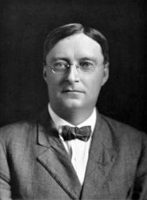
|
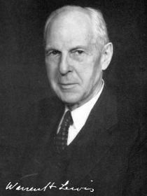
|
| Charles Russell Bardeen, M.D. | and Warren Harmon Lewis, M.D. |
From the Anatomical Laboratory of the Johns Hopkins University , Baltimore Md.
With 9 Plates and 27 Text Figures.
Introduction
The purpose of the following paper is a description of various typical stages in the development of the back, the limbs, and the body-wall in man. The work is based primarily upon reconstructions, according to the method of Born,[1] of parts of five human embryos; it has been extended and controlled by a study of the external form and of serial sections of several other human embryos. Dr. Lewis has devoted special study to the formation of the arm, Dr. Bardeen to that of the leg, the body-wall and the back.
In the accompanying table a list is given of the embryos utilised those marked with an asterisk have been reconstructed.
We shall consider the early stages in the development of the limbs, the body-wall and the back, first, from the point of view of the external form and, secondly, from that of internal structural ditferentiation.
I. External Form
The external form of the embryos we have used has been compared with that of embryos of a corresponding stage of development pictured in the His Atlas.[2] Figs. 1-15, on pages 3 to 9 represent a series of embryos belonging some to the Mall collection and some to the His collection. The general relation of the limbs and body-wall in embryos between two and seven weeks of age,[3] and between 2.1 and 20 mm. in length, are here represented by simple outline diagrams, based in part upon published drawings and in part upon photographs and upon sketches of the embryos indicated. On Plate I the photographs utilized are reproduced. On Plates II to IX are represented several typical stages in the general development of the body-wall and limbs. Figs. A and B, Plate II, are drawn from wax-plate reconstructions. Figs. C to E, Plates III to Y, are based, in the main, upon a reconstruction of the regions of the arm, abdomen and leg of embryos CLXIII and CIX, and upon excellent photographs. Figs. F to I, Plates VI to IX, are based upon wax-plate reconstructions of Embryo XXII.
Table 1
Template:BardeenLewis1901 table1
The Roman numerals refer to embryos in the collection of human embryos belonging to Prof. Mall, in the Anatomical Laboratory of the Johns Hopkins University. To Dr. Mall we are greatly indebted for the use of these embryos.
Reference to the embryos given in Table I will be found in the following articles by Dr. Franklin P. Mall
- No. II, A Human Embryo Twenty-six Days Old, Jour, of Morph., Vol. V;
- Nos. II, XII, XXII and XLIII, Development of the Human Coelom, Jour, of Morph., Vol. XII; Nos. II, XII and XXII, Ueber die Bntwickelung des Menschlichen Darmes, Arch, fiir Anat. und Phys., Special Bd., 1897;
- Nos. II, XLIII and LXXVI, Development of the Internal Mammary and Deep Epigastric Arteries in Man, Johns Hopkins Hospital Bulletin, 1898; Nos. II, XH, XXII, XLIII, LXXVI and CIX, Development of the Ventral Abdominal Walls in Man, Jour, of Morph., Vol. XIV, 1898;
- Nos. II, XII, XXII. LXXX CVI. CIX and CXLVIII, A Contribution to the Study of Pathology of Early Human Embryos, Johns Hopkins Hospital Reports, Vol. IX, 1901.
Fig. 1. X about 10 d.
Fig. 2.
Fig. 3.
Fig. 4. X about .5 d.
Fig. 5. Embryo XII.
The development of the neuro-muscular apparatus begins in the human embryo in the cervical region. In Fig. 1 is represented Embryo XII, 2.1 mm. in length and about two weeks of age. The axis of the embryo is curved in a semicircle about the heart and the umbilical vesicle. The axis contains neural tube, notochord, myotomes, dorsal-aortas, and mesenchyme (see Fig. 16). There are fourteen myotomes on each side. Mall considers three of them occipital, eight cervical, and three thoracic. The first cervical and the first thoracic myotomes are numbered " 1 " in Fig. 1. Caudal to the fourteenth myotome, an unsegmented band of tissue extends along each side of the spinal cord. The neural tube is open dorsally anterior to the fourth and posterior to the fourteenth myotome. Opposite the twelfth myotome a solid band of cells, the "' neurenteric canal," unites spinal-cord and entoderm. The notochord extends from a point opposite the cephalic margin of the heart to the region of the neurenteric canal. The dorsal aortae run a course parallel with the notochord, but extend further than the myotomes caudally. A considerable amount of mesenchyme is formed at the cephalic extremity of the axis of the body, in the region of the heart, but toward the caudal extremity little exists. The heart and the pericardial and pleural cavities are developed in the cephalic region of the wall of the umbilical vesicle. Between the region of the heart and the neural tube the pharynx extends forwards. Trom it project the hrst and second branchial pockets, Seessel's pocket, and the thyroid diverticulum. Into the caudal end of the embryo the hind-gut extends. The umbilical vesicle projects forwards from the region opposite the 1-6 cervical myotomes (see Fig. 1). At this period the amnion arises on each side along the length of the axis of the embryo as far forward as the region of the heart (Fig. 1). Externally and internally the amnion is covered by a layer of epithelial cells. From epithelium lining the coelom, several layers of cells have arisen (Fig. 16). There is, however, as yet, no true body-wall caudal to the region of the heart. There are no external visible signs of limb buds.
Embryo Lr
In Fig. 2 is represented the His embryo Lr; length, neck-breach, 4.2 mm.; age, about three weeks. The back of this embryo presents a slight concavity opposite the ninth (first thoracic) myotome. It is probable that this is an artefact, due to the removal of the embryo from the ovum, and that in the natural condition the back curved about the viscera as it does in the embryos represented in Figs. 1, 3, 4 and 5. "Lr" shows externally thirty-one myotomes (8c, 12t, 51, 5s, Ic). The ninth (first thoracic) and twenty-first (first lumbar) myotomes are designated by the numeral " 1. Lateral to the region of the myotomes lies the Wolffian ridge, a band of tissue which represents the anlage of the limbs and body-wall. The arm is represented by a slight swelling opposite the 5th to 8th cervical and 1st thoracic myotomes. The leg is represented by a slight swelling opposite the 1st to 5th lumbar and 1st sacral myotomes. The amnion was probably attached, in this embryo, to the umbilical cord. Between the Wolffian ridge and the umbilical cord the menibrana reuniens extends, at this period, so as to cover over the thoracic and abdominal viscera. It is represented as torn along the heavy irregular line.
Embryo CXLVIII
In Fig. 3 is represented Embryo CXLVIII; length, neck-breach, 4.3 mm.; age, about three weeks. A photograph of the embryo is given on Plate I. Though more advanced in development than Lr, but twenty-seven myotomes are present (2o, 8c, lOt, 51, 2s). This has been determined by careful counting of the myotomes in serial sections of the embryo. The base of the arm-bud appears to lie opposite the seventh to the eleventh myotomes. It is, therefore, probable that two occipital myotomes are present. But nine myotomes lie in the area between the arm-bud and the leg-bud. The base of the latter lies opposite 31st to the 25th or 86th myotomes. If two myotomes be considered occipital myotomes, the leg, in this instance, lies two segments nearer the head than usual. It is therefore probable that this embryo has an unusually short body-wall.
4 See Mall, Human Coelom, op. cit., p. 421.
Embryo LXXVI (length, 4.5 mm.; age, about three weeks) is of essentially the same stage of development as CXLVTII. It has thirty five myotomes (3o, 8c, 13t, 51, 5s, 2c). The base of the arm lies opposite the eighth (fifth cervical) to the twelfth (first thoracic) myotomes. The base of the leg lies opposite the twenty-fourth (first lumbar) to the twenty-ninth (first sacral) myotomes. Eleven myotomes lie between the regions of the arm and leg-buds. In CXLVIII the limb buds protrude more than in LXXVI and the body-wall extends further ventrally.
Embryo LXXX (length, 5 mm.; age, about three weeks), a photograph of which is given in Plate I, is similar, though slightly more advanced in development than Embryo CXLVIII.
Embryo a
In Fig. 4 is represented the His embryo a ; length, neck-breach, 4 mm.; age, about 23 days. The back of this embryo is very greatly flexed. Thirty-five myotomes are present (8c, 12t, 51, 5s, 5c). The armbud lies opposite the 5th to 8th cervical and 1st thoracic myotomes; the leg-bud opposite the 1st to 5th lumbar and 1st sacral myotomes. Both protrude further than in CXLVIII. The arm-bud projects in a caudal direction. The leg is represented, not as in the original His drawing, but instead in the more normal position shown in the His drawing of the right side of the same embryo.
Embryo R
In Fig. 5 is represented the His embryo E; length, 5 mm.; age, about 3^ weeks. Thirty-five myotomes are pictured (8c, 12t, 51, 5s, 5c). The arm-bud lies opposite the 5th to 8th cervical and 1st thoracic myotomes; the leg-bud opposite the 1st to 5th lumbar and 1st sacral. Both point somewhat caudally.
Embryo II
In Fig. G is represented Embryo II; length, neck-breach, 7 mm.; vertex-breach, 6 mm.; age, about 4 weeks. Thirty-eight myotomes are present (3o, 8c, 12t, 51, 5s, 5-6c). The extensions of the myotomes within the body Avail are pictured. The base of the arm-bud lies opposite the 5th to 8th cervical and 1st thoracic myotomes; that of the legbud opposite the 1st to 5th lumbar and 1st sacral myotomes. The armbud projects caudally, the leg-bud outwards and slightly caudally.
Embryo A
In Fig. 7 is represented the His embryo A; length, 7.5 mm.; age, about 4 weeks. Thirty-five myotomes are pictured (8c, 12t, 51, 5s, 5c). The arm-bud lies opposite the 5th to 8th cervical and 1st thoracic; the leg-bud lies opposite the 1st to 5th lumbar and 1st sacral myotomes.^ Both project caudally. Both show a slight division into segments. This, however, is much more marked in the following embryo.
Fig. 7. X about 5 d.
Embryo CLXIII
In Fig. 8 is represented Embryo CLXIII; length, 9 mm.; age, about 4.5 weeks. Two photographs of this embryo are shown on Plate I. Thirty-three myotomes are present (8c, 12t, 51, 5s, 3c). The base of the arm lies opposite the 4th to 8th cervical and 1st thoracic, and that of the leg opposite the 1st to 5th lumbar and 1st to 2nd sacral myotomes. The arm projects nearly caudally. A constriction on the cephalic and caudal borders separates the rounded upper arm from the flattened lower arm and hand. The constriction on the caudal border is close to where the arm joins the body-wall, while that on the cephalic border is at a point some distance from the body-wall. This difference on the two borders is to be correlated with the caudal projection of the arm.
5 This statement is based on the drawing given in Fig. 3, Plate I* of the Atlas.
The medio-lateral flattening of the distal portion of the arm-bud is especially well marked. Proximal to this flattened portion swellings on both medial and lateral surfaces indicate -where pre-muscle tissue is developed. A constriction may likewise be seen dividing the leg-bud into two distinct divisions. Owing to a slight torsion the lower portion of the leg-bud presents to view the anterior margin instead of the flattened lateral surface.
Fig. 9. X about 5 d.
Fig. 10.
Embryo Br
In Fig. 9 is represented the His embryo Br,; length, 11 mm.; age, about 4.5 weeks. Thirty-five myotomes are pictured (8c, 12t, 51, 5s' 5c).The base of the arm lies opposite the 4th to 8th cervical and 1st thoracic spinal ganglia. The division of the arm into its main segments is advanced beyond that pictured in Embryo CLXIII. The upper arm still projects caudally. The lower arm, owing to flexion at the elbow projects caudo-ventrally. The hand is flattened and can be distinguished from the forearm. Swellings of the digits are visible. The first indications of the shoulder are present. The posterior limb shows a differentiation of foot, leg and thigh regions.
- Externally visible segmentation at this stage is due to the spinal ganglia not to myotomes. The latter have lost their identity.
Embryo CIX
Fig. 10 represents Embryo CIX; length, 11 mm.; age, about 5 weeks Two photographs of this embryo are reproduced in Plate I. The base of the arm lies opposite the 3d to 8th cervical and 1st thoracic spinal ganglia. The upper arm still projects caudally. The forearm is more flexed and projects ventrally; it is now quite well marked off from the upper arm and hand. The digital swellings have increased and are now visible on the margin as well as on the flattened surface of the hand. The shoulder is more marked. The base of the arm is larger and extends higher in the cervical region than in the younger stages. The posterior limb shows a distinct differentiation of the foot. The kneebend may be distinguished. The hip region is not clearly marked externally.
Fig. 11.
Fig. 12. X about 2^ d.
Fig. 13.
Embryo CLXXV
Fig. 11 represents Embryo CLXXV; length, 13 mm.; age, about 5-| weeks. Two photographs of this embryo are given in Plate I. The various regions of the arm and the swellings of the digits are well marked. The forearm has a more caudal projection than in Fig. 10, In the posterior limb the various regions are more or less distinctly indicated. In the foot digitation has begun. The body wall has advanced half-way across the surface of the liver.
Embryo CVI
Fig. 12 represents Embryo CVI; length, vertex-breach, 17 mm., neckbreach, 15.5 mm.; age, about 5^ weeks. A photograph is reproduced on Plate I. The limbs and body-wall are similar in development to those of Embryo CLXXV, but the flexion at the elbow and knee is more marked, and the body-wall lias advanced further across the abdomen.
Embryo CLXVII
Fig. 13 represents Embryo CLXVII; length vertex-breach 14.5 mm., neck-breach 13.5 mm.; age, 5-|- weeks. A photograph is shown on
Plate I. While the embryo is similar in general differentiation to Embryos CLXXV (Fig. 11) and CVI (Fig. 13), the development of the digits of the hands and feet is further advanced. The flexion of the forearm is also more marked.
Fig:. 14. X about 3^ d.
Fig. 15.
Embryo XLIII
Fig. 14 represents Embryo XLIII; length, vertex-breach 16 mm., neck-breach 14 mm.; age, about 6 weeks. The forearm and leg have grown considerably beyond the stage shown in Figs. 11-13, but without marked alteration in external form.
Embryo XXII
Fig. 15 represents Embryo XXII; length, vertex-breach 20 mm., neck-breach 18 mm.; age, about 7 weeks. A photograph is reproduced on Plate I. The limbs have begun to resemble those of the adult. In the hand the clefts between the digits are well marked. The forearm and hand are somewhat pronated. The tips of the digits now reach nearly to the ventral mid-line. In the hind-limb the foot still lies in the same plane with the leg. The twist at the ankle which brings the foot into adult position has not begun. The toes are fairly distinct.
- A number of embryos have been pictured which correspond essentially in external form to those above described. The following list gives reference to the articles in which several of these have been described and pictured. To the His embryo R. Fig. 5, corresponds :
Foil's 5.6 mm. embryo, Fig. 217, p. 386, Minot's Embryology. To No. II, Fig. 6, corresponds :
Keibel's embryo H. S., length 8 ram., Arch, fiir Anat. und Phys., 1891, Taf. TvIX, Fig. 2.
Relations of the Limbs and Body-Wall to the Spinal Segments
In noting the main features of development in early human embryos, the most convenient landmarks for describing relative positions of structures are the myotomes. By the end of the second week of embryonic development about fourteen myotomes have been distinctly differentiated in the human embryo (see Fig. 1). The formation of myotomes continues until by the end of the fourth week about thirty eight have been differentiated (see Embryo II, Fig. 6). Anterior to the eight cervical myotomes, three occipital myotomes seem usually to be developed; posterior to the cervical region, twelve thoracic, five lumbar, five sacral, and five to six coccygeal myotomes are formed. The division of the myotomes into occipital, cervical, thoracic, luinbar, sacral and coccygeal groups depends upon the nerves and skeletal structures related to the body segments in which the myotomes lie.
The occipital myotomes are transient structures developed in conjunction with roots of the Twelfth Cranial Nerve. Before the spinal nerves have appeared they cannot with certainty be distinguished. No account of occipital myotomes is given in the description of the embryos pictured in the His Atlas. In the Mall embryos, LXXVI. CXLVIII and II, the occipital myotomes have been determined by a careful study of serial sections.
To the His embryo A, Fig. 7, correspond : His's embryo Pr, length 10 mm., Atlas, Taf. XIII, Fig. 4, and Taf. X, Fig. 10. His's embryo B, length 7 mm.. Atlas, Taf. I, Fig. 1.
To No. CLXIII, Fig. 8, correspond: Kollmann's 10.3 mm. embryo, Arch, of Anat. and Phys., 1891, Taf. Ill, Fig. 1. Ruge's 9.1 mm. embryo, His's Atlas, Taf. XIII, Fig. 5, and Taf. X, Fig. 12.
To the His embryo Br, Fig. 9, corresponds : Keibel's embryo, H. S. Bui., length 11. .5 mm.. Arch, fiir Anat. und Phys., 1891, Taf. XIX, Fig. 13a.
To No. CIX, Fig. 10, corresponds : His's embryo S, length 13..5 mm.. Atlas, Taf. XIII, Fig. 7, and Taf. X, Fig. 16.
To No. CLXXV, Fig. 11, corresponds: His's embryo Schj, length 13.8 mm., Atlas, Taf. XIV, Fig. 3, and Taf. X, Fig. 18.
To No. CVI, Fig. 13, correspond : His's embryo Br, length 13.3 mm.. Atlas, Taf. XIV, Fig. 1. Ruge's 13.6 mm., embryo, His Atlas, Taf. XIV, Fig. 4, and Taf. X, Fig. 19.
To No. CLXVII, Fig. 13, corresponds : His's embryo Pr, length 14..5 mm.. Atlas, Taf. XIV, Fig. .5, and Taf. X, Fig. 30.
To No. XLIII, Fig. 14 corresponds : His's embyro 8^, length 1.5..5 mm., Atlas, Taf. X, Fig 31.
To No. XXII, Fig. 1.5 correspond : His's embryo XCI, length 16 mm., Atlas, Taf. X, Fig. 33. His's embryo Ltj, length 17..5 mm., Atlas, Taf. X, Fig. 33. His's embryo Z. W., Atlas, Taf. X, Fig. 34.
The cervical myotomes are those developed in conjuction with the cervical spinal nerves. In the vast majority of instances these are eight in number. The occasional absence in the adult of a cervical vertebra indicates that in the embryo less than the normal number of cervical spinal segments sometimes develop. In all of the embryos we have studied, the arm-bud begins its development opposite the fifth to the eighth cervical segments and opposite the following spinal segment. The most caudal myotome lying opposite the arm-bud may, therefore, be taken to represent the first thoracic segment.
Between the base of the arm-bud and that of the leg-bud, as a rule, eleven myotomes intervene. A marked exception to this is found in Embryo CXLVIII, where but nine myotomes seem to lie in this region. In other instances, twelve myotomes have been pictured. Such is apparently the case in the His embryo B (Atlas, Taf. I, Fig. 1) and in the His embryo Pr (Atlas, Taf. XIII, Fig. 4). It is difficult to determine this with certainty from the figures. Variation in the number of myotomes intervening between, the regions of the arm and leg corresponds with well-known variations in the length of the spinal axis in the adult.[4]
In the His wax-models of young human embryos, Embryo Lr (No. 6) shows nine myotomes between the regions of the swellings which indicate the arm- and leg-buds. Embryo A shows fifteen myotomes between the arm and leg areas. The number of the myotomes in the thoraco-abdominal region in each of the models is probably incorrect. Fig. .5, Plate XX, of the His Atlas seems to show the usual number of thoraco-abdominal myotomes in Lr. Fig. 2, Plate I*, shows twelve instead of fifteen thoraco-abdominal myotomes in A. The model of A presents, in case of the thoracoabdominal myotomes, conditions characteristic of the pig. Out of twelve young pig embryos of various sizes, in eleven instances we have found sixteen myotomes intervening on each side between the arm and leg regions, and in one instance fifteen. In the pig, therefore, five more body segments than in man lie between the arm and leg areas. This is also to be seen in the adult. In the pig there are two more thoracic segments than in man, and, as indicated by the nerve distribution to the abdominal wall, three more abdominal segments. The third lumbar nerve in the pig corresponds in distribution somewhat to the first lumbar in man, but it has a more extensive abdominal distribution. While therefore in man the first lumbar segment is counted as belonging to the leg area, in the pig the third lumbar segment may be considered to belong to the thoraco-abdominal region. There are six lumbar vertebrie in the pig. The furcal nerves are the fifth and sixth lumbar usually, but sometimes the fifth, probably sometimes also the sixth, may be the sole furcal nerve.
In the Keibel " Normentafeln " of the pig the number of myotomes pictured between the arm and leg areas varies from fifteen to eighteen in different embryos. It
The leg-bud arises opposite six myotomes. These usually are the twenty-fourth to the twenty-ninth, corresponding to the five lumbar and 1st sacral myotomes. Caudal to this region arise the remaining sacral and the coccygeal myotomes.
The myotomes give rise to the dorsal musculature, to the thoracoabdominal musculature, and to the musculature of the neck, tongue (?) and caudal region. When differentiation of body musculature takes place the distinction between the myotomes becomes lost, and they can no longer be used as landmarks. This occurs by the time the embryo has reached a length of 11 mm. and an age of five weeks. Hereafter segmental skeletal structures and spinal ganglia may be used as landmarks. The former present the more stable relative conditions.
General Summary of the Most External Features noted in the Early Development
Development of the spinal axis begins in the cervical region. At the end of the second week fourteen myotomes are present (3o, 8c, 3t); at the end of the fourth week, thirty-eight (3o, 8c, 12t, 51, 5s and 5c). During the fifth week the myotomes, owing to fusion of their dorsal surfaces, cease to be externally visible. The spinal ganglia, however, give rise to a segmentation externally visible for a somewhat longer period.
The limbs and body-wall arise in the region where the amnion joins the axis of the embryo. By the end of the second week, at the stage represented in Embryo XIT, the amnion is attached directly to the axis along a line extending from a region anterior to the heart to the caudal extremity (Fig. 1). The amniotic cavity rapidly enlarges. That part of the amnion near the axis of the embryo is carried ventrally so that it closes in the viscera which have previously protruded free into the general coelomic cavity. Finally the amnion reaches the allantoic stalk (or umbilical cord) and becomes attached to this. That part of the amnion extending from the umbilical cord to the axis of the embryo is now known as the menibrana reuniens. It forms the chief covering of the pericardial, pleural and peritoneal cavities. Fig. 2 probably, and Fig. 3 certainly, represent stages in which the viscera are completely inclosed by the menibrana reuniens.
is possible tliat no great care was taken in determining accurately the number of myotomes in tbis region. In the " Normentafeln " of the chick the number of myotomes pictured in this area varies from seven to ten. Apparently seven in the normal number.
During the second half of the third week the membrana reuniens becomes markedly thickened along its line of attachment between, usually, the fourth and twenty-sixth spinal segments. This thickening is known as the WoMan ridge. In Figs. 2-7 the ventral margin of this ridge is emphasized by a heavy line. Opposite the fifth to the ninth and the twenty-first to the twenty-sixth spinal myotomes this thickening is especially marked. The two latter areas represent the inception of the limb-buds; the intervening area represents the rudiment of the lateral body-wall. These three areas first become well marked toward the end of the third week (Figs. 2-3).
The limb-buds increase very rapidly in size. At first the limb-buds extend directly laterally from the Wolffian ridge (Figs. 2 and 3), but as development proceeds they project more and more in a caudal direction (Figs. 4, 5 and 6). Meanwhile two distinct divisions appear in each limbbud. The part which arises directly from the Wolffian ridge we may call the basal division. This portion continues to grow in the ventrolateral direction taken by the limb-bud originally. Beyond this basal part the limb-bud bends in a ventro-median direction. The part of the limb-bud beyond the bend we may call the " distal part " of the limbbud. For some time the basal portion of the limb-bud continues to grow in a ventro-lateral direction, while the distal part grows in a ventro-median direction. The limb as a whole meanwhile points distinctly in a caudal direction.
Dorsally the base of the limb becomes continuous with the dorsal margin of the Wolffian ridge, ventrally with its ventral margin. The dorso-lateral surface of the base of the limb-bud is therefore extensive; the ventro-median surface is small in area. Owing to the caudal direction assumed by the limb, the anterior (cephalic) surface of the base is extensive, while the posterior (caudal) surface is limited in extent.
The distal part of each limb-bud is flattened so that it presents median and lateral surfaces and anterior (cephalic), ventral and posterior (caudal) margins. A constriction may be seen immediately beyond the region where the distal flattened portion of the limb joins the thicker rounded base. In Fig. 8 the constriction is emphasized by heavy lines. In the arm it is seen as one looks at the limb directly from the side. In the leg, which is here slightly twisted, one may see the constriction as it appears when the limb is viewed on the anterior margin. The constriction is just beginning to appear in the leg-bud shown in Fig. B, Plate II, and in Fig. C, Plate III.
From the basal portion of each limb-bud are developed the limbgirdle (shoulder and pelvic-girdles), and the upper limb (upper arm and thigh), from the distal portion are developed forelimb (forearm and leg) and extremity (hand and foot). The place where the distal part of the limb joins the base represents the future elbow or knee. These two joints are formed in a flexed position (see especially Figs. F, G, H and I, Plates VI-IX).
The constriction mentioned above serves to designate the differentiation of the distal part of the limb-bud into the forelimb and extremity. Opposite the constriction the forelimb is formed; immediately distal to it the extremity (Figs. 8, 9, 10, 11, 12, 13, 14, 15).
The hand and foot are at first flattened, disk-shaped bodies. Digitation is first marked on the lateral surface (see the hand in Fig. 9), and then in the free margin also (Fig. 10).
Differentiation of the base of the limb-bud is somewhat less easy to follow than that of the distal part. This is due mainly to the fact that many distinguishing structures are at first deep-seated. As the musculature, however, becomes distinctly differentiated, the various main groups of muscles give rise to distinctive external characteristics.
The limbs at the stage shown in Fig. 15, p. 9, and in Figs. F, Gr, H and I, Plates VI-IX, exhibit rudiments of most of the structures characteristic of the adult limbs. Much growth and shifting of parts, however, is necessary before adult conditions are reached.
The great curvature of the axis of the body opposite the limbs during their formation has been noticed by His.[5]
The arm-bud at first lies opposite the 5th to 8th cervical and 1st thoracic myotomes. As it grows in size the area of attachment to the body enlarges and extends on its cephalic side to the level of the 3rd cervical neural process (see Embryo CIX, Fig. 10). We find also that the shoulder comes to lie closer and closer to the precervical sinus until the embryo reaches a length of about 20 mm. As development still further proceeds the shoulder gradually migrates caudally and the distance between the shoulder and precervical sinus increases imtil the adult position is attained. Simultaneous with this caudal migration of the arm, the lower portion of the ventral wall of the neck appears.
The posterior limb arises slightly later than the anterior. Throughout its development the leg lingers somewhat behind the arm. The base of the leg-bud is at first opposite the 1st to 5th lumbar and the 1st sacral myotomes. Gradually the base extends so as to include the region opposite the second and third sacral myotomes and the upper extremity of the base ceases to extend over the region of the first lumbar segment. As adult condition is approached, the leg assumes a much more caudal position.
During the growth of the limbs the body-wall grows forwards at the expense of the membrana reuniens. In the- various figures the ventral line of the body-wall is indicated. The abdominal wall has closed in against the umbilical cord by the time the embryo has reached a length of 6 cm. and an age of about 3 months.
Having thus considered the more general external features presented in the early development of the limbs and body-wall, let us turn to a consideration of the formation of the main structures within these areas.
Fig. 16. Cross section through the fourth cervical segment of Embryo XII. X 55 d.
II. Internal Structural Differentiation
Second Week
Fig. 17. Cross section through the fifth thoracic segment of embryo LXXVI. x 55 d. The right side of the section passes through the middle, the left side through the posterior third of the segment.
In an embryo of two weeks, No. XII, Fig. 1, the viscera are attached to the ventral surface of the spinal axis of the embryo, and the amnion is attached to its lateral margin. Fig. 16 shows the conditions which are seen in a transverse section through the fourth cervical segment. The spinal cord is, in this region, a closed tube, the lateral walls of which are composed of five or six layers of epithelial cells. On each side lie the myotomes. These are oval in cross-section, square with rounded corners when viewed from the lateral surface. Each has a well-marked myocoel, surrounded on all sides by four or five layers of epithelial cells. On the left in Fig. 16 the section passes through the centre, at the right it passes through the anterior margin of a myotome. Ventral to the spinal-cord lies the notochord; on each side of this a dorsal aorta. Between notochord, aortse, spinal-cord and myotomes lies a certain amount of mesenchyme, which arises apparently from the ventro-median surface of the myotomes.
In the region between the dorsal margin of the myotomes, the spinal cord and the ectoderm lie spinal ganglia cells.
Ventral to the aorta on each side the splanchnopleure is attached to the axis of the embryo; ventral to the myotomes on each side, the somatopleure. The latter is continuous with the amnion. The coelom is surrounded by epithelium, and this is surmounted by several layers of mesenchyme cells which apparently have arisen from the epithelium lining the coelom.
Third Week
During the third week of embryonic development marked changes take place in the spinal axis of the embryo and in the adjoining somatopleure. A section through the fifth thoracic segment of an embryo of the third week. No. LXXVI, is shown in Fig. 17. The spinal ganglia are definite, well-developed groups of cells on each side of the spinal-cord. In the spinal-cord the ventral-root zone is well marked, and the first ventral-root fibres have appeared. The myotomes have become flattened and elongated. The median surface of each of these has become converted into muscle cells, shown in cross-section in the figure. There is a single dorsal aorta, on each side of which a Wolfl&an body has developed. Dorsal to the Wolffian body lies the cardinal vein. The axial mesenchyme, which arises, at first at least, apparently from the myotomes, and the mesenchyme which springs from the coelomic wall have increased very greatly in amount, and have fused so as to form a common mass of tissue which surrounds- the other structures. None, however, intervenes between the dorso-lateral surface of the myotomes and the ectoderm.
A vascular plexus is developing in the mesenchyme. The axial mesenchyme of the caudal third of each segment has become dense. This is shown at the left in Fig. 17. It represents the anlage of the intervertebral disc and of the vertebral arch and costal processes. The somatopleure is considerably thicker than at the stage shown in Fig. 16. This increased thickness is due to an increase in the amount of the mesenchyme. This mesenchyme extends for a short distance dorsally between the ventral tip of the myotome and the ectoderm. The myotomes have not yet entered the body-wall.
In the region of the posterior limb the increase of mesenchyme between coelom and ectoderm precedes the formation of muscle on the median layer of the myotomes, the formation of the spinal ganglia, and the appearance of the ventral nerve roots. This is shown in Fig. 18, taken through the leg region of Embryo LXXVI.
Fig. 18. Cross section tbrougli tlie lumbar region of Embryo LXXVI. x 55 d.
Fig. 19, taken through the leg region of Embryo CXLVIII, shows an older stage in which the limb-bud is considerably more advanced in development. The spinal ganglia are distinct. Formation of muscle cells has begun on the median surface of the myotomes.
Fig. 19. Cross section tbrough the lumbar region of Embryo CXLVIII. x 55 d.
The Wolffian ridge and limb-buds, which appear during the third week, as shown in Figs. 2, 3 and 4, consist, therefore, at the end of this period, merely of a mass of mesenchyme which intervenes between the coelom and the ectoderm lateral to the axis of the body. This mesenchyme contains a vascular plexus. Along the free edge of the limbbuds the ectoderm consists of several layers of epithelial cells (Fig. 19).
Fourth Week
During the fourth week certain structures extend from the spinal axis into the Wolffian ridge and the limb-buds. Let us consider first the relation of these axial structures to the body-wall and then their relation to the limb-buds.
Fig. 20. Cross section through the fifth thoracic segment of Embryo II. x .55 d. The right half of the section passes through the middle, the left half through the posterior third of the segment.
Fig. 21. Diagrammatic cross section through the mid-thoracic region of Embryo II. About 2.5 d. In looking at the section the si)ectator is supposed to be facing towards the head of the embryo. In the background one sees the fourth thoracic scleromere with arch and costal process, and at the right the intersegmental artery and the distal edge of the fourth myotome. In the foreground the spinal cord and spinal ganglia in section, and the spinal nerves of the fifth thoracic segment are shown. At the left the fifth thoracic myotome is shown in section.
Fig. 20 shows the general appearance of a typical thoracic segment (the fifth) in an embryo at the end of the fourth week. In Fig. 21 similar structures are shown somewhat more diagrammatically. Fig. A, Plate II, shows a reconstruction of the axis of the body and a part of the right lateral wall and the leg in the same embryo.
The skeletal portion of each axial segment consists of a condensation of the mesenchyme at the caudal third of the segment as shown at the left in Fig. 20. To this skeletal tissue the term scleromere may be applied. It represents the intervertebral disc, the arch or neural process and the costal process of a segment of the future spinal column. The general form of the scleromeres at this stage may be readily seen in Fig. A. The scleromeres do not as yet extend into the body-wall.
The general form of the myotomes may be seen in Fig. A. At the right the 11th and 13th thoracic and the 1st and 3d lumbar myotomes are viewed from the side and slightly in front. At the left a portion of the median surface of several myotomes may be seen. In Fig. 20 two myotomes are shown in cross-section. The myocoel has disappeared. The median surface of each myotome has been entirely converted into musculature. The lateral surface and ventral and dorsal tips are covered by epithelium. A certain portion of the dorsal surface of the thoracic myotomes is, however, converted into musculature. This is shown in the myotomes at the right in Fig. A. The thoracic myotomes extend for a short distance into the body-wall. In Fig. 20 this is shown in a cross-section. In Fig. A the projecting tips of the myotomes may be seen through tlie membrane lining the coelom.
The thoracic spinal nerves project for a shorter distance into the bodywall than do the myotomes. Each is divided at the dorsal margin of the coelom into two portions, a median which becomes the sympathetic ramus, and a lateral which extends outwards between the median surface of the myotome and the lining membrane of the coelom and becomes the ventral trunk of the spinal nerve (see Figs. A, 20 and 21).
From the aorta intersegmental arteries arise and send branches dorsally toward the spinal ganglia, laterally between the myotomes and ventrally into the body-wall (see Fig. 21). A considerable vascular plexus is developed in the mesenchyme. The general relations of the latter are shown in Fig. 20. The venous blood is collected in the cardinal veins and in branches of the umbilical vein.
The general relation of the formed structures of the axis of the body to the leg-bud at the end of the fourth week are shown in Fig. A, Plate II. The limb-bud lies opposite the five lumbar and the first sacral segments. The coelom extends to a point opposite the first sacral segment, but in the region of the limb it does not extend as far dorsally as in the thoracic region. From the model represented in Fig. A, several of the myotomes of the left side, the axial mesenchyme, the aorta, the left cardinal vein, the intestines and urino-genital organs have been removed. A ^^ortion of the right cardinal vein and a portion of the right umbilical artery are shown, reduced in size for the sake of clearness. The umbilical artery curves about the distal extremity of the coelom. From the umbilical artery a branch passes into the limb-bud. Veins pass from the limb-bud into the cardinal vein. The blood-vessels of the limb exist at this time in the form of an irregular sinusoidal plexus.
The second, third and fourth lumbar nerves may be seen sending spreading bundles of nerve fibres into the dense tissue of the limb, dorsal to the cardinal vein. They extend, however, for no considerable distance into the limb-bud. The myotomes do not extend into the limbbud.
By following the successive spinal segments from the fourth sacral to the first lumbar in Fig. A, an idea may be gained of the development of segmental structures in the axial region. The scleromeres are represented at the caudal third of each segment. On the left side of the body the myotomes are omitted from the 3d sacral to the 3d lumbar segments. The spinal nerves first appear in the first and second sacral segments.
Fig. 33. Tangential section tlirougli the leg region of Embryo II. 35 d.
Fig. 22 shows the general conditions existing in the limb region of Embryo II when seen in section. At the left the leg-bud is shown cut through an area near the distal extremity of the coelom. At the right the cut is more dorsal and extends through the tips of the lumbar spinal nerves.
In the arm region of Embryo II, the cervico-brachial plexus is formed and limb nerves extend into the limb-bud. The conditions are essentially similar to the conditions found in the leg region of Embryo CLXIII and described below.
Fifth Week
During the fifth week of development a considerable amount of organization occurs in the spinal axis, the body-wall and the limbs. The nature of the processes taking place are indicated in Embryo CLXIII (length, 9 mm.; probable age, 4^ weeks).
The structure of the back, the limbs and body-wall in this embryo is shown in Fig. B, Plate II, and Fig. C, Plate III. The areas mentioned are drawn from reconstructions. The remaining parts of Pig. C are drawn from an excellent photograph.
Fig. 28. Diagrammatic cross section through the 5th-6th thoracic segments of Embryo CLXIII. x 25 d. The general arrangement of the structures represented is like that described for Fig. 21.
Fig. 23 shows diagrammatically the general nature of the structures in a typical thoracic segment (the 6th) of Embryo CLXIII. The changes taking place in the thoracic region during the first half of the 5th week may be readily followed by comparing Fig. A with Fig. B, and Fig. 21 with Fig. 23.
The skeletal portion of the segment consists in Embryo CLXIII, as in Embryo II, of a condensed mesenchyme at the distal third of the segment, but the scleromere is far more definitely outlined. Xeural and costal processes are well developed. The latter present something of the general form of ribs in the thoracic region (see Fig. 23 and Fig. B). A sheet of condensed mesenchyme connects the neural and costal process of the scleromeres of neighboring segments. That connecting the neural processes is shown in Fig. B at the right. That portion of each scleromere representing an intervertebral disc is likewise united ventrally and dorsally with its neighbors by a dense sheet of tissue at the periphery of the disc. The ventral portions of these sheets of tissue are represented in Fig. B. Within the space lying between the scleromeres is developed the chondrogenous tissue which gives rise to the vertebral bodies. Marked alterations have taken place in the myotomes. In the thoracic region the two layers of the myotome have, with the exception of the dorsal and ventral tips in the more distal segments, become converted into musculature (see Fig. 23 and Fig. B). As shown at the left in Fig. 23, the myogenous tissue which has arisen from the myotomes is being divided into three portions — a dorsal, a lateral and a ventral — by the ingrowth of a vascular mesenchyme. The finer changes taking place during this period are essentially similar to those previously described as taking place in the body-wall of the pig.[6]
The tissue of the superficial lateral layers of the myotomes has formed into a continuous layer (see Fig. C, Plate III), Segmentation, however, is still visible.
Both the costal processes of the scleromeres and the myotomes have extended well into the body-wall (Fig. 23).
The thoracic spinal nerves likewise have kept pace in growth with the myotomes. The ventral extremities of the spinal nerves are caught between the tips of the myotomes (see Fig. B, Plate II). From the spinal nerves, dorsal and lateral branches have arisen as well as the sympathetic. A sympathetic cord has arisen from the extremities of the sympathetic rami.
The segmental arteries (see Fig. 23) are similar in nature to those of the stage shown in Fig. 21, but the branching is more extensive. The mesenchyme is much more developed and now surrounds all formed structures in each spinal segment. It has begun to invade the myotomes.
In the region of the posterior limb bundles of fibres from the five lumbar and first two sacral nerves have become anastomosed into a plexus, from which in turn four nerves have sprung. These represent the femoral, obturator, tibial and peroneal nerves (Fig. B). Within the legbud the central mesenchyme, near the axis of the embryo, has become condensed. This condensed mesenchyme represents the femur and hip bone of the adult limb. In the drawing the outline of this sclerogenous mass is made dia grammatically sharp. The femoral portion of the skeletal mass fades gradually into the undifferentiated mesenchyme of the distal portion of the limb. It. is this skeletal mass which seems to divide the bundles of nerve fibres of the plexus into the four main divisions which form the origin of the four chief nerves of the limb. The main artery and vein of the limb are represented, but smaller in proportion to the other structures than in nature. The border vein at this period is well developed. The conditions just described are well shown in I'ig. C, Plate III, which represents the conditions seen in the limb from the lateral side after removing the ectoderm and the undifferentiated mesenchyme. It is to be noted that the myotomes do not extend into the limb-bud.
The arm is somewhat more advanced in development than the leg. The following description applies to the conditions represented in Fig. C, Plate III. A detailed account of the structure of the arm at this stage is reserved for a later paper.
Lateral to the myotome system in the arm region is an ill-defined mass of mesenchyme extending from the upper cervical to the 7th thoracic myotomes. At the level of the 1st and 2d ribs it divides into several masses. The first passes ventral to the arm and brachial plexus. The main portion of it joins the arm pre-muscle sheath. From this mass the pectoral muscles develop, hence we may designate it the pectoral pre-muscle mass. It is continuous ventrally with an in-egular mass of condensed tissue, the ventral neck pre-muscle mass. The second division of the lateral pre-muscle mass passes dorsal to the arm and brachial plexus and joins the arm pre-muscle sheath. It constitutes the latissimus dorsi pre-muscle mass. The third division, parallel to the ventral portion of the cervical myotome column, represents the levator scapulge and serratus anterior pre-muscle mass. Lateral to it is an ill-defined mass of pre-muscle tissue. The fourth and most dorsal division is thinner and less clearly marked than the others. We may call it the rhomboid pre-muscle mass. The caudal limit of the trapezius pre-muscle mass appears at the upper end of the arm region. It is at the level of the 4th cervical neural processes, and from here the muscle mass extends to the occipital region. Most of the arm premuscle sheath which surrounds the skeletal core has been dissected away. Toward the distal end of the arm the skeletal and pre-muscle tissues blend (Fig. C). Beyond this point differentiation is less advanced. The proximal portion of the arm sheath is continuous with that around the scapula.
Part of the skeletal core is seen projecting caudally beyond the cut edge of the pre-muscle sheath. The lowej end of the humerus, ulna and radius are represented. The ulna and radius are continuous with the hand plate, which is composed of less differentiated tissue. There is a slight bend at the elbow. The upper end of the humerus is continuous with the scapula. The scapula is a flattened oval mass embedded in the scapular pre-muscle tissue. No coracoid or arromiom processes are present. The clavicula is not present. The skeletal core is composed of dense mesenchyme. It shades oft", however, into the surrounding pre-muscle sheath.
The brachial plexus is formed by the 5th to 8th cervical and 1st thoracic nerves. The spinal nerves, as well as the plexus they form, have scarcely any caudal inclination, but pass ventrolaterally into the arm. The plexus is fairly well formed. The main nerves arising from it are present. The presence of the condensed skeletal core separates these into two groups. The musculo-spiral and circumflex on the dorsal and the ulnar, median and musculo-cutaneous on the ventral side. In Fig. C only the tips of the musculo-spiral and median nerves are shown. At this stage they have scarcely grown to the elbow.
Sixth Week
During the sixth week the limbs and the body-wall and the back begin to approach the structural conditions characteristic of the adult.
The conditions present at the end of the fifth week are shown in Embryo CIX; length, 11 mm.; age, about 5 weeks. This embryo is pictured in Figs. D and E, Plates IV and V. These figures, with the exception of the head in Fig. D, are drawn from wax-plate reconstructions. Fig. 24 represents a cross-section through the 6th to 7th thoracic segment of a slightly older embryo, CVI (length, 15.5 mm.; age, about 5-| weeks).
The typical thoracic segment during the first half of the sixth week exhibits the following conditions:
The skeletal structures have begun to assume adult form. Between the intervertebral discs the bodies of the vertebrae are now formed of chondrogenous tissue. This may be seen in the dotted area of the scleromere in Fig. 24 and in the darker areas of the spinal column in Fig. E. The chondrogenous tissue extends into the neural and transverse processes of the scleromere (see the left side of Fig. 24). Each costal process extends considerably further ventrally than at the stage shown in Fig. 23. Within the dense tissue of the costal process chondrogenous tissue is formed similar to, but without direct connection with, that of the vertebral body and transverse process. A lateral view of the neural and transverse processes of the vertebrae is shown in the cervical region in Fig. D. The extent of development of the ribs is shown in Fig. E.
Fig. 24. Diagrammatic cross section through the 6th-7th thoracic segments of Embryo CVI. x 35 d. This figure is made to correspond with Figs. 31 and 23. See legend for Fig. 31.
The musculature has undergone most marked changes. The dorsal musculature is slightly separated from the ventro-lateral by vascular mesenchyme. The dorsal musculature is further subdivided into three muscle columns. These correspond to the ileo-costal, longissimus dorsi and spinalis groups of dorsal muscles in the adult. The ventral musculature is likewise subdivided into two main portions, the rectus muscle and the lateral musculature. The last is further subdivided into external oblique, the intercostals, the internal oblique, and the transversalis muscles. In the intercostal muscles alone is the segmentation characteristic of the myotomes fully maintained.
In Fig. D the separation of dorsal from ventro-lateral musculature is clearly shown. The dorsal musculature is shown divided into three columns in the upper thoracic region; caudally differentiation is not so extensive. The musculature of the external oblique may be seen, in Fig. D, covering the external intercostal muscle, the ribs and in part the rectus musculature. In Fig. E the rectus musculature, internal intercostal and internal oblique may be seen. Differentiation, however, is not so far advanced at the stage shown in Figs. D and E as it is at the stage shown in Fig. 34. The ventro-lateral abdominal musculature of Embryo CTX is connected by an irregular dense band of tissue Avith the pubic process of the anlage of the pelvic bone. This band of tissue is represented with diagrammatic distinctness in the draAvings.
The neural apparatus has undergone rapid development. Fig. 21 shows clearly the main branches arising from the typal thoracic nerve. Muscle twigs are arising. The general appearance of the spinal nerves at this stage is shown in Fig. E.
The mesenchyme is extensive in amount. The blood-vessels are similar in general distribution to those described in Embryo CLXIII, but the vascular plexus is more extensively developed.
In the posterior limb the central skeletal mass has assumed somewhat definite outlines. Fairly good views of it are presented in Figs. D and E. The pelvic portion of the skeleton of the limb consists of a central region continuous with the head of the femur. From this central acetabular portion spring iliac, ischial and pubic processes. The femur is short and thick. It is indistinctly shown in Fig. D. The tibia and fibula are of fairly definite form (Figs. D and E). The skeleton of the foot has the form shown in Figs. D and E. It is composed of condensed mesenchyme. No accurate division into parts can be distinguished.
The main nerve trunks have continued their growth into the limb. From them many of the principal muscular and cutaneous branches have sprung. The general distribution of the lateral nerves of the limb, the femoral and peroneal, and their branches, may be seen in Fig. D. That of the median nerves of the limb, the obturator and tibial, in Fig. E, In both figures the anterior border nerves (the ilio-hypogastric and genito-crural) and the posterior border nerves (the pudic and posteriorcutaneous) are shown.
About the main branches of the nerves of the limb a differentiation of musculature has begun. This is indicated in Figs. D and E. In Fig. 25 are shown the appearances in cross-section presented by the early developing musculature of the leg. The blood-vessels of the limb are shown in Figs. D and E. The sciatic artery is still the chief source of supply, but the femoral and obturator arteries also have appeared. The blood is carried into the cardinal (iliac) vein partly by the femoral vein and partly by the sciatic. The formed structures of the limb are surrounded by a vascular mesenchyme.
Fig-. 25. Section through Embryo CIX. x 25 d.
The conditions existing in the arm are considerably in advance of those in the leg. The following description of the conditions in the arm region can be followed from Figs. D and E, Plates IV and V.
The lateral pre-muscle mass has become completely divided into several groups. The tissue of these groups is now fibrillated. The first division, the one which passes ventral to the brachial plexus, is seen in Plate V. It constitutes the pectoral muscle mass, representing both pectoralis major and minor muscles. This mass extends from the level of the third rib to the humerus and clavicle. There is no attachment to the ribs. The intercostal muscles have been dissected away in Plate V to show its costal end. The second division of the lateral pre-muscle mass has developed into the latissimus dorsi and teres major muscle mass. It extends, from the level of the 4th rib to the humerus, where it blends with the scapulo-humeral mass. Its development and differentiation have not proceeded so far as the pectoral mass. The third division of the lateral pre-muscle mass has developed into a long muscle extending from the 1st cervical vertebra to the 9th rib, Digitations extend to the transverse processes of the cervical vertebra and to the cephalic 9 ribs. The muscle lies in a more median plane than the scapula and has as yet no attachment to it. It represents the levator scapula? and serratus anterior muscles. The trapezius mass has extended to a lower level than found in CLXIII. There is no scapular attachment. Only the ventral half of the mass pictured in Plate IV consists of muscle fibres, the remaining connective tissue portion connects with the dorsal ends of the neural processes.
Considerable differentiation in the pre-muscle sheath has taken place. The scapulo-humeral mass is with difficulty separated into the various muscles. These are more blended into a single mass than would appear from Plates IV and V. Here portions of the deltoid and trapezius have been dissected away.
The extensor mass of the forearm can be separated into three groups which are more or less blended however. The larger superficial mass has been partially dissected away. It represents the extensor digitorum communis, extensor carpi ulnaris, and extensor digiti quinti proprius muscles. The second group arises beneath the first group, taking a course at nearly right angles to it. It represents the deep extensor muscle of the forearm. These two groups fuse distally with the general condensed mesenchyme of the digits, where all traces between premuscle and pre-cartilage are lost. The third group represents the brachio-radialis and the extensor carpi radiates longus et brevis. A • portion of the brachialis can be seen in Plate IV. The flexor surface of the arm is shown in Fig. E, Plate V. The biceps and coracobrachialis are obscured by the pectoral mass. The flexor mass of the forearm has split into two layers. It shows less differentiation than the extensor mass.
The arm skeleton shows considerable advance. The shape of the scapula has changed, both coracoid process and acromion are present and of large size. The clavicle has begun to develop and consists of an ill-defined mass of condensed tissue projecting about one-half the distance from the acromion to the tips of the first rib. The humerus is fairly well defined and is continuous with the scapula as well as the ulna and radius. The elbow bend is well marked. The ulna and radius are farther advanced than in Embryo CLXIII. They end distally in the carpal plate. In the carpal plate indications of formation of the carpal elements are seen. The digits consist of undifferentiated tissue in which are blended skeletal muscle and tendinous elements. The humerus, ulna and radius have a core of hyaline cartilage. The rest of the skeletal tissue consists of condensed mesenchyme and pre-cartilage.
In Plate IV are shown the circumflex, radial and musculo-cutaneous nerves. The brachial plexus with portions of the nerves arising from it are seen in Plate V. The plexus is well formed. It has only a slight caudal inclination. The spinal accessory nerve is seen on the median surface of the trapezius. Branches from the 3d and 4th cervical nerves join it.
Seventh Week
By the end of the seventh week most of the structures characteristic of the adult back, body-wall and limbs have appeared. Subsequent development depends in the main upon growth and upon relative shifting of parts.
The structures of the abdominal wall in a seven-week embryo (XXII, length 20 mm.) are shown in the Figs. F, G, H and I, Plates VI, VII, VIII and IX. The vertebra are composed of embryonic hyaline cartilage. Each presents a neural and a transverse process on each side. The cartilaginous portions of the vertebrae are shown in the upper thoracic region of Fig. H. The. ribs are likewise composed of embryonic cartilage, shown in the same portion of the figure. The cartilage of the ribs is at no time continuous with that of the vertebrae. The ribs and vertebras are surrounded by a dense mesenchyme. This is continuous from the ribs to the transverse process of the vertebra. Between the bodies of the vertebrae it forms the intervertebral discs (Fig. I). It is continuous over the surface of the spinal column. From it are developed the ligaments characteristic of the thorax and spinal column together with the perichondrium and periosteum. No thirteenth rib is present in this embryo or in any of the other young embryos we have studied. Fig. 26 shows at the right the portion of the skeleton composed of embryonic cartilage, at the left the covering of dense mesenchyme.
The musculature of the back and abdominal walls has a general resemblance to that of the adult. Fig. I shows the musculature of the abdomen and thorax as seen from within, Figs. F and G that of the more superficial layers of the abdomen and thorax, and Fig. H that of the deeper layers of the abdomen and thorax. The dorsal muscles are not clearly shown in any of the figures, but they are divisible into the three distinct groups, the ileo-costal, longissimus dorsi, and spinalis muscles, characteristic of the adult.
Fig. 26. Skeleton of distal half of Embryo XXII. At the left side the covering of dense embryonic connective tissue is shown, at the right the parts composed of embryonic cartilage, x 10 d.
The nerves, like the muscles, have a distribution essentially similar to that found in the adult. The figures indicate with sufficient clearness the distribution of the thoraco-abdominal and border nerves.
The main blood-vessels are those characteristic of the adult. â– In the posterior limb the skeletal tissue has undergone extensive differentiation. The rudiments of all of the bones of the leg may be seen in the form of cartilage except that the terminal phalanges of the three onter toes have not yet appeared (see Fig. 26, right side). The cartilaginous skeleton of the limb, like that of the spine, is covered by a dense mesenchyme. Torsion has not yet begun at the ankle-joint.
The musculature of the posterior limb is so far differentiated that all of the individual muscles characteristic of the adult may be distinguished except the lumbricales. The muscles lie in distinct groups, as is shown
in the various figures.
The femoral or extensor group of muscles is shown in Figs. F and H. The groups of muscles belonging to the peroneal nerve and its branches, the gluteal, peroneal, and pedal extensor muscles may be seen in Figs. F and H. The adductor or obturator group of muscles is best seen in Fig. G. The groups of muscles belonging to the tibial nerve may be seen in Fig. I. A detailed account of these muscles is reserved for a subsequent article.
The nerves of the posterior limb, like the muscles, are so well developed that most of them may be readily compared with those of the adult. To reach the adult position a considerable amount of shifting, however, must take place.
At the period under consideration the blood-vessels of the limb are those characteristic of the adult. The main artery and the chief vein are the femoral.
An idea of the general condition of the tissues of the limb at this stage may be obtained from Fig. 27, which represents a photograph taken through a section passing through the limb region.
The anterior limb presents similar conditions of structure.
The muscular system shows well-marked fibrillation. All of the muscles of the adult arm are present and in about the relative adult positions.
The bones of the arm are represented by hyaline cartilage except the distal row of phalanges of the 2d to 5th digits. This row is represented by masses of undifferentiated condensed tissue, into which the long extensor and flexor tendons merge.
The clavicle is well developed and extends to the 1st rib, where it comes in contact with the sternal anlage. The sternal anlage is composed of condensed mesenchyme. The ribs, vertebrse and their neural processes are composed of cartilage. All of the cartilages of the arm are surrounded by a condensed mesenchymal sheath, the perichondrium.
The nervous system presents nearly the adult conditions. It has not been possible, however, to resolve the brachial plexus into its usual distinct cords. They appear from study of sections and the reconstruction to be fused into one mass.
Fig. 37. Photograph of a section through the leg region of Embryo XXIL
In an embryo of two weeks, XII, Figs. 1 and IG, fourteen myotomes are present, three occipital and eleven spinal (8c, 3t). In an embryo of four weeks, II, Figs. 6, 20, 21 and A, thirty-eight myotomes have been counted, three occipital and thirty-five spinal (8c, 12t, 51, 5s and 5c). At this stage the formation of myotomes ceases. Soon hereafter the occipital myotomes disappear. From the thoracic myotomes processes enter the hody wall. During the fifth week the myotonies give rise to a dorso-ventral muscle-mass in which the segmentation characteristic of the myotomes mainly disappears. This muscle-mass becomes divided longitudinally into two great divisions — a dorsal and a ventro-lateral. Into the composition of the dorsal division all of the spinal myotomes enter. From it is derived the dorsal musculature of the adult. The ventro-lateral muscle-mass is formed from the processes which extend from the thoracic myotomes into the body wall. From it are derived the intrinsic muscles of the thorax and abdomen. The differentiation of these muscles takes place during the fifth, sixth and seventh weeks.
From the median surface of the myotomes near the ventral margin mesenchyme springs to surround the intrinsic structures of the spinal axis (Fig. 16). This mesenchyme is at first non-segmental in distribution. Gradually, however, it becomes denser at the posterior third of each spinal segment. This condensed tissue forms the scleromeres. From the scleromeres are developed the intervertebral discs, the arches and transverse processes of the vertebrae, and the ribs. Between the scleromeres the bodies of the vertebras are formed. The vertebral column at first surrounds only the ventral half of the spinal-cord. It is at a comparatively late period that the vertebral arches from each side meet dorsally to form the vertebral spines.
Owing to the accurate studies by His, the main stages in the development of the spinal-cord and early formation of the spinal nerves are too well known to demand further description. We find, however, that the dorsal divisions of the spinal nerves are given off after the division of the spinal nerve into somatic and sympathetic branches, a period later than that described by His. When the dorsal musculature begins to be formed from the tissues derived from the myotomes, the dorsal divisions appear and pass into the differentiating musculature, where they give rise to the characteristic median and lateral trunks from which muscular and cutaneous branches spring. The ventral trunks of the spinal nerves in the cervical and lumbo-sacral regions unite to form plexuses from which in turn the nerves of the neck and limbs arise. In the thoracic region the ventral trunks pass inio the body wall as intercostal nerves. Sympathetic branches are given off at the end of the fourth week, at the period when the thoracic nerves reach the dorsal margin of the coelom. The lateral and ventral cutaneous branches are given off during the fifth, the main muscular branches during the sixth week.
Soon after the two dorsal aortas fnse into a single dorsal aorta intersegmental arteries are given off. From these, main branches pass between the myotomes, between the spinal ganglia and to the ventral surface of the spinal-cord. Longitudinal anastomosing branches are formed between these vessels and an extensive vascular plexus arises. The blood is collected again in the cardinal veins and into the abdominal branches of the umbilical vein.
The limbs and body-wall are developed in the Wolffian ridge. This first appears as a thickening of the membrana reuniens along its attachment to the axis of the body between the 4th and 26th spinal segments. The limbs are developed from special bud-like projections from the Wolffian ridge, the anterior extremity appearing opposite the 5th to 9th spinal segments, the posterior extremity opposite the 21st to 26th. At the end of the third week the Wolffian ridge and limb-buds are well marked, but are without special internal differentiation.
The body-wall is developed by an ingrowth into the Wolffian ridge of processes from the myotomes, scleromeres, nerves and blood-vessels belonging to the twelve thoracic spinal segments, and a gradual differentiation of adult structures from embryonic. Ingrowth begins during the fourth week, structures essentially similar to those characteristic of the adult are differentiated by the end of the sixth week. The body-wall does not complete its growth to the midline, however, until toward the end of the third month.
Into the limb-buds blood-vessels and nerves extend from the axis of the embryo, but neither myotomes nor scleromeres send processes into the limbs. The skeletal and muscular structures of the limb are differentiated from the mesenchyme of the limb-bud. Ingrowth of bloodvessels precedes ingrowth of nerves. The brachial plexus is formed and nerves grow into the anterior limb during the latter half of the fourth week. The lumbo-sacral plexus is formed, and nerves grow into the posterior limb during the first half of the fifth week. Skeletal differentiation begins in the region of the shoulder or hip, and extends distally and proximally. This differentiation immediately precedes ingrowth of the nerves of the limbs. The skeletal structures serve in part to guide the nerves in their distribution. Muscle differentiation immediately follows the entrance of a motor nerve into a given region. From this, however, it must not be concluded that a causal connection exists between the nerves and differentiation of muscles.[7]
The structures of the upper arm and thigh are differentiated before those of the forearm and leg, and the latter before the hand and foot. Differentiation in the anterior limb precedes that in the posterior limb. Most of the main structures of the anterior limb may be distinguished at the end of the sixth week; most of those of the posterior limb at the end of the seventh week.
During the first two months of embryonic life, therefore, are developed the rudiments of the muscles, nerves, blood-vessels, and skeletal structures characteristic of the back, the body-wall and the limbs. Adult conditions are reached by an increase in size and complexity of the various organs and by a relative shifting of parts.
Plates
Plate I
A series of photographs of human embryos in the collection belonging to Dr. Mall in the Anatomical Laboratory of the Johns Hopkins University The Roman numerals refer to the numbers by which these embryos are designated. The Arabic numbers indicate the ratio between the size of the photographic image and the size of the embryo.
Plate II
Figure A. Magnification about 20 d.
Drawing from a reconstruction of the region of the posterior extremity in Embryo II; length, 7 mm.; age, 26 days. The xvii (9th thoracic) to the xxix (4th sacral) spinal segments, and, at the left, the right leg and a portion of the body-wall are represented. From the region ventral to the spinal-cord unformed mesenchyme, the aorta and left cardinal vein, and the smaller blood-vessels, the intestines and urino-genital organs have been removed. From the spinal segments the myotomes of the left side have been removed with the exception of the last two thoracic, the first two lumbar and the fourth sacral. Half encircling the spinal-cord the scleromeres may be seen at the distal third of each spinal segment. The chorda dorsalis runs ventral to the midline of the spinal-cord. The 9th to the 12th right thoracic myotomes and nerves of the right side may be seen extending for a short distance behind the lateral surface of the coelom. The thoracic nerves give off sympathetic branches at the dorsal margin of the ccelom. The first four lumbar nerves give off spreading branches towards the limb bud. The 5th lumbar and 1st sacral nerves are but slightly developed. The umbilical artery curves about the posterior tip of the coelom and sends an arterial branch into the leg-bud. Below this the cardinal vein sends a branch into the limb.
Figure B. Magnification about 20 d.
Drawing from a reconstruction of the region of the posterior extremity in Embryo CLXIII; length, 9 mm.; age, about 4% weeks. The xvi (8th thoracic) to the xxx (fifth sacral) spinal segments are represented. The right leg and a portion of the right body-wall are shown. From the region ventral to the spinal-cord unformed mesenchyme, the blood-vessels, the lining membrane of the coelom, the intestine, and the uro-genital organs have been removed. The 11th and 12th thoracic myotomes only are represented on the left side. The 9th to the 12th myotomes, costal processes and spinal nerves extend ventrally in the right body-wall. Lateral and dorsal branches have arisen from the spinal nerves. The sympathetic cord receives branches from the thoracic and the first two lumbar nerves. The five lumbar and the first two sacral nerves combine to form a plexus. From this the four main nerve trunks of the posterior limb are beginning to spring. The sciatic artery and vein are represented entering the limb. At the centre of the base of the limb-bud the femur and hip-bone are beginning to be differentiated by the formation of a dense mass in the mesenchyme. The mesenchyme lying median to the limb-bud has been removed so as to expose this skeletal mass and the lumbo-sacral plexus.
Plate III
Figure C. Magnification about 15 d.
Lateral view of Embryo CLXIll; length, 9 mm.; age, 41/0 weeks. The areas from which the skin has been removed are drawn from reconstructions, the remaining portions are drawn from excellent photographs. The myotomes hide from view most of the deeper structures of the back and body-wall. The superficial tissue of the myotomes has to a certain extent fused, so that segmentation is becoming indistinct. In the region of the arm, certain dense masses of tissue are represented in which later the musculature of the arm is differentiated (see p. 23). In the region of the forearm and hand this " premuscle " tissue has been removed so as to disclose the dense mass of mesenchyme which at the centre of the limb-bud represents the forerunner of the skeleton. The skeletal tissue is represented with sharper outlines than in nature. Ulna, radius and hand plate are shown. The musculo-spiral and median nerves may be seen reaching about to the elbow.
In the region of the leg the superficial tissue has been moved so as to disclose the border vein, the sciatic artery, the skeletal rudiment of the femur and hip-bone and the lumbo-sacral plexus. Into the formation of the latter enter the five lumbar nerves and the first two sacral.
Plate IV
Figure D. Magnification about 12 d.
Lateral view of Embryo CIX; length, 11 mm.; age, about 5 weeks. The areas from which the skin has been removed are drawn from wax-plate reconstructions, the remaining portions are drawn partly from photographs, partly from an embryo of corresponding age. The arches and transverse processes of the 4th to 8th cervical vertebrae have been exposed by the removal of the dorsal musculature in that region. The embryonic cartilage of which these structures are formed has been exposed by the removal of the perichondrial mesenchyme. The dorsal musculature has likewise been removed from the 5th lumbar and first three sacral segments. In this region, however, is shown the dense mesenchyme which incloses the cartilaginous portions of the spinal column.
The heads of the first three ribs may be seen median to the transverse processes of the first three thoracic vertebrae. The third to the eleventh ribs may be seen through the lateral musculature of the body-wall.
The dorsal musculature is distinctly separated from the ventro-lateral. In the thoracic region little evidence remains of segmentation in the dorsal musculature. In the lumbar, sacral and coccygeal regions myomeric structure is still visible. The ventro-lateral musculature, which has developed from processes from the twelve thoracic myotomes, is beginning to assume a differentiation into the muscles characteristic of the thorax and abdomen.
The dorsal divisions of the spinal nerves are shown in the regions where the vertebrae are exposed. The lateral branches of the ventral divisions are shown in the thoracic region.
In the region of the anterior limb superficial tissues have been removed so as to expose the main structural features. Near the spinal column the trapezius and serratus anticus muscles are shown, the former being represented as semi-transparent. From the shoulder the greater portion of the deltoid muscle has been removed, from the upper arm the greater portion of the triceps, and from the forearm the greater portion of the extensor digitorum communis.
In the region of the leg the more superficial tissue has been removed so as to expose the skeletal, muscular, nervous and vascular apparatus.
The skeleton consists of hip-bone, femur, tibia and fibula, which are composed of embryonic pre-cartilage covered by a dense mesenchyme, and of a dense mass of tissue which represents the anlage of the ankle and foot.
The five lumbar and the first three sacral nerves enter into the formation of the lumbo-sacral plexus. From this arise the femoral nerve, which enters a mass of tissue that represents the extensor muscles of the thigh; the peroneal nerve, which gives off gluteal branches to the gluteal muscle mass, a posterior cutaneous branch, branches to the extensor musculature of the foot, and a peroneal branch, which extends a short distance along the fibula. The peroneal musculature has not yet become differentiated from the mesenchyme. At the posterior extremity of the plexus the pudic nerve may be seen.
The border vein empties into the femoral and sciatic veins. The sciatic artery is shown terminating in the extensor musculature of the foot.
Plate V
Figure E. Magnification about 15 d.
Drawing from a reconstruction of the regions of the arm, leg and bodywall of Embryo CIX; length, 11 mm.; age, about 5 weeks. In the arm region the cervico-brachial plexus, and the nerves and muscles of the arm and hand are exposed (see text, page 27); in the region of the body-wall the ribs, the spinal nerves, and the thoracic musculature (see text, page 26); ' and in the region of the leg the lumbo-sacral plexus and the main nerves of the limb, the anlage of the limb musculature, the skeletal rudiment and the blood-vessels.
The five lumbar and the first three sacral nerves enter into the formation of the lumbo-sacral plexus. The twelfth thoracic nerve, however, sends a communicating branch to the ileo-hypogastric. From the first lumbar nerve arise the ileo-hypogastric nerve and a communicating branch to the lumbar plexus. From the lumbar plexus the femoral nerve may be seen passing behind the pubic process of the hip-bone into the extensor muscle mass. Just above the femoral nerve the genito-crural nerve arises from the plexus. Between the pubic and ischial processes of the hip-bone the obturator nerve passes forward into a mass of tissue which represents the adductor musculature. Below the ischial process the sciatic nerve passes into the limb, and from this the tibial nerve extends distally on the median surface of the skeleton of the leg and terminates in the flexor musculature of the foot. Along the course of the tibial nerve several muscle masses may be distinguished. These represent the perineal, obturator internus and quadratus femoris, ham-string and soleus-gastroenemus muscle masses. Posterior to the tibial nerve the pudic nerve arises. The sciatic artery passes in company with the tibial nerve, the obturator artery with the obturator nerve, and the femoral with the femoral nerve. The border, sciatic and femoral veins may be distinguished.
A portion of the interosseal musculature has been removed near the tips of the first three ribs.
Plate VI
Figure F. Magnification about 10 d.
Drawing- from a reconstruction of Embryo XXII; length, 20 mm.; age, about 7 weeks (see also Figs. G, H and I). From the arm, leg, body-wall, and the adjacent dorsal region the ectoderm and the superficial tissues have been removed. The muscles and nerves of the body-wall may be recognized readily from their likeness to adult structures. The shoulder muscles and the brachialis and triceps muscles of the upper arm are likewise plain. In the forearm the following muscles may be distinguished from above downwards: brachio-radialis, extensor carpi radialis longus et brevis, abductor pollicis longus, extensor pollicis brevis, extenso;r pollicis longus and extensor indicis proprius, extensor digitorura communis, extensor carpi ulnaris, and flexor carpi ulnaris; and in the hand the abductor minimi digiti. Branches from the circumflex and radial nerves may be seen.
In the posterior limb the sartorius and the extensor muscles (the vastus internus, rectus and vastus externus) may be seen above the femur. Between the femur and ilium the tensor vaginae femoris and the gluteus minimus, medius and maximus muscles may be seen. The biceps curves below the knee-joint. In the leg the tibialis anticus, extensor hallucis longus, extensor digitorum communis, and peroneus tertius muscles may be distinguished, and below the last the peroneal muscles. The middle and lateral cutaneous nerves lie over the thigh; the long saphenus, musculocutaneous and lateral saphenus lie exposed in the region of the leg and foot.
The perichondrium has been dissected away from the phalanges of the hand, leaving the cartilaginous cores visible. In the leg and foot the condensed mesenchyme or perichondrium surrounding the cartilages has been left intact.
Plate VII
Figure G. Magnification about 10 d.
Drawing- from a reconstruction of Embryo XXII; length, 20 mm.; age, about 7 weeks (see also Figs. F, H and I). The left arm was not reconstructed, but has been drawn in part from the reconstruction of the right arm, in part from a photograph. In the region of the right arm the pectoralis major, biceps, coraco-brachialis, brachialis, brachio-radialis, extensor carpi radialis longus et brevis, the extensor communis digitorum and extensor carpi ulnaris; and the interossenis muscles may be seen. In the abdominal region the external oblique and rectus muscles are exposed. In the region of the posterior limb the adductor and ham-string muscles, and the tibialis anticus, the extensor pollicis longus and extensor digitorum communis muscles may be distinguished. The ventral tips of the thoracic nerves and the long saphenus nerve, and the tip of the anterior tibial nerve are shown.
The dense mesenchyme covering the cartilaginous parts of the skeleton is pictured intact.
Plate VIII
Figure H. Magnification aboiit 10 d.
Drawing from a reconstruction of Embryo XXII; length, 20 mm.; age, about 7 weeks (see also Figs. F, G and I). The dorsal musculature has been removed. From the thoracic region all intrinsic muscles lateral to the internal intercostal muscles have been removed down to the 8th rib. The serratus anterior, however, has been left in position. The attachments of the external oblique musculature are shown. The rest of the external oblique muscle and a considerable portion of the internal oblique muscles have been removed. The thoraco-abdominal nerves are exposed. The most anterior nerve shown is the 7th cervical. The 7th cervical vertebra bears a rib-like process. The cartilaginous portions of the vertebrae and ribs of the first eight thoracic segments are represented. The ribs and vertebrae distal to this point are shown covered with a dense embryonic connective tissue.
The skeletal portions of the arm are drawn without the condensed mesenchyme or perichondrium which surrounds all the cartilages. Portions of the cartilages of the scapula, clavicle, humerus, ulnacarpus, metacarpus and phalanges are shown.
Most of the superficial muscles of the arm have been partially dissected away. Most of the deltoid, the infraspinatus and teres minor, the teres major and latissimus dorsi, the triceps and anconeus, the extensor digitorum communis and the extensor carpi ulnaris muscles have been dissected away except for their attached ends which can be readily recognized. The senatus anterior, supraspinatus brachialis, brachioradialis, extensor carpi radialis longus et brevis, supinator brevis, abductor pollicis longus, extensor pollicis brevis, extensor pollicis longus and extensor indicis muscles are left intact. The intrinsic muscles of the hand as well as the insertions there of the muscles of the forearm have been entirely cut away.
The suprascapular, circumflex and radial nerves with their main branches are shown.
In the posterior limb portions of the rectus and sartorius muscles have been removed so as to expose the chief branches of the femoral nerve; and portions of the tensor vaginae femoris, gluteus medius and gluteus maximus muscles, so as to expose the chief branches of the gluteal nerves. In the leg a portion of the extensor digitorum communis muscle has been removed so as to expose the extensor hallucis longus and the extensor digitorum brevis muscles, and the distribution of the anterior tibial nerve. Portions of the peroneal muscles have been removed so as to expose the distribution of nerves to these muscles.
In the anterior limb the cartilaginous portions of the skeleton are represented except at the joints, where some condensed tissue is pictured. In the posterior limb is shown the condensed tissue which incloses the cartilaginous portions of the skeleton.
Plate IX
Figure I. Magnification about 12 d.
Drawing from a reconstruction of Embryo XXII; length, SO mm.; age, about 7 weeks (see also Figs. F, G and H). The thoracic, abdominal and pelvic viscera have been removed. The attachment of the diaphragm to the body-wall is shown. The intrinsic muscles and nerves of the thorax, abdomen and pelvis are shown intact, and may be readily distinguished by their relative positions. The ventral ends of the upper four ribs and the median end of the clavicle are not shown.
In the region of the shoulder and upper arm the deltoid, biceps, brachialis, coracobrachialis and subscapular muscles are shown intact. The attached ends of the pectoral muscles may be seen. In the forearm the following muscles may be distinguished from above downwards: brachio-radialis, pronator teres, flexor carpi radialis, palmaris longus, flexor digitorum sublimis and flexor carpi ulnaris. The brachial plexus is shown together with the main nerves of the arm. Some of the interossei muscles are shown in the hand.
The brachial plexus arising- from the 5th to 8th cervical and 1st thoracic nerves is seen. The relatively large size of the nerves and plexus is at once noticed. The plexus itself forms a closely packed mass of fibers in which it is just possible to distinguish the position of the three main cords from which the principal nerves of the arm arise. The posterior cord is not visible in this figure. The suprascapular, small nerve to the subclavious, branch to the pectoral muscles, musculocutaneous, median, ulnar, and internal cutaneous nerves are seen arising from the plexus.
In the region of the posterior limb the psoas muscle is shown cut away over the lumbar plexus. The sartorius and vastus medius muscles may be seen above the femur. In the region of distribution of the obturator nerve the belly of the gracilis muscle has been removed so as to expose the adductor muscles. The bellies of the semi-membranous and semi-tendinosus muscles have been removed so as to expose the sciatic nerve, below which The long head of the biceps may be seen. In the leg the gastroenemeus, soleus, popliteus muscles, and the flexors of the toes may be distinguished. The lumbo-sacral plexus arises from the 12th thoracic to the 3d sacral spinal nerves. The inguinal nerve arises from in front of the first lumbar nerve, the genito-crural from in front of the second, and the lateral cutaneous from a point just above the region where the obturator and femoral nerves are given off. From the lumbar plexus a large nerve bundle passes into the psoas muscle. After passing Pouparts ligament the main trunk of the femoral nerve may be seen below the sartorius muscle. The long saphenus nerve may be traced to the ankle. The main branches of the obturator nerve are shown. The pudic nerve may be seen passing out between the great sacro-iliac ligament and the levator ani muscle, the posterior cutaneous nerve is given ofE on the lateral side of sacro-iliac ligament. The main branches of the tibial nerve in the leg and foot may be readily followed.
Footnotes
- ↑ See Bardeen : Wax plate reconstruction according to the method of Born as utilized in the Anatomical Laboratory of the Johns Hopkins University. The Johns Hopkins Bulletin, April-May-June, 1901.
- ↑ Anatomic menschlicher Embryonen, Leipzig, 188.5.
- ↑ The ages given are for the most part only roughly approximate.
- ↑ Bardeen, Costo-vertebral Variation in Man, Anat. Anz., 1900.
- ↑ Zur Geschichte des Gehirns etc., Bd. XIV, der Abhandl. der Mathematisch-pbysiclien Classe der Konigl. Sachs. Gesellschaft der Wissenschaften, p. 381.
- ↑ Bardeen, Development of the musculature of the body wall in the pig, including its histogenesis, and its relations to the myotomes and to the skeletal, and nervous apparatus. Vol. IX, Johns Hopkins Hospital Reports, 1900.
- ↑ See: Leonowa. Ein Fall von Anencephalle combinirt mit totaler Amyelie Neurol. Centralbl. Leipsic. Bd. XII (1893), s. 218, 363.
| Historic Disclaimer - information about historic embryology pages |
|---|
|
Cite this page: Hill, M.A. (2024, April 26) Embryology Paper - The development of the limbs, body-wall and back. Retrieved from https://embryology.med.unsw.edu.au/embryology/index.php/Paper_-_The_development_of_the_limbs,_body-wall_and_back
- © Dr Mark Hill 2024, UNSW Embryology ISBN: 978 0 7334 2609 4 - UNSW CRICOS Provider Code No. 00098G