File:Sensenig1951 plate01.jpg
Original file (1,979 × 2,591 pixels, file size: 1.35 MB, MIME type: image/jpeg)
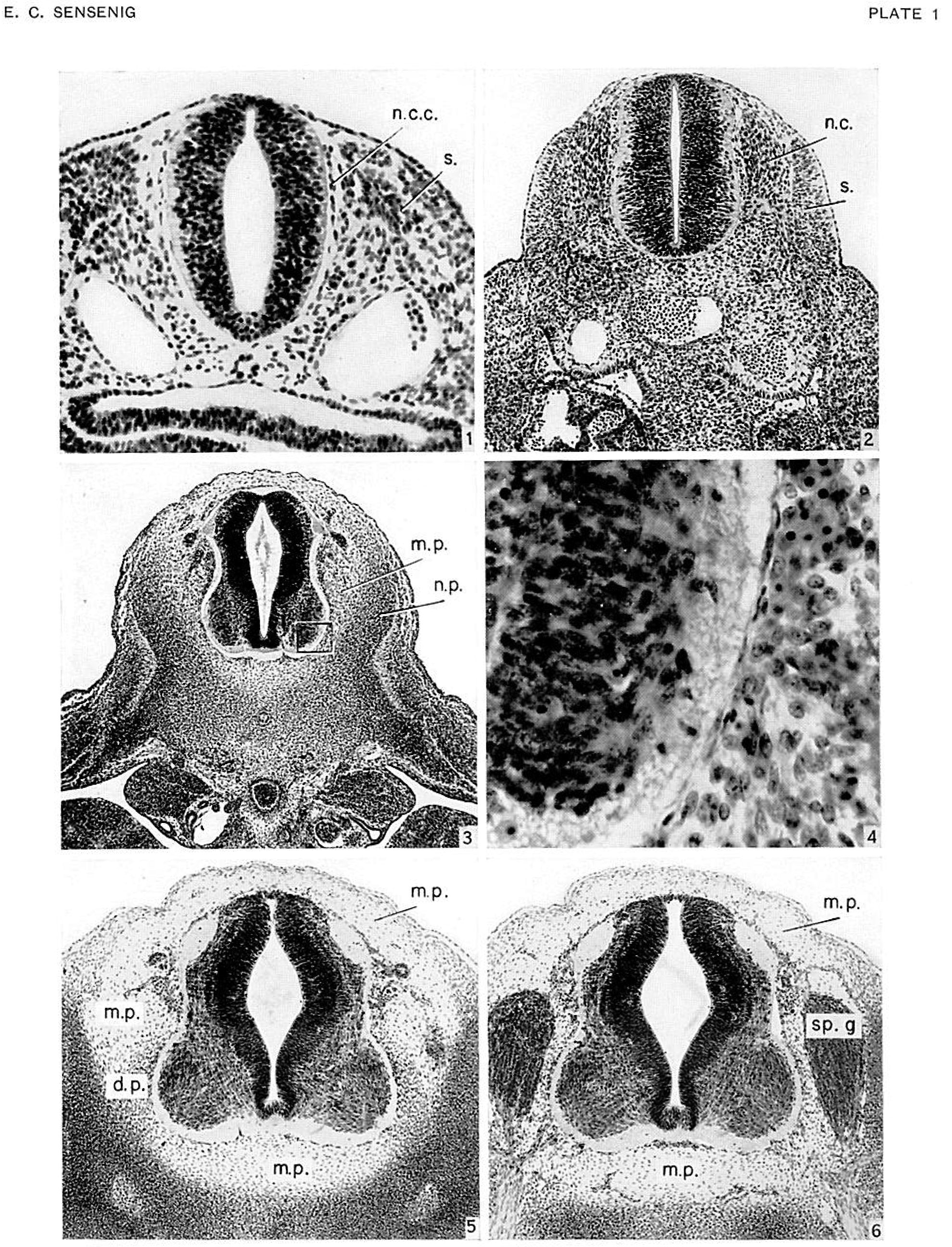
Plate 1
Fig. 1. Transverse section of cervical region of embryo no. 2053, 3.1 mm., age group xi, cut 10 microns. X:-.50. Shows distribution of paraxial mesodcrm about neural tube, and a single-layered strand of cells (n.c.c.) along periphery of neural tube which originates dorsally from the neural crests.
Fig. 2. Transverse section of upper thoracic region of embryo no. 8119, 5.3 mm., age group xiii, cut 8 X 100. Shows initial vascularization about periphery of neural tube. Differentiation between neural- crest and mesodermal cells still distinct.
Fig. 3. Transverse section of upper thoracic region of embryo no. 721, 9.0 mm., age group xv, cut 15 microns. X60. Shows intermediate zone (meninx primitiva) between denser vertebral rudiments and periphery of neural tube, and advanced vascularization about neural tube. microns.
Fig. 4. Outlined area in figure 3. X500. Ventrolateral part of neural tube and adjacent tissue of meninx primitiva. Single layer of cells of meninx primitiva lying on periphery of neural tube represents pia mater and can be regarded as of neural-crest origin.
Figs. 5, 6. Transverse sections of 4th thoracic segment of embryo no. 6521, 13.2 mm., age group xvii, cut 8 microns. X75. Shows distribution of meninx primitiva between neural tube and inner boundary of rudimentary vertebral canal.
Fig. 5, an interganglionic section, demonstrating the rudimentary dentate processes.
Fig. 6, a ganglionic section, the ganglia separated from canal wall by vascular channels. Tissue of meninx primitiva separates ganglia from neural tube.
Abbreviations used in Plates: d.1., dentate ligament d.m., dura mater d.p., dentate process m.p., meninx primitiva n.c., neural crest n.c.c., neural-crest cells n.p., neural process p., perichondrium pm., rudimentary posterior longitudinal ligament s., somite :p.c., spinal cord :p.g., spinal ganglion
| Historic Disclaimer - information about historic embryology pages |
|---|
 |
- Links: plate 1 | plate 2 | plate 3 | plate 4 | Sensenig 1951 | Spinal Cord | Meninges
Reference
Sensenig EC. The early development of the meninges of the spinal cord in human embryos. (1951) Contrib. Embryol., Carnegie Inst. Wash. Publ. 611.
Cite this page: Hill, M.A. (2024, April 27) Embryology Sensenig1951 plate01.jpg. Retrieved from https://embryology.med.unsw.edu.au/embryology/index.php/File:Sensenig1951_plate01.jpg
- © Dr Mark Hill 2024, UNSW Embryology ISBN: 978 0 7334 2609 4 - UNSW CRICOS Provider Code No. 00098G
File history
Click on a date/time to view the file as it appeared at that time.
| Date/Time | Thumbnail | Dimensions | User | Comment | |
|---|---|---|---|---|---|
| current | 14:57, 5 June 2016 | | 1,979 × 2,591 (1.35 MB) | Z8600021 (talk | contribs) | ==Plate 1== {{Sensenig1951 images}} {{Historic Disclaimer}} ===Reference=== {{Ref-Sensenig1951}} {{Footer}} |
You cannot overwrite this file.
File usage
The following 2 pages use this file: