File:Ramsey1972 fig01.jpg: Difference between revisions
From Embryology
(Z8600021 uploaded a new version of File:Ramsey1972 fig01.jpg) |
mNo edit summary |
||
| Line 9: | Line 9: | ||
{{Ramsey1972 figures}} | |||
[[Category:Monkey]] | |||
{{ | |||
Revision as of 07:17, 23 February 2017
Fig. 1. Camera lucida drawings of the vascular bed at three stages of the menstrual cycle in the rhesus monkey
| Left. postmenstrual; | Center. postovulatory; | Right. late secretory. |
Myometrium stippled. (Reprinted with permission from Bartelmez. Contrib. Embryol. 361153-182, 1957.)
- Links: fig 1 | fig 2 | fig 3 | fig 4 | fig 5a | fig 5b | fig 6 | fig 7 | fig 8 | fig 9 | 1972 Ramsey | Historic Papers | Placenta Development
Reference
Ramsey EM. Placental Circulation. (1972) MCV Quarterly, 8(1): 61-68.
© VCU. Licensed under a Creative Commons Attribution-Noncommercial-Share Alike 3.0 Unported License. http://creativecommons.org/licenses/by-nc-sa/3.0 Acknowledgement of the Virginia Commonwealth University Libraries as a source is required.
Cite this page: Hill, M.A. (2024, April 26) Embryology Ramsey1972 fig01.jpg. Retrieved from https://embryology.med.unsw.edu.au/embryology/index.php/File:Ramsey1972_fig01.jpg
- © Dr Mark Hill 2024, UNSW Embryology ISBN: 978 0 7334 2609 4 - UNSW CRICOS Provider Code No. 00098G
File history
Click on a date/time to view the file as it appeared at that time.
| Date/Time | Thumbnail | Dimensions | User | Comment | |
|---|---|---|---|---|---|
| current | 18:44, 21 February 2017 |  | 824 × 1,000 (187 KB) | Z8600021 (talk | contribs) | |
| 18:42, 21 February 2017 | 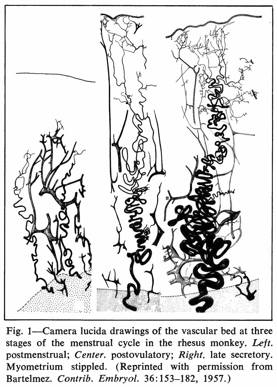 | 958 × 1,338 (290 KB) | Z8600021 (talk | contribs) | ===Reference=== {{Ref-Ramsey1972}} |
You cannot overwrite this file.
File usage
The following page uses this file: