File:Placenta anchoring villi.jpg
Placenta_anchoring_villi.jpg (600 × 450 pixels, file size: 167 KB, MIME type: image/jpeg)
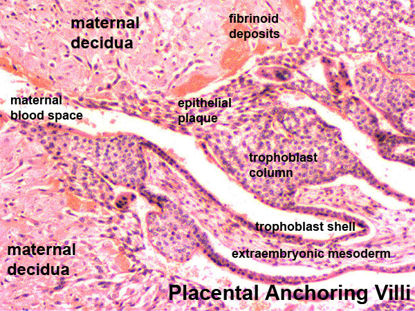
Placenta Anchoring Villi
Histological image showing the junctional region between the trophoblast shell of the conceptus and the maternal decidua.
In week 2, the trophoblast shell cells proliferate and form a syncitiotrophoblast and cytotrophoblast layer around he conceptus. Syncitiotrophoblast cells migrate into the uterine wall, forming maternal blood-filled spaces (lacunae).
Placentation begins once the conceptus begins to implant in the uterine wall and the placenta will have both a fetal and a maternal component. The fetal component begins as villi form. The fetal chorion will form two regions: smooth chorion (chorion laeve) and villous chorion (chorion frondosum).
The maternal component is formed by the decidualization of the endometrium.
- Links: Implantation | Placenta Development
Image Source: UNSW Embryology, no reproduction without permission.
File history
Click on a date/time to view the file as it appeared at that time.
| Date/Time | Thumbnail | Dimensions | User | Comment | |
|---|---|---|---|---|---|
| current | 14:37, 3 August 2009 | | 600 × 450 (167 KB) | MarkHill (talk | contribs) | Placenta anchoring villi Histological image showing the junctional region between the trophoblast shell of the conceptus and the maternal decidua. In week 2, the trophoblast shell cells proliferate and form a syncitiotrophoblast and cytotrophoblast lay |
You cannot overwrite this file.
File usage
The following 31 pages use this file:
- 2009 Lecture 4
- 2009 Lecture 8
- 2010 BGD Lecture - Development of the Embryo/Fetus 1
- 2010 Lab 4
- 2010 Lecture 4
- 2010 Lecture 8
- 2011 Lab 4
- A
- ACPS Seminar 2014 - Implantation
- ANAT2341 Lab 4 - Decidua and Cord
- ANAT2341 Lab 4 - Implantation and Villi Development
- ASA Meeting 2013 - Placenta
- BGDA Lecture - Development of the Embryo/Fetus 1
- BGDA Practical Placenta - Maternal Decidua
- BGDA Practical Placenta - Villi Development
- D
- F
- Implantation
- Lecture - Placenta Development
- Lecture - Week 1 and 2 Development
- Lecture - Week 3 Development
- Placenta - Histology
- Placenta - Maternal Decidua
- Placenta - Membranes
- Placenta Development
- Trophoblast - Protein Expression
- Week 2
- Yolk Sac Development
- Talk:ANAT2341 Lab 4 - Implantation and Villi Development
- Talk:Lecture - Week 3 Development
- User:Z5014754