File:Human neural crest cell migration-in vitro.jpg: Difference between revisions
m (→Reference) |
|||
| Line 17: | Line 17: | ||
<pubmed>18689800</pubmed>| [http://hmg.oxfordjournals.org/cgi/content/full/17/21/3411 Hum Mol Genet.] | <pubmed>18689800</pubmed>| [http://hmg.oxfordjournals.org/cgi/content/full/17/21/3411 Hum Mol Genet.] | ||
====Copyright==== | |||
© 2008 The Author(s) | © 2008 The Author(s) | ||
This is an Open Access article distributed under the terms of the Creative Commons Attribution Non-Commercial License (http://creativecommons.org/licenses/by-nc/2.0/uk/) which permits unrestricted non-commercial use, distribution, and reproduction in any medium, provided the original work is properly cited. | This is an Open Access article distributed under the terms of the Creative Commons Attribution Non-Commercial License (http://creativecommons.org/licenses/by-nc/2.0/uk/) which permits unrestricted non-commercial use, distribution, and reproduction in any medium, provided the original work is properly cited. | ||
{{Footer}} | |||
[[Category:Human Embryo]] [[Category:Carnegie Stage]] [[Category:Carnegie Stage 11]] [[Category:Carnegie Stage 12]] [[Category:Carnegie Stage 13]] [[Category:Week 4]] [[Category:Neural]] [[Category:Neural Crest]] | [[Category:Human Embryo]] [[Category:Carnegie Stage]] [[Category:Carnegie Stage 11]] [[Category:Carnegie Stage 12]] [[Category:Carnegie Stage 13]] [[Category:Week 4]] [[Category:Neural]] [[Category:Neural Crest]] | ||
Revision as of 02:41, 29 May 2017
Primary Human Embryo Neural Crest Cell Migration (in vitro)
- A Human neural tube (NT) from an embryo at Carnegie stage (C)13.
- B After 16 h, most hNCC have migrated away from the dorsal NT.
- C The intact NT is detached from the culture dish.
- D An enriched hNCC population remains, phenotypically similar to murine or avian NCC.
- E Empirical evaluation of hNCC migration (none to few, some and many) from 31 explanted neural tubes. Third-order polynomial regressions reflect the rostral-to-caudal temporal maturation gradient of the human NT and maturation of NCC. Peaks occur during C11 at cephalic levels, and during late C12 at rostral trunk levels (segments extending from somites 5 through the last-formed somite pair).
Original File: Figure 1. http://hmg.oxfordjournals.org/cgi/content/full/17/21/3411/DDN235F1
Reference
<pubmed>18689800</pubmed>| Hum Mol Genet.
Copyright
© 2008 The Author(s) This is an Open Access article distributed under the terms of the Creative Commons Attribution Non-Commercial License (http://creativecommons.org/licenses/by-nc/2.0/uk/) which permits unrestricted non-commercial use, distribution, and reproduction in any medium, provided the original work is properly cited.
Cite this page: Hill, M.A. (2024, April 26) Embryology Human neural crest cell migration-in vitro.jpg. Retrieved from https://embryology.med.unsw.edu.au/embryology/index.php/File:Human_neural_crest_cell_migration-in_vitro.jpg
- © Dr Mark Hill 2024, UNSW Embryology ISBN: 978 0 7334 2609 4 - UNSW CRICOS Provider Code No. 00098G
File history
Click on a date/time to view the file as it appeared at that time.
| Date/Time | Thumbnail | Dimensions | User | Comment | |
|---|---|---|---|---|---|
| current | 13:29, 31 August 2010 | 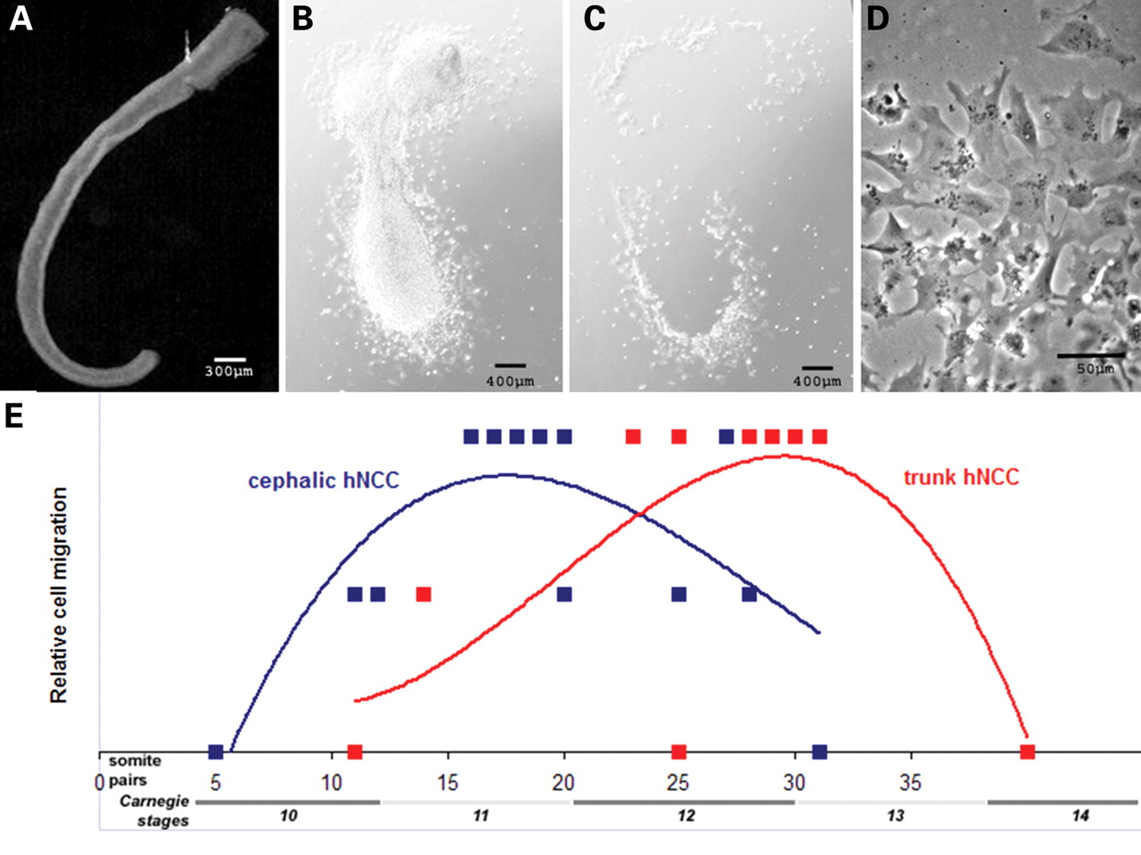 | 1,280 × 959 (163 KB) | S8600021 (talk | contribs) | ==Primary Human Embryo Neural Crest Cell Migration (in vitro)== (A) Human neural tube (NT) from an embryo at Carnegie stage (C)13. (B) After 16 h, most hNCC have migrated away from the dorsal NT. (C) The intact NT is detached from the culture dish. |
You cannot overwrite this file.
File usage
The following 3 pages use this file: