File:Bardeen1906-plate02.jpg: Difference between revisions
mNo edit summary |
mNo edit summary |
||
| Line 3: | Line 3: | ||
Four sections through the base of the posterior limb to illustrate different stages in the development of the nerves and musculature. | Four sections through the base of the posterior limb to illustrate different stages in the development of the nerves and musculature. | ||
'''Fig. 1.''' Section passing through the right limb-bud in embryo | '''Fig. 1.''' Section passing through the right limb-bud in embryo {{CE2}}, length 7 mm., age 26 days. The tips of the neighboring myotomes do not extend into the mass of tissue of which the limb-buds are composed and in which as yet no specific differentiation is visible. 33 diam. | ||
into the mass of tissue of which the limb-buds are composed and in which as yet no specific differentiation is visible. 33 diam. | |||
'''Fig. 2.''' Secton passing transversely through the base of the right limb-bud of embryo | '''Fig. 2.''' Secton passing transversely through the base of the right limb-bud of embryo {{CE109}}, length 11 mm., age about five weeks. At the center of the limb-bud the acetabular region of the skeleton appears as a condensed mass of tissue. About this the femoral, obturator, and sciatic nerves may be seen extending into the limb bud. Myogenous tissue is fairly well marked near the femoral and sciatic nerves. 25 diam. | ||
the limb-bud the acetabular region of the skeleton appears as a condensed | |||
mass of tissue. About this the femoral, obturator, and sciatic nerves may be | |||
seen extending into the limb bud. Myogenous tissue is fairly well marked near the femoral and sciatic nerves. 25 diam. | |||
'''Fig. 3.''' Transverse section passing through the acetabular region of left | '''Fig. 3.''' Transverse section passing through the acetabular region of left leg of embryo {{CE144}}, length 14 mm., age about five and" one-half weeks. The femoral, obturator, gluteal, and sciatic nerves may be seen extending into the limb bud, and in the vicinity of these nerves the anlages of the iliopsoas, pectineus, adductor, hamstring, and gluteal muscles. 25 diam. | ||
leg of embryo | |||
femoral, obturator, gluteal, and sciatic nerves may be seen extending into | |||
the limb bud, and in the vicinity of these nerves the anlages of the iliopsoas, | |||
pectineus, adductor, hamstring, and gluteal muscles. 25 diam. | |||
'''Fig. 4.''' Transverse section passing through the acetabular region of embryo | '''Fig. 4.''' Transverse section passing through the acetabular region of embryo {{CE145}}, length 33 mm., age about two months. The femoral, obturator, inferior gluteal, and sciatic nerves may be seen entering the limb. The chief fasciculi of the iliopsoas, pectineus, adductor, and gluteus maximus muscles are separated by an amount of connective tissue relatively greater than in the adult. 10 diam. | ||
inferior gluteal, and sciatic nerves may be seen entering the limb. The | |||
chief fasciculi of the iliopsoas, pectineus, adductor, and gluteus maximus | |||
muscles are separated by an amount of connective tissue relatively greater | |||
than in the adult. 10 diam. | |||
{{Bardeen1906 figures}} | {{Bardeen1906 figures}} | ||
[[Category:Carnegie Embryo 2]][[Category:Carnegie Embryo 109]] | |||
[[Category:Carnegie Embryo 144]][[Category:Carnegie Embryo 145]] | |||
Latest revision as of 20:40, 13 March 2019
Plate II. Four sections through the base of the lower limb Embryo 7 to 33 mm
Four sections through the base of the posterior limb to illustrate different stages in the development of the nerves and musculature.
Fig. 1. Section passing through the right limb-bud in embryo 2, length 7 mm., age 26 days. The tips of the neighboring myotomes do not extend into the mass of tissue of which the limb-buds are composed and in which as yet no specific differentiation is visible. 33 diam.
Fig. 2. Secton passing transversely through the base of the right limb-bud of embryo 109, length 11 mm., age about five weeks. At the center of the limb-bud the acetabular region of the skeleton appears as a condensed mass of tissue. About this the femoral, obturator, and sciatic nerves may be seen extending into the limb bud. Myogenous tissue is fairly well marked near the femoral and sciatic nerves. 25 diam.
Fig. 3. Transverse section passing through the acetabular region of left leg of embryo 144, length 14 mm., age about five and" one-half weeks. The femoral, obturator, gluteal, and sciatic nerves may be seen extending into the limb bud, and in the vicinity of these nerves the anlages of the iliopsoas, pectineus, adductor, hamstring, and gluteal muscles. 25 diam.
Fig. 4. Transverse section passing through the acetabular region of embryo 145, length 33 mm., age about two months. The femoral, obturator, inferior gluteal, and sciatic nerves may be seen entering the limb. The chief fasciculi of the iliopsoas, pectineus, adductor, and gluteus maximus muscles are separated by an amount of connective tissue relatively greater than in the adult. 10 diam.
| Historic Disclaimer - information about historic embryology pages |
|---|
 |
- Links: Fig. 2 | Fig. 3 | Plate 1 | Plate 2 | Plate 3-1 | Plate 3-2 | Plate 4-1 | Plate 4-2 | Plate 5-1 | Plate 5-2 | Plate 6 | Bardeen 1906 | Historic Papers
| Online Editor |
|---|
Note that not all plates described in the paper are currently available online. |
Reference
Bardeen CR. Development and variation of the nerves and the musculature of the inferior extremity and of the neighboring regions of the trunk in man. Am J Anat. 1906;6:259–390.
Cite this page: Hill, M.A. (2024, May 21) Embryology Bardeen1906-plate02.jpg. Retrieved from https://embryology.med.unsw.edu.au/embryology/index.php/File:Bardeen1906-plate02.jpg
- © Dr Mark Hill 2024, UNSW Embryology ISBN: 978 0 7334 2609 4 - UNSW CRICOS Provider Code No. 00098G
File history
Click on a date/time to view the file as it appeared at that time.
| Date/Time | Thumbnail | Dimensions | User | Comment | |
|---|---|---|---|---|---|
| current | 21:46, 7 September 2015 | 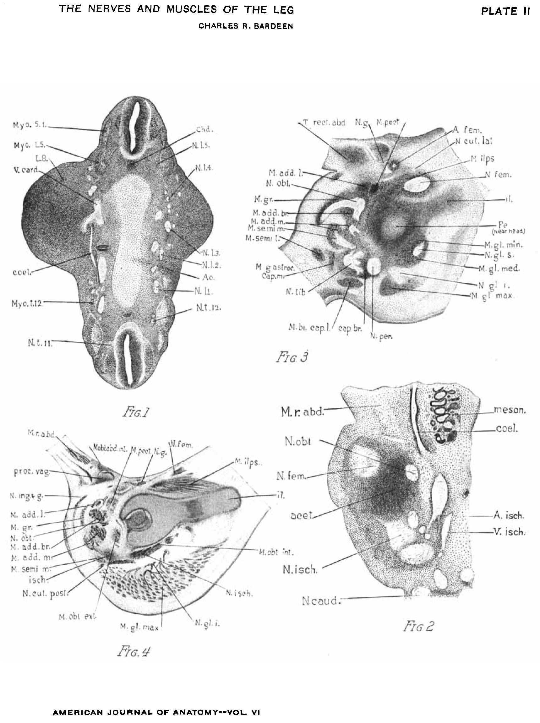 | 1,719 × 2,302 (512 KB) | Z8600021 (talk | contribs) |
You cannot overwrite this file.
File usage
The following page uses this file: