File:BakerHookSeveringhaus1944 plate01.jpg

Original file (1,534 × 2,295 pixels, file size: 228 KB, MIME type: image/jpeg)
Plate 1
Unless otherwise stated, the figures are from 3 μm sections of Champy-fixed material stained by the Severinghaus Altmann—Masson technique; Golgi preparations were osmicated by the Sevoringliefus (Nassonov-Kolatschev) method before staining.
1 Drawing of cytotrophoblast cell showing two granules in the Golgi region with associated mitochondria. (black). A large vacuole is present at the right of the sgvncytial nucleus. Case 6. About x 4300.
2 Drawing of the syncytium showing minute granules in the subnuclear zone in association with clear curved channels, the negative image of the Golgi apparatus. The granules increase in Size towards the surface of the syneytium and show varied degrees of affinity for aniline blue. The majority are homogeneous; one large granule at upper right shows definite structure. Superficial vacuoles at upper left. Mitochondria. have accumulated in the supranuclear zone and about the negative image of the Golgi apparatus. Case 12. About x 3200.
3 Accumulation of mitochondria in the superficia.l- zone of the syllcytium in the absence of vacuoles and granules. Mitochondria (black) are shown in the cytotrophoblast cell, especially in the Golgi region at the left of the nucleus. The syncytial nuclei are more chromatic than those of the cytotrophoblast. Case 12. x 1160.
4 Secretory granules in the superficial zone apparently in various stages of liquefaction, with some eecuxnuluted mitochondria. A cytotrophoblast cell (arrow) appears above the capillary probably in process of transformation to syncytium. The cytoplasm is dark, and the mitochondria at left of nucleus, large and intensely stained. Contrast cytoplasm to that of cytotrophoblast cells in figures 3 and 5. Case 12. x1160.
5 Reguklar brush border and even distribution of mitochondria throughout syncytium. Some lipid droplets (black) in the syncytium. 01min of mitochondria above nucleus of middle cytotrophoblast cell. Case 12. x 1160.
6 Brush border after Bouin fixation. Case 13. x 1160.
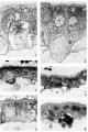
plate 1
plate 2
plate 3
plate 4


Reference
Baker BL. Hook SJ. and Severinghaus AE. The cytological structure of the human chorionic villus and decidua parietalis. (1944) Amer. J Anat. 73(3): 291-325.
Cite this page: Hill, M.A. (2024, April 27) Embryology BakerHookSeveringhaus1944 plate01.jpg. Retrieved from https://embryology.med.unsw.edu.au/embryology/index.php/File:BakerHookSeveringhaus1944_plate01.jpg
- © Dr Mark Hill 2024, UNSW Embryology ISBN: 978 0 7334 2609 4 - UNSW CRICOS Provider Code No. 00098G
File history
Click on a date/time to view the file as it appeared at that time.
| Date/Time | Thumbnail | Dimensions | User | Comment | |
|---|---|---|---|---|---|
| current | 09:41, 2 February 2018 | | 1,534 × 2,295 (228 KB) | Z8600021 (talk | contribs) | |
| 09:30, 2 February 2018 |  | 1,585 × 2,389 (215 KB) | Z8600021 (talk | contribs) |
You cannot overwrite this file.