File:Accessory renal artery.jpg: Difference between revisions
m (→Reference) |
mNo edit summary |
||
| Line 2: | Line 2: | ||
* More than one artery supplying a kidney is the most common arterial variation, and this is seen in about 24% of cases. | * More than one artery supplying a kidney is the most common arterial variation, and this is seen in about 24% of cases. | ||
* These arteries are divided into two groups: hilar (accessory) and polar (aberrant) arteries. | * These arteries are divided into two groups: hilar (accessory) and polar (aberrant) arteries. | ||
* Image - Accessory renal artery and early branching in 50-year-old female voluntary kidney donor. | |||
* Image - Accessory renal artery and early branching in 50-year-old female voluntary kidney donor. | |||
** Anterior volume rendered image shows early branching of main left renal artery (black arrowhead) and presence of accessory renal artery arising from aorta (black arrow). | ** Anterior volume rendered image shows early branching of main left renal artery (black arrowhead) and presence of accessory renal artery arising from aorta (black arrow). | ||
** Right inferior phrenic artery is seen arising from right main renal artery (white arrow). | ** Right inferior phrenic artery is seen arising from right main renal artery (white arrow). | ||
{{Renal Vascular Anomalies}} | {{Renal Vascular Anomalies}} | ||
===Reference=== | ===Reference=== | ||
{{#pmid:20461189}} | |||
====Copyright==== | ====Copyright==== | ||
This is an Open Access article distributed under the terms of the Creative Commons Attribution Non-Commercial License (http://creativecommons.org/licenses/by-nc/3.0) which permits unrestricted non-commercial use, distribution, and reproduction in any medium, provided the original work is properly cited. | This is an Open Access article distributed under the terms of the Creative Commons Attribution Non-Commercial License (http://creativecommons.org/licenses/by-nc/3.0) which permits unrestricted non-commercial use, distribution, and reproduction in any medium, provided the original work is properly cited. | ||
Original file name: Fig. 5 kjr-11-346-g005.jpg | |||
{{Footer}} | {{Footer}} | ||
[[Category:Human]] [[Category:Adult]] [[Category:Renal]] [[Category:Cardiovascular]] [[Category:Arterial]] [[Category:Computed Tomography]] | [[Category:Human]] [[Category:Adult]] [[Category:Renal]] [[Category:Cardiovascular]] [[Category:Arterial]] [[Category:Computed Tomography]] | ||
Latest revision as of 22:39, 21 March 2018
Accessory Renal Artery
- More than one artery supplying a kidney is the most common arterial variation, and this is seen in about 24% of cases.
- These arteries are divided into two groups: hilar (accessory) and polar (aberrant) arteries.
- Image - Accessory renal artery and early branching in 50-year-old female voluntary kidney donor.
- Anterior volume rendered image shows early branching of main left renal artery (black arrowhead) and presence of accessory renal artery arising from aorta (black arrow).
- Right inferior phrenic artery is seen arising from right main renal artery (white arrow).
Renal Vascular Anomalies: Multiple renal arteries | Accessory renal artery | Supernumerary right renal vein 1 | Supernumerary right renal vein 1 | Multiple right renal veins 2 | Multiple right renal veins 2 | Cardiovascular System Development
Reference
Kumar S, Neyaz Z & Gupta A. (2010). The utility of 64 channel multidetector CT angiography for evaluating the renal vascular anatomy and possible variations: a pictorial essay. Korean J Radiol , 11, 346-54. PMID: 20461189 DOI.
Copyright
This is an Open Access article distributed under the terms of the Creative Commons Attribution Non-Commercial License (http://creativecommons.org/licenses/by-nc/3.0) which permits unrestricted non-commercial use, distribution, and reproduction in any medium, provided the original work is properly cited.
Original file name: Fig. 5 kjr-11-346-g005.jpg
Cite this page: Hill, M.A. (2024, May 16) Embryology Accessory renal artery.jpg. Retrieved from https://embryology.med.unsw.edu.au/embryology/index.php/File:Accessory_renal_artery.jpg
- © Dr Mark Hill 2024, UNSW Embryology ISBN: 978 0 7334 2609 4 - UNSW CRICOS Provider Code No. 00098G
File history
Click on a date/time to view the file as it appeared at that time.
| Date/Time | Thumbnail | Dimensions | User | Comment | |
|---|---|---|---|---|---|
| current | 12:13, 3 September 2011 | 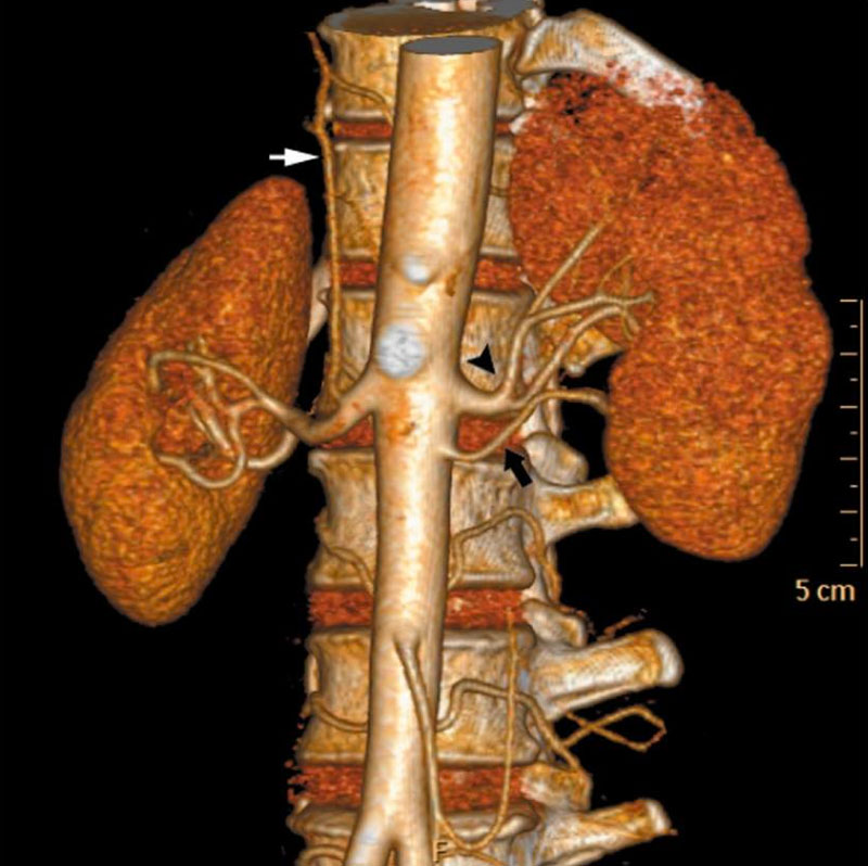 | 800 × 798 (103 KB) | S8600021 (talk | contribs) | ==Accessory Renal Artery== * More than one artery supplying a kidney is the most common arterial variation, and this is seen in about 24% of cases. * These arteries are divided into two groups: hilar (accessory) and polar (aberrant) arteries. * Image - A |
You cannot overwrite this file.
File usage
The following 4 pages use this file: