Endocrine - Adrenal Development: Difference between revisions
mNo edit summary |
mNo edit summary |
||
| Line 31: | Line 31: | ||
<pubmed limit=5>Adrenal Embryology</pubmed> | <pubmed limit=5>Adrenal Embryology</pubmed> | ||
|} | |||
==Adrenal Movies== | |||
{| | |||
| valign="bottom"|{{Adrenal movie}} | |||
| Cartoon showing migration of neural crest cells from original location to form the fetal medulla cells. | |||
|- | |||
| valign="bottom"|{{Adrenal GA32 movie}} | |||
| Surface rendering of human fetal adrenal glands in the third trimester (week 30 {{GA}} week 32). | |||
|} | |} | ||
==Adrenal Overview== | ==Adrenal Overview== | ||
Revision as of 08:49, 26 November 2014
| Embryology - 27 Apr 2024 |
|---|
| Google Translate - select your language from the list shown below (this will open a new external page) |
|
العربية | català | 中文 | 中國傳統的 | français | Deutsche | עִברִית | हिंदी | bahasa Indonesia | italiano | 日本語 | 한국어 | မြန်မာ | Pilipino | Polskie | português | ਪੰਜਾਬੀ ਦੇ | Română | русский | Español | Swahili | Svensk | ไทย | Türkçe | اردو | ייִדיש | Tiếng Việt These external translations are automated and may not be accurate. (More? About Translations) |
Introduction
The developing adrenal gland has both an interesting origin and an intruiging fetal role. Furthermore recent studies suggest that the adrenal cortex share a common embryonic origin with the early gonad. The adrenal gland and placenta also act in synergy, and the notes endocrine placenta should also be read.
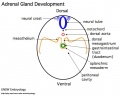
The 2 adrenal glands (suprarenal gland, glandulæ suprarenales) are named by their anatomical postion sitting above the 2 kidneys (renal). The 2 main parts of the adrenals have different embryonic origins. The inside core adrenal medulla is neural crest in origin. Mesenchyme surrounding these cells differentiates to form a fetal cortex. This fetal cortex is later replaced by the adult cortex. The outside adrenal cortex is derived from mesothelium and can be further divided into 3 distinct layers (zona reticularis, zona fasiculata, zona glomerulosa) each with distinct hormonal functions.
During fetal development, adrenal hormones are involved with the maturation of the lung and other developing systems.
| Lecture - Neural Crest Development
Some Recent Findings
The morphometry of fetal adrenal gland is rarely described with MRI of high magnetic field. The purpose of this study is to assess the normal fetal adrenal gland length (AL), width (AW), height (AH), surface area (AS) and volume (AV) in the second half of gestation with 3.0T post-mortem MRI. Fifty-two fetal specimens of 23-40 weeks gestational age (GA) were scanned by 3.0T MRI. Morphological changes and quantitative measurements of the fetal adrenal gland were analyzed. Asymmetry and sexual dimorphism were also obtained. The shape of the fetal adrenal gland did not change substantially from 23 to 40 weeks GA. The bilateral adrenal glands appeared as a 'Y', pyramidal or half-moon shape after reconstruction. There was a highly linear correlation between AL, AW, AH, AS, AV and GA. AW, AH, AS and AV were larger for the left adrenal gland than the right. No sexual dimorphism was found."
|
| More recent papers |
|---|
 This table allows an automated computer search of the external PubMed database using the listed "Search term" text link.
More? References | Discussion Page | Journal Searches | 2019 References | 2020 References Search term: Adrenal Embryology <pubmed limit=5>Adrenal Embryology</pubmed> |
Adrenal Movies
|
Cartoon showing migration of neural crest cells from original location to form the fetal medulla cells. | |||
|
Surface rendering of human fetal adrenal glands in the third trimester (week 30 GA week 32). |
Adrenal Overview
- Richly vascularized - arterioles passing through cortex, capillaries from cortex to medulla, portal-like circulation
- Fetal Cortex - produces a steroid precursor (DEA), converted by placenta into estrogen
- Adult Medulla - produces adrenalin (epinephrine), noradrenaline (norepinephrine)
- Fetal adrenal hormones - influence lung maturation
Cortical Hormones
(steroids) Cortisol, Aldosterone, Dehydroepiandrosterone
- zona glomerulosa - regulated by renin-angiotensin-aldosterone system controlled by the juxtaglomerular apparatus of the kidney.
- zona fasciculata - regulated by hypothalamo-pituitary axis with the release of CRH and ACTH respectively.
Medullary Hormones
(amino acid derivatives) Epinephrine, Norepinephrine
Adrenal Development
Adrenal Cortex
Adrenal Medulla
|
|

|
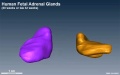
| Human fetal adrenal gland morphology and size.[1] |
Development Overview
Medulla - Neural crest cells migrate toward the coelomic cavity wall and form the adrenal medulla. These chromaffin (chromaphil) cells originally named because of their staining (yellow) with chromium salts.
Cortex - Week 4 celomic epithelium (mesothelium) cells proliferate initially forming small buds that separate from the epithelium. Week 6 these now mesenchymal cells first form the fetal adrenal cortex which will be later replaced by the adult cortex.
Adrenal Cortex


|

|
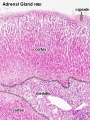
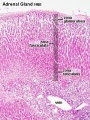
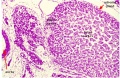
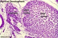
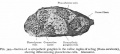
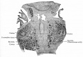
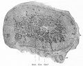
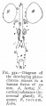
| Human Embryo (7 weeks, stage 22) adrenal gland showing the fetal and permanent adrenal cortex. Note that the medulla of the adrenal gland is not yet encapsulated by the cortex. | Human Fetus (10 week, 40mm, parasagittal section) shows location of the developing adrenal gland. The spongy appearance at the centre of the adrenal is the degenerating fetal cortex. The dense region around the outside of the adrenal is the developing adult cortex.) |
Week 4 - coelomic epithelium (mesothelium) cells proliferate initially forming small buds that separate from the epithelium.
Week 6 - these now mesenchymal cells surrounding the developing medulla cells differentiate first form the fetal adrenal cortex which will be later replaced by the adult cortex.
Week 8 to 9 - fetal adrenal cortex synthesizes cortisol and is maximal at 8-9 weeks post conception (wpc) under the regulation of ACTH (also stimulates androstenedione and testosterone secretion).[6]
Adult cortex - mesothelium mesenchyme encloses fetal cortex.
Late Fetal Period - differentiates to form cortical zones.
Birth - zona glomerulosa, zona fasiculata present.
Year 3 - zona reticularis present.
Fetal Cortex

Fetal adrenal cortical growth involves several cellular processes: hypertrophy, hyperplasia, apoptosis, and migration.
In the second and third trimesters a steroid precursor dehydroepiandrosterone (DHEA) and sulphated derivative (DHEAS) which is converted by placenta into estrogen.
Three functional zones:
- Fetal zone - throughout gestation expresses enzymes required for DHEA-S synthesis.
- Transitional zone - initially identical to the fetal zone but later (after 25-30 weeks) expresses enzymes that suggest glucocorticoid synthesis.
- Definitive zone - after 22-24 weeks expresses enzymes that suggest mineralocorticoid synthesis.
Neonatal
- human males produce high levels of DHEA) and DHEAS
- decline within a few months of birth
- due to regression of the adrenal fetal zone
Adult
- zona reticularis (ZR) source for production of DHEA and DHEAS
Adult Cortex


Early Adult Cortex (week 12)
- Reticularis - narrow band, many small cells and capillaries androgens. source for production of DHEA and DHEAS
- Fasiculata - high lipid content, pale foamy cells cortisol, corticosterone, cortisone.
- Glomerulosa - small cells, cords or oval groups, aldosterone.
Species Difference
- rat - zona glomerulosa and zona fasciculata separated by an undifferentiated zone (ZU, or Zona Intermedia)
- mouse - no undifferentiated zone separation.
- capsule mesenchyme cells have properties of adrenocortical stem/progenitor cells.
Adult Histology
Adrenal - Cortex and Medulla
Adrenal - Cortical Zones
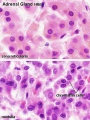
Adrenal - Zona Reticularis and Medulla
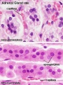
Adrenal - Zona Glomerulosa and Fasciculata


- Adrenal Histology: Cortex and Medulla | Unlabelled Overview | Cortical Zones | Zona Glomerulosa and Fasciculata | Zona Glomerulosa | Zona Fasciculata | Zona Reticularis and Medulla | Zona Reticularis | Medulla | Fetal Cortex | Developing Adult Cortex | BGD - Endocrine Histology | Histology Stains | Adrenal Development
Links: Histology | Histology Stains | Blue Histology images copyright Lutz Slomianka 1998-2009. The literary and artistic works on the original Blue Histology website may be reproduced, adapted, published and distributed for non-commercial purposes. See also the page Histology Stains.
Cite this page: Hill, M.A. (2024, April 27) Embryology Endocrine - Adrenal Development. Retrieved from https://embryology.med.unsw.edu.au/embryology/index.php/Endocrine_-_Adrenal_Development
- © Dr Mark Hill 2024, UNSW Embryology ISBN: 978 0 7334 2609 4 - UNSW CRICOS Provider Code No. 00098G
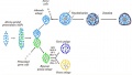
Molecular
Steroidogenic Factor 1

Adrenal and gonad steroidogenic factor 1 (SF-1) expression in different species [7]
- 53 kDa protein called Ad4BP (Adrenal 4 Binding Protein) or SF-1 (Steroidogenic Factor 1)
- classical DNA-binding domain (DBD) characterized by two Cys2-Cys2 zinc fingers in the N-terminal region
- SF-1 binds DNA as a monomer
- high homology with the drosophila Ftz-F1 transcription factor that controls fushi tarazu homeotic gene expression
Steroidogenic Factor 1 Mutation Effects
| Organism | Human | ||
| Genotype | SF-1 -/- | SF-1 +/- | SF-1 -/+ or SF-1 -/- |
| Adrenal | Agenesis | Histological defects
Hyporesponse to stress Compensatory growth defects |
Insufficiency (agenesis or dysgenesis) |
| Testis | Agenesis
Sex reversal |
Sex reversal | |
| Ovary | Agenesis | Normal | |
| Ventro-Medial Hypothalamus | Agenesis
Obesity caused by absence of the VMH (8 weeks) |
||
| Pituitary | Defects of gonadotrope cells | ||
Table modified from review.[7]
SoxE
Sry-box (Sox) 8, and Sox10 are expressed in the neural crest and in neural crest cells migrating to the adrenal gland.[8]
DAX1
CYP17
Abnormalities
Congenital Adrenal Hyperplasia
(CAH) A family of inherited disorders of adrenal steroidogenesis enzymes which impairs cortisol production by the adrenal cortex.
Enzymes most commonly affected: 21-hydroxylase (21-OH), 11beta-hydroxylase, 3beta-hydroxysteroid dehydrogenase.
Enzymes less commonly affected: 17alpha-hydroxylase/17,20-lyase and cholesterol desmolase.
Classical CAH - androgen excess leads newborn females with external genital ambiguity and postnatal progressive virilization in both sexes.
Pheochromocytomas
(PCC) Catecholamine-producing (neuro)endocrine tumor located in the adrenal medulla. Similar catecholamine-producing tumors outside the adrenal gland are called paragangliomas (PGL).
Cushing's Syndrome
(hypercortisolism) A relatively rare metabolic hormonal disorder caused by prolonged exposure of the body’s tissues to high levels of the adrenal hormone cortisol, most commonly affects adults aged between 20 to 50 and also the obese with type 2 diabetes.
Adrenocortical Tumour

Adrenocortical tumours (ACT) can occur at all ages and have a bimodal distribution with peaks of incidence at about 5 years of age and again at 40 to 50 years of age. Clinically, a routine hormonal profile for suspected patients includes measurements of serum (8am, 11pm) cortisol, testosterone, DHEA-S, androstenedione, 17-hydroxyprogesterone, aldosterone, and plasma renin activity.[9]
References
- ↑ 1.0 1.1 <pubmed>24116052</pubmed> Cite error: Invalid
<ref>tag; name 'PMID24116052' defined multiple times with different content - ↑ <pubmed>22430763</pubmed>
- ↑ <pubmed>20715567</pubmed>
- ↑ <pubmed>19723922</pubmed>
- ↑ <pubmed>21051591</pubmed>
- ↑ <pubmed>16585961</pubmed>
- ↑ 7.0 7.1 <pubmed>14594453</pubmed>| Nucl Recept.
- ↑ <pubmed>18272785</pubmed>
- ↑ 9.0 9.1 <pubmed>20223012</pubmed>
Online Textbooks
Endocrinology: An Integrated Approach Nussey, S.S. and Whitehead, S.A. Oxford, UK: BIOS Scientific Publishers, Ltd; 2001. 4.7. Embryology of the adrenal gland | The Adrenal Gland | Anatomical and functional zonation in the adrenal cortex
Developmental Biology (6th ed) Gilbert, Scott F. Sunderland (MA): Sinauer Associates, Inc.; c2000. Figure 13.6. Final differentiation of a trunk neural crest cell committed to become either an adrenomedullary (chromaffin) cell or a sympathetic neuron
Molecular Biology of the Cell (4th Edn) Alberts, Bruce; Johnson, Alexander; Lewis, Julian; Raff, Martin; Roberts, Keith; Walter, Peter. New York: Garland Publishing; 2002. table 15-1. Some Hormone-induced Cell Responses Mediated by Cyclic AMP | Cells Can Respond Abruptly to a Gradually Increasing Concentration of an Extracellular Signal
Health Services/Technology Assessment Text (HSTAT) Bethesda (MD): National Library of Medicine (US), 2003 Oct. Adrenal Gland search Results
Search NLM Online Textbooks- "adrenal development" : Endocrinology | Molecular Biology of the Cell | The Cell- A molecular Approach
Reviews
<pubmed>18670886</pubmed> <pubmed>18493131</pubmed> <pubmed>17046275</pubmed> <pubmed>16928368</pubmed> <pubmed>16807499</pubmed> <pubmed>9888597</pubmed> <pubmed>9183569</pubmed>| Endocrine Reviews
Articles
<pubmed>20010965</pubmed> <pubmed>19723922</pubmed> <pubmed>17537799</pubmed> <pubmed>16585961</pubmed> <pubmed>16093324</pubmed> <pubmed>11319516</pubmed> <pubmed>9888597</pubmed>"The rapid growth of the human fetal adrenal gland, which is primarily a reflection of the growth of the unique fetal zone, is regulated by ACTH acting indirectly to stimulate the expression of locally produced growth factors, of which IGF-II and bFGF appear to play key roles. Through most of gestation, the outer definitive zone appears to function as a reservoir of progenitor cells which move centripetally to populate the rest of the gland. At the end of pregnancy, the fetal zone undergoes senescence through an apoptotic process. Activin and TGF-beta are capable of inducing apoptosis in the fetal zone. Corticotropin-releasing hormone, which is produced by the placenta in markedly increased amounts at the end of gestation, may orchestrate a variety of processes, including direct stimulation of fetal adrenal steroidogenesis, culminating in the initiation of parturition."
Search PubMed
Search April 2010
- Adrenal Development - All (646) Review (52) Free Full Text (84)
- Congenital Adrenal Hyperplasia - All (2091) Review (211) Free Full Text (314)
Search Pubmed: adrenal development | Congenital Adrenal Hyperplasia
Additional Images
Human Embryo Stage 22
Human Embryo Stage 22
Adrenal and gonad early development
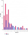
Childhood adrenocortical tumours graph


Historic Images
| Historic Disclaimer - information about historic embryology pages |
|---|
|
Historic image
Historic image
Historic image
Historic image


Terms
Glossary Links
- Glossary: A | B | C | D | E | F | G | H | I | J | K | L | M | N | O | P | Q | R | S | T | U | V | W | X | Y | Z | Numbers | Symbols | Term Link
Cite this page: Hill, M.A. (2024, April 27) Embryology Endocrine - Adrenal Development. Retrieved from https://embryology.med.unsw.edu.au/embryology/index.php/Endocrine_-_Adrenal_Development
- © Dr Mark Hill 2024, UNSW Embryology ISBN: 978 0 7334 2609 4 - UNSW CRICOS Provider Code No. 00098G