Embryology Models
Introduction
The very size and dynamic nature of embryonic development has always made it a difficult topic to teach. The structures are always so small and the changing nature of development means that a feature you identify at any one time is called, and becomes something else, at a later stage.
Models made from many different media (papier-mâché, ceramic, wax or plastic) not the "animal models" described elsewhere on this site, are very useful in that they make tangible the microscopic structures to the students. Models have been in use for some time in the medical teaching of human development with the original plaster models now replaced by commercial modern plastic versions. Many of the early teaching models were not about development, but about the process of childbirth for doctor and midwife education.
A historic alternative to the use of teaching models, were series of plates and drawings found in textbooks and papers from the late 19th century onwards. At the beginning of the 20th century photography also came into play, seen mainly associated with embryos within the Carnegie collection.
Some links on this page are to both historic non-commercial and modern commercial suppliers of some education models for embryology. In some cases these are museum exhibits only available for viewing. This list is provided for information purposes only and does not indicate an endorsement of these commercial products.
Historic Models
Galletti Models
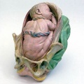
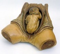
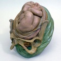
Giuseppe Galletti (? - 1819) these models are currently available for viewing at the Institute and Museum of the History of Science (Italy) "Specimens of obstetric models: the wax models are life-sized; the terracotta versions are reduced to a 1:3 scale. Together with the anatomical waxes in the Specola Museum in Florence, these models are among the most significant examples of the use of artistic techniques for teaching medicine and obstetrics to midwives and surgery students in Florentine hospitals at the end of the eighteenth century."
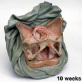
Week 10
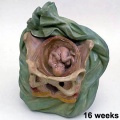
Week 16
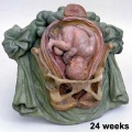
Week 24
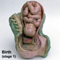
Birth
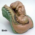
Birth
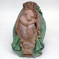
Birth (breech)
Birth (breech)
Birth (breech)
Birth (breech)
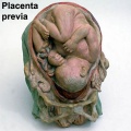
Birth (placenta previ)


- Galletti Links: Birth | Week 10 Fetus Model | Week 16 Fetus Model | Week 24 Fetus Model | Stage 1 Terracotta Model 1 | Stage 1 Terracotta Model 2 | Stage 1 Wax Model | Breech Birth 1 | Breech Birth 2 | Breech Birth 3 | Breech Birth Wax Model | Placenta Previa | Category:Galletti1770 | 17th and 18th Century Anatomies | Embryology History
Ziegler Models

Adolf Ziegler (1820 - 1889) a German modeller developed these models at his Studio for Scientific Teaching Models (Atelier für wissenchaftliche Unterrichtsmodelle) from the 1860's onwards of both human and animal embryos. Ziegler's models were first used in Germany, but were also later exhibited in Paris (1867) and Vienna (1873).
His son, Friedrich Ziegler (- 1936), continued the modelling company.


- Links: Ziegler Models
Carnegie Models
A description of the early modelling method can be found in the 1921 Contributions to Embryology Volume XII - No.56.
- "At first our record of sections was made by drawing them with the camera lucida or, better still, in a dark room by the aid of a magic lantern. This, however, is a very laborious and time-consuming task, and has a tendency to check the mental activity of the investigator, for unfortunately the work can not be done successfully by a technical assistant. When the collection was transferred to the Carnegie Institution we availed ourselves of the opportunity to install an accurate projection apparatus patterned somewhat after the one used by His (1892). With this apparatus sections of embryo No. 460, previously referred to, were photographed on glass negatives with an enlargement of 50 diameters. Thus it was possible to make several prints. As the photographs were taken with a 50-mm. planar lens, they show all the details wonderfully well.
- As everything which we wished to reconstruct had to be transferred to wax, it was highly desirable that the final model should be composed of some temperature resisting substance. A method has therefore been devised whereby all the structures desired are eliminated from the wax. Thus, in a block of wax the model is represented in outline by a hole or a series of holes, according to the number of structures to be reproduced, and these holes are subsequently filled with plaster of pans. When this hardens and the wax is removed, the finished plaster model is liberated.
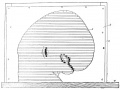
Fig. 3. Method of piling with cardboard outline as guide.
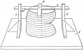
Fig. 4. Method of making guide lines.
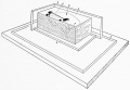
Fig. 5. Method of piling wax mold.


- This process of modeling depends solely upon absolute accuracy in superposing the wax plates to correspond to the original sections of the embryo. Hence a perpendicular line must be established, and this is done as follows: The wax plates of the sections are placed one upon another until a complete model of the embryo is built up (fig. 3), the construction being guided by photographs of the embryo made before it was cut. Thus it is possible to duplicate accurately the external form of an embryo on an enlarged scale. After this is done it is a simple matter to mark the wax plates in a perpendicular direction that is, by rightangle lines drawn upon every plate through its axis. These constitute what we call guide-lines, the instrument for marking which was invented by Dr. W. H.
- Lewis (1915) and is known as the Lewis guide liner (fig. 4). After the guide-lines have been drawn on each wax plate they are transferred to the photographs or drawings. This is done by superposing each wax plate upon its photograph or drawing and marking the end of the guide-lines. When the wax plate is removed, these points are connected by lines similar to those on the plates. After the two principal guide-lines have been established, it has been found convenient to use secondary guide-lines, 5 cm. apart, which run parallel to the primary ones over the entire surface of the photograph..
- In order to make a reconstruction of any portion of an embryo it is necessary first to transfer the outlines of that structure from the photograph to the wax plates by means of carbon paper and a glass point used as a pencil. These outlines are then cut and removed from the wax and the plates squared off along the secondary guide-lines sufficiently far outside of the proposed model not to conflict with its casting.
- In order that these finished plates, which we call mold plates, may be kept in exact apposition, they are piled in a rectangular corner made of plate glass (fig. 5). While this is being done it is necessary to cut certain artificial channels as vents, so that the air will escape from the openings when the plaster is poured in."
Osborne O. Heard

One of the first to be hired, in 1913, was modeler Osborne O. Heard, who spent 42 years at the department and made over 700 wax-based reconstructions. The results of this team effort still stand as the international standard by which human embryos are described and classified. The models were mainly made by the lost-wax casting process and his models were also more detailed than the earlier (1880's) Ziegler embryo models.
- Links: Early Carnegie Models | Carnegie_Stages | Osborne O. Heard
Smithsonian Museum
- 21 models showing the development of an egg to the thirtieth day of gestation Établissements du docteur Auzoux (about 1895)
- Links: Human Anatomical Model
Modern Models
Nucleus
- Giant Embryo Model
- 2nd Month Embryo
- Fetal Skull, without stand
- Classic Pregnancy Series, 8 Models
- Classic Pregnancy Pelvis 2-part
- Deluxe Pregnancy Pelvis 3-part
- 5 stage Birth Process Model
The Evolution Store
Lippincott Williams and Wilkins
Shanghai Honglian Medical Instrument Development
- Fertilization and Early Development Shows the development from ovum to the fetus in third month.
- Early Development Fecundation to the embryo of fourth week.
- Embryo The embryo model is about 4 weeks old, and mounted on a stand.
- Embryonic Development The model includes 8 parts showing the uterus with embryo and fetus from the first to the seventh month pregnancy.
Glossary Links
- Glossary: A | B | C | D | E | F | G | H | I | J | K | L | M | N | O | P | Q | R | S | T | U | V | W | X | Y | Z | Numbers | Symbols | Term Link
Cite this page: Hill, M.A. (2024, April 30) Embryology Embryology Models. Retrieved from https://embryology.med.unsw.edu.au/embryology/index.php/Embryology_Models
- © Dr Mark Hill 2024, UNSW Embryology ISBN: 978 0 7334 2609 4 - UNSW CRICOS Provider Code No. 00098G