Embryology Education: Difference between revisions
(→Movies) |
|||
| Line 1: | Line 1: | ||
==Introduction== | ==Introduction== | ||
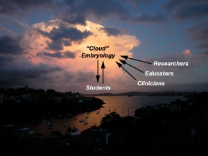
[[File:Clouds 02.jpg|thumb|300px|The "Cloud" Embryology Concept]] | |||
The very size and dynamic nature of embryonic development has always made it a difficult topic to teach. The structures are always small and the changing nature of development means that a feature you identify at any one time changes and becomes something else at a later stage. Students take some time to come to grips with all the new terminology and the sequence of events. Interestingly, the discipline of embryology was quick utilise each new technology as as they came along to help with understanding development. The internet today is the latest technology which can allow a wide distribution to students to be used at any time and quickly linked to other related topics. This page provides a broad introduction to some of these educational concepts that are covered in detail elsewhere, identified by internet links. I have also included on this page links to meeting presentations that cover specific topics drawing upon the many available resources on this site. | |||
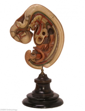
Historically models made from many different media (papier-mâché, ceramic, wax or plastic) not the [[Animal_Development|"animal models"]] described elsewhere on this site, were very useful in that they make tangible the microscopic structures to the students. Models have been in use for some time in the medical teaching of human development with the original plaster models now replaced by commercial modern plastic versions. Many of the early teaching models were not about development, but about the process of childbirth for doctor and midwife education. | |||
An alternative to the use of teaching models, was the use of plates and drawings found in historic textbooks and papers from the late 19th century onwards. These often beautifully drawn images gave a semi-realistic view of embryo anatomy, often with sections removed to show anatomical relationships or systems drawn in isolation. These early papers often included drawings of histological sections from specific embryos, providing a prelude to later photomicrographs. | |||
At the beginning of the 20th century photography also came into play and can be seen associated with embryo collections, such as the Carnegie and Kyoto collection. These photographic embryo images even today are unique tools in understanding human embryonic development. Animal embryos were far more easy to both obtain and to characterise. The realisation that most developmental processes are conserved across species, just differing in timing, led to growth in [[Animal_Development|"animal models"]]. In particular establishing a staging sequence allowed more consistent comparison of research findings in the literature. | |||
Today there are many teaching and research movies developed to show specific developmental processes. This use of dynamic media most closely links to the dynamics seen in development. | Today there are many teaching and research movies developed to show specific developmental processes. This use of dynamic media most closely links to the dynamics seen in development. | ||
{{Educational Links}} | |||
{{History Links}} | {{History Links}} | ||
==Meeting Presentations== | ==Meeting Presentations== | ||
Revision as of 14:19, 8 December 2012
Introduction
The very size and dynamic nature of embryonic development has always made it a difficult topic to teach. The structures are always small and the changing nature of development means that a feature you identify at any one time changes and becomes something else at a later stage. Students take some time to come to grips with all the new terminology and the sequence of events. Interestingly, the discipline of embryology was quick utilise each new technology as as they came along to help with understanding development. The internet today is the latest technology which can allow a wide distribution to students to be used at any time and quickly linked to other related topics. This page provides a broad introduction to some of these educational concepts that are covered in detail elsewhere, identified by internet links. I have also included on this page links to meeting presentations that cover specific topics drawing upon the many available resources on this site.
Historically models made from many different media (papier-mâché, ceramic, wax or plastic) not the "animal models" described elsewhere on this site, were very useful in that they make tangible the microscopic structures to the students. Models have been in use for some time in the medical teaching of human development with the original plaster models now replaced by commercial modern plastic versions. Many of the early teaching models were not about development, but about the process of childbirth for doctor and midwife education.
An alternative to the use of teaching models, was the use of plates and drawings found in historic textbooks and papers from the late 19th century onwards. These often beautifully drawn images gave a semi-realistic view of embryo anatomy, often with sections removed to show anatomical relationships or systems drawn in isolation. These early papers often included drawings of histological sections from specific embryos, providing a prelude to later photomicrographs.
At the beginning of the 20th century photography also came into play and can be seen associated with embryo collections, such as the Carnegie and Kyoto collection. These photographic embryo images even today are unique tools in understanding human embryonic development. Animal embryos were far more easy to both obtain and to characterise. The realisation that most developmental processes are conserved across species, just differing in timing, led to growth in "animal models". In particular establishing a staging sequence allowed more consistent comparison of research findings in the literature.
Today there are many teaching and research movies developed to show specific developmental processes. This use of dynamic media most closely links to the dynamics seen in development.
| Education Links: Educational Presentations | Embryology Education | Embryology iBooks | Embryology Models | Textbooks | Embryology History | Category:Education |
Meeting Presentations
- 2013 Australian Sonographers Association (ASA) Meeting
- 2013 Australian Chapter, American Academy of Craniofacial Pain (AACP) Meeting
- 2012 Australian and New Zealand Association of Clinical Anatomists (ANZACA) Meeting
- Medicine Learning and Teaching Forum 2012 - Online Projects
- 2012 Fetal ECHO Meeting
- 2012 Learning & Teaching Seminar
Awards - 2011 ALTC Citation for Outstanding Contributions to Student Learning
Embryology Models
Models made from many different media (papier-mâché, ceramic, wax or plastic) not the "animal models" described elsewhere on this site, are very useful in that they make tangible the microscopic structures to the students. Models have been in use for some time in the medical teaching of human development with the original plaster models now replaced by commercial modern plastic versions. Many of the early teaching models were not about development, but about the process of childbirth for doctor and midwife education.
A historic alternative to the use of teaching models, were series of plates and drawings found in textbooks and papers from the late 19th century onwards. At the beginning of the 20th century photography also came into play, seen mainly associated with embryos within the Carnegie collection.
- LInks: Embryology Models
Movies
- Movie LInks: Movies | Quicktime Movies | Flash Movies | Animation | Category:Quicktime | Category:Flash
Student Teaching Projects
Mobile Access
External Links
External Links Notice - The dynamic nature of the internet may mean that some of these listed links may no longer function. If the link no longer works search the web with the link text or name. Links to any external commercial sites are provided for information purposes only and should never be considered an endorsement. UNSW Embryology is provided as an educational resource with no clinical information or commercial affiliation.
Glossary Links
- Glossary: A | B | C | D | E | F | G | H | I | J | K | L | M | N | O | P | Q | R | S | T | U | V | W | X | Y | Z | Numbers | Symbols | Term Link
Cite this page: Hill, M.A. (2024, May 19) Embryology Embryology Education. Retrieved from https://embryology.med.unsw.edu.au/embryology/index.php/Embryology_Education
- © Dr Mark Hill 2024, UNSW Embryology ISBN: 978 0 7334 2609 4 - UNSW CRICOS Provider Code No. 00098G

