Chicken stages: Difference between revisions
mNo edit summary |
mNo edit summary |
||
| (21 intermediate revisions by the same user not shown) | |||
| Line 1: | Line 1: | ||
{{Header}} | |||
== Introduction == | == Introduction == | ||
[[File: | [[File:HHstage11.jpg|thumb|Hamburger & Hamilton Stage 11 (40-45 hr; 13 somites)]] | ||
Hamburger | {| | ||
| width=110px valign=top|[[File:Viktor Hamburger.jpg|100px|alt=Viktor Hamburger]] | |||
Viktor Hamburger (1900 – 2001) | |||
| valign=top|[[Hamburger Hamilton Stages|Hamburger & Hamilton]] staged the chicken embryo in 1951.{{#pmid:1304821|PMID1304821}} The original paper had approx 25 citations between 1955 - 59, while in the year 1991 alone there were over 300 citations. '''Series of Embryonic Chicken Growth.''' J. Morphology, '''88''' 49 - 92 (1951). Atlas recently republished by J.R. Sanes in Developmental Dynamics '''195''' 229-275 (1992). There was also an earlier [[Chicken_Stages_-_Witschi|Witschi staging]] though the [[Hamburger Hamilton Stages]] most commonly currently used. | |||
|- | |||
| [[File:Franz Keibel.jpg|100px|alt=Franz Keibel|link=Embryology History - Franz Keibel]] | |||
Franz Keibel (1861 - 1929) | |||
| valign=top|An even older 1900 staging<ref name=Keibel1900>{{Ref-Keibel1900}}</ref> in the Normentafeln zur Entwicklungsgeschichte der Wirbeltiere - ''Gallus domestics'' ([[Book_-_Normal_Plates_of_the_Development_of_Vertebrates_2|Normal Plates of the Development of the Chicken Embryo]] by [[Embryology_History_-_Franz_Keibel|Franz Keibel]] and Karl Abraham. This was part of a series of embryo stagings in different species designed by [[Embryology_History_-_Franz_Keibel|Franz Keibel]]. | |||
See also the 1910 description of chicken somitogenesis staging.<ref name=Williams1910>{{Ref-Williams1910}}</ref> | |||
|} | |||
The table overview below describes feature of the chicken that can be seen at specific times (mean of a range) after fertilization, see [[Hamburger Hamilton Stages]] page for complete details and stage images. Some of the stages can be clicked to see sample embryo pictures on another page. There is also a [[Chicken_stages#Standard_Stage_Comparison|stage comparison]] with the alternative [[Chicken_Stages_-_Witschi|universal Witschi staging]]. | |||
{{Bird incubation period table}} | {{Bird incubation period table}} | ||
{{Chicken links}} | |||
== Hamburger Hamilton Stages == | == Hamburger Hamilton Stages == | ||
| Line 53: | Line 62: | ||
| 6 - 7 hr | | 6 - 7 hr | ||
| Initial primitive streak, 0.3-0.5 mm long | | Initial primitive streak, 0.3-0.5 mm long | ||
| | | [[Gastrulation]] | ||
|-bgcolor="F5FAFF" | |-bgcolor="F5FAFF" | ||
| <center>3</center> | | <center>3</center> | ||
| Line 63: | Line 72: | ||
| 18 - 19 hr | | 18 - 19 hr | ||
| Definitive primitive streak, 1.88 mm long | | Definitive primitive streak, 1.88 mm long | ||
| | | [[Gastrulation]] | ||
|-bgcolor="F5FAFF" | |-bgcolor="F5FAFF" | ||
| <center>5</center> | | <center>5</center> | ||
| Line 78: | Line 87: | ||
| 23 - 26 hr | | 23 - 26 hr | ||
| 1 somite; neural folds | | 1 somite; neural folds | ||
| [[Somitogenesis]] | |||
|- | |- | ||
| <center>7 to 8-</center> | | <center>7 to 8-</center> | ||
| Line 143: | Line 152: | ||
| <center>16</center> | | <center>16</center> | ||
| 51 - 56 hr | | 51 - 56 hr | ||
| 26 - 28 somites; wing bud; posterior amniotic fold | | 26 - 28 somites; wing bud; posterior amniotic fold; trigeminal ganglion (CN V) close lateral head ectoderm. | ||
| [[:File:Heart chicken embryo stage 16.jpg|Heart]] | | [[:File:Heart chicken embryo stage 16.jpg|Heart]] | ||
| Line 636: | Line 645: | ||
|} | |} | ||
Modified from: '''GROWTH''' Altman, P.L. and Dittmer, D.S. (ed) Biological Handbooks, FASEB (1962) | Modified from: '''GROWTH''' Altman, P.L. and Dittmer, D.S. (ed) Biological Handbooks, FASEB (1962) | ||
==Images== | |||
<gallery> | |||
File:Chicken_antero-posterior_endoderm_patterning_01.jpg|HH4 and HH10 endoderm | |||
File:Chicken HH20 MyoR expression 01.jpg|HH20 head muscle MyoR | |||
File:Chicken HH20 MyoR expression 02.jpg|HH20 head muscle MyoR and MyoD | |||
</gallery> | |||
==References== | ==References== | ||
| Line 645: | Line 664: | ||
** This is an ATLAS (no description of development) , asically reprinted from the original 1963 edition. | ** This is an ATLAS (no description of development) , asically reprinted from the original 1963 edition. | ||
** Photos with labelled diagrams covering Amphioxus (worm) Frog, Chicken. | ** Photos with labelled diagrams covering Amphioxus (worm) Frog, Chicken. | ||
* | * {{Ref-ButlerJuurlink1987}} | ||
** This ATLAS is not a complete series of development but has interesting comparisons of species. | ** This ATLAS is not a complete series of development but has interesting comparisons of species. Mostly photos of embryos with a few drawn diagrams and a series of staging correlation graphs. | ||
{{Glossary}} | |||
{{ | {{Footer}} | ||
[[Category:Animal Development]] [[Category:Chicken]] | [[Category:Animal Development]] [[Category:Chicken]] | ||
Latest revision as of 08:13, 24 April 2018
| Embryology - 27 May 2024 |
|---|
| Google Translate - select your language from the list shown below (this will open a new external page) |
|
العربية | català | 中文 | 中國傳統的 | français | Deutsche | עִברִית | हिंदी | bahasa Indonesia | italiano | 日本語 | 한국어 | မြန်မာ | Pilipino | Polskie | português | ਪੰਜਾਬੀ ਦੇ | Română | русский | Español | Swahili | Svensk | ไทย | Türkçe | اردو | ייִדיש | Tiếng Việt These external translations are automated and may not be accurate. (More? About Translations) |
Introduction


Viktor Hamburger (1900 – 2001) |
Hamburger & Hamilton staged the chicken embryo in 1951.[1] The original paper had approx 25 citations between 1955 - 59, while in the year 1991 alone there were over 300 citations. Series of Embryonic Chicken Growth. J. Morphology, 88 49 - 92 (1951). Atlas recently republished by J.R. Sanes in Developmental Dynamics 195 229-275 (1992). There was also an earlier Witschi staging though the Hamburger Hamilton Stages most commonly currently used. |

Franz Keibel (1861 - 1929) |
An even older 1900 staging[2] in the Normentafeln zur Entwicklungsgeschichte der Wirbeltiere - Gallus domestics (Normal Plates of the Development of the Chicken Embryo by Franz Keibel and Karl Abraham. This was part of a series of embryo stagings in different species designed by Franz Keibel.
|
The table overview below describes feature of the chicken that can be seen at specific times (mean of a range) after fertilization, see Hamburger Hamilton Stages page for complete details and stage images. Some of the stages can be clicked to see sample embryo pictures on another page. There is also a stage comparison with the alternative universal Witschi staging.
| Bird | Days |
|---|---|
| Budgerigar | 18 |
| Chicken | 21 |
| Duck | 28 |
| Finch | 14 |
| Goose | 28 |
| Guinea fowl | 28 |
| Muscovy duck | 35 |
| Parrot | 26 |
| Pheasant | 24 |
| Pigeon | 18 |
| Quail | 16 |
| Swan | 35 |
| Turkey | 28 |
Hamburger Hamilton Stages
| 3.5 - 4.5 hr | Shell membrane of egg formed in isthmus of oviduct | |||
| Germ wall formed from marginal periblast | ||||
| 4.5 - 24.0 hr | Shell of egg formed in uterus | |||
| Preprimitive streak (embryonic shield) | ||||
| 6 - 7 hr | Initial primitive streak, 0.3-0.5 mm long | Gastrulation | ||
| 12 - 13 hr | Intermediate primitive streak | |||
| 18 - 19 hr | Definitive primitive streak, 1.88 mm long | Gastrulation | ||
| 19 - 22 hr | Head process (notochord) | |||
| 23 - 25 hr | Head fold | |||
| 23 - 26 hr | 1 somite; neural folds | Somitogenesis | ||
| ca. 23 - 26 hr | 1-3 somites; coelom | |||
| 26 - 29 hr | 4 somites; blood islands | |||
| 29-33 hr | 7 somites; primary optic vesicles | |||
| ca. 33 hr | 8-9 somites; anterior amniotic fold | |||
| 33-38 hr | 10 somites; 3 primary brain vesicles | |||
| 40 - 45 hr | 13 somites; 5 neuromeres of hindbrain | |||
| 45 - 49 hr | 16 somites; telencephalon | Heart | ||
| 48-52 hr | 19 somites; atrioventricular canal | |||
| ca. 50 - 52 hr | 20-21 somites; tail bud | |||
| 50 - 53 hr | 22 somites; trunk flexure; visceral arches I and II, clefts 1 and 2 | |||
| ca. 50 - 54 hr | 23 somites; premandibular head cavities | |||
| 50 - 55 hr | 24 - 27 somites; visceral arch III, cleft 3 | |||
| 51 - 56 hr | 26 - 28 somites; wing bud; posterior amniotic fold; trigeminal ganglion (CN V) close lateral head ectoderm. | Heart | ||
| 52 - 64 hr | 29-32 somites; leg bud; epiphysis | |||
| 3 da | 30 - 36 somites extending beyond level of leg bud; allantois | |||
| 3.0 - 3.5 da | 37 - 40 somites extending into tail; maxillary process | |||
| 3.0 - 3.5 da | 40 - 43 somites; rotation completed; eye pigment | |||
| 3.5 da | 43-44 somites; visceral arch IV, cleft 4 | Heart | ||
| 3.5 - 4.0 da | Somites extend to tip of tail | |||
| 4 da | Dorsal contour from hindbrain to tail is a curved line | |||
| 4.5 da | Toe plate, eye primordium reaches optic cup stage. | |||
| 4.5 - 5.0 da | Elbow and knee joints | Heart | ||
| 5 da | 1st 3 toes | |||
| 5.0 - 5.5 da | Beak | |||
| 5.5 - 6.0 da | 3 digits, 4 toes | |||
| 6.0 - 6.5 da | Rudiment of 5th toe | |||
| 6.5 - 7.0 da | Feather germs; scleral papillae; egg tooth | |||
| 7.0 - 7.5 da | Web between 1st and 2nd digits | |||
| 7.5 da | Anterior tip of mandible has reached beak | |||
| 7.5 - 8.0 da | Web on radial margin of wing and 1st digit | |||
| 8 da | Nictitating membrane | |||
| 8.5 - 9.0 da | Phalanges in toes | |||
| 10 da | Length of 3rd toe from tip to middle of metatarsal joint = 5.4±0.3mm; length of beak from anterior angle of nostril to tip of bill = 2.5mm; primordium of comb; labial groove; uropygial gland | |||
| 11 da | Length of 3rd toe = 7.4±0.3mm; length of beak = 3.0 mm | |||
| 12 da | Length of 3rd toe = 8.4±0.3 mm; length of beak = 3.1 mm | |||
| 13 da | Length of 3rd toe = 9.8±0.3 mm; length of beak = 3.5 mm | |||
| 14 da | Length of beak = 4.0 mm; length of 3rd toe = 12.7±0.5 mm | |||
| 15 da | Length of beak from anterior angle of nostril to tip of upper bill = 4.5 mm; length of 3rd toe = 14.9±0.8 mm | |||
| 16 da | Length of beak = 4.8 mm; length of 3rd toe = 16.7±0.8 mm | |||
| 17 da | Length of beak = 5.0 mm; length of 3rd toe = 18.6±0.8 mm | |||
| 18 da | Length of beak = 5.7 mm; length of 3rd toe = 20.4±0.8 mm | |||
| 19 - 20 da | Yolk sac half enclosed in body cavity; chorio-allantoic membrane contains less blood and is "sticky" in living embryo | |||
| 20 - 21 da | Newly-hatched chick | |||
Standard Stage Comparison
| Hamburger Hamilton Stages | |||
| 3,4 | Early cleavage | 3.5-4.5 hr2 | Shell membrane of egg formed in isthmus of oviduct |
| 5,6 | During cleavage | Germ wall formed from marginal periblast | |
| 7 | Late cleavage | 4.5-24.0 hr2 | Shell of egg formed in uterus |
| 8,9 | 1 | Preprimitive streak (embryonic shield) | |
| 10 | 2 | 6-7 hr | Initial primitive streak, 0.3-0.5 mm long |
| 11 | 3 | 12-13 hr | Intermediate primitive streak |
| 12 | 4 | 18-19 hr | Definitive primitive streak, ±1.88 mm long |
| 13a | 5 | 19-22 hr | Head process (notochord) |
| 13b | 6 | 23-25 hr | Head fold |
| 14a | 7 | 23-26 hr | 1 somite; neural folds |
| 14b | 7 to 8- | ca. 23-26 hr | 1-3 somites; coelom |
| 14c | 8 | 26-29 hr | 4 somites; blood islands |
| 15a | 9 | 29-33 hr | 7 somites; primary optic vesicles |
| 15b | 9+ to 10- | ca. 33 hr | 8-9 somites; anterior amniotic fold |
| 15c | 10 | 33-38 hr | 10 somites; 3 primary brain vesicles |
| 16a | 11 | 40-45 hr | 13 somites; 5 neuromeres of hindbrain |
| 16b | 12 | 45-49 hr | 16 somites; telencephalon |
| 16c | 13 | 48-52 hr | 19 somites; atrioventricular canal |
| 17a | 13+ to 14- | ca. 50-52 hr | 20-21 somites; tail bud |
| 17b | 14 | 50-53 hr | 22 somites; trunk flexure; visceral arches I and II, clefts 1 and 2 |
| 17c | 14+ to 15- | ca. 50-54 hr | 23 somites; premandibular head cavities |
| 17d | 15 | 50-55 hr | 24-27 somites; visceral arch III, cleft 3 |
| 18 | 16 | 51-56 hr | 26-28 somites; wing bud; posterior amniotic fold |
| 19 | 17 | 52-64 hr | 29-32 somites; leg bud; epiphysis |
| 20 | 18 | 3 da | 30-36 somites extending beyond level of leg bud; allantois |
| 21 | 19 | 3.0-3.5 da | 37- 40 somites extending into tail; maxillary process |
| 22 | 20 | 3.0-3.5 da | 40-43 somites; rotation completed; eye pigment |
| 23 | 21 | 3.5 da | 43-44 somites; visceral arch IV, cleft 4 |
| 24 | 22 | 3.5-4.0 da | Somites extend to tip of tail |
| 25 | 23 | 4 da | Dorsal contour from hindbrain to tail is a curved line |
| 26 | 24 | 4.5 da | Toe plate |
| 27 | 25 | 4.5-5.0 da | Elbow and knee joints |
| 28 | 26 | 5 da | 1st 3 toes |
| 29 | 27 | 5.0-5.5 da | Beak |
| 30 | 28 | 5.5-6.0 da | 3 digits, 4 toes |
| 31 | 29 | 6.0-6.5 da | Rudiment of 5th toe |
| 32 | 30 | 6.5-7.0 da | Feather germs; scleral papillae; egg tooth |
| 33a | 31 | 7.0-7.5 da | Web between 1st and 2nd digits |
| 33b | 32 | 7.5 da | Anterior tip of mandible has reached beak |
| 34a | 33 | 7.5-8.0 da | Web on radial margin of wing and 1st digit |
| 34b | 34 | 8 da | Nictitating membrane |
| 34c | 35 | 8.5-9.0 da | Phalanges in toes |
| 34d | 36 | 10 da | Length of 3rd toe from tip to middle of metatarsal joint = 5.4±0.3mm; length of beak from anterior angle of nostril to tip of bill = 2.5mm; primordium of comb; labial groove; uropygial gland |
| 34e | 37 | 11 da | Length of 3rd toe = 7.4±0.3mm; length of beak = 3.0 mm |
| 34f | 38 | 12 da | Length of 3rd toe = 8.4±0.3 mm; length of beak = 3.1 mm |
| 35a | 39 | 13 da | Length of 3rd toe = 9.8±0.3 mm; length of beak = 3.5 mm |
| 35b | 40 | 14 da | Length of beak = 4.0 mm; length of 3rd toe = 12.7±0.5 mm |
| 35c | 41 | 15 da | Length of beak from anterior angle of nostril to tip of upper bill = 4.5 mm; length of 3rd toe = 14.9±0.8 mm |
| 35d | 42 | 16 da | Length of beak = 4.8 mm; length of 3rd toe = 16.7±0.8 mm |
| 35e | 43 | 17 da | Length of beak = 5.0 mm; length of 3rd toe = 18.6±0.8 mm |
| 35f | 44 | 18 da | Length of beak = 5.7 mm; length of 3rd toe = 20.4±0.8 mm |
| 36a | 45 | 19-20 da | Yolk sac half enclosed in body cavity; chorio-allantoic membrane contains less blood and is "sticky" in living embryo |
| 36b | 46 | 20-21 da | Newly-hatched chick |
Modified from: GROWTH Altman, P.L. and Dittmer, D.S. (ed) Biological Handbooks, FASEB (1962)
Images
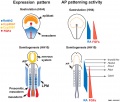
HH4 and HH10 endoderm

HH20 head muscle MyoR
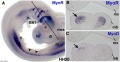
HH20 head muscle MyoR and MyoD


References
- ↑ Hamburger V & Hamilton HL. (1992). A series of normal stages in the development of the chick embryo. 1951. Dev. Dyn. , 195, 231-72. PMID: 1304821 DOI.
- ↑ Keibel F. and Abraham K. Normal Plates of the Development of the Chicken Embryo (Gallus domesticus). (1900) Vol. 2 in series by Keibel F. Normal plates of the development of vertebrates (Normentafeln zur Entwicklungsgeschichte der Wirbelthiere) Fisher, Jena., Germany.
- ↑ Williams LW. The somites of the chick. (1910) Amer. J Anat. 11(1): 56 -100.
Other Chicken Atlases
- Vertebrate and Invertebrate Embryos (7th Edition) G.C. Schoenwolf, Prentice Hall, New Jersey
- An Atlas of Embryology (1975) W.H. Freeman and B. Bracegirdle, Heinemann Educational Books, UK.
- This is an ATLAS (no description of development) , asically reprinted from the original 1963 edition.
- Photos with labelled diagrams covering Amphioxus (worm) Frog, Chicken.
- Butler H. and Juurlink BHJ. An atlas for staging mammalian and chick embryos. (1987) CRC. Boca Raton, Florida.
- This ATLAS is not a complete series of development but has interesting comparisons of species. Mostly photos of embryos with a few drawn diagrams and a series of staging correlation graphs.
Glossary Links
- Glossary: A | B | C | D | E | F | G | H | I | J | K | L | M | N | O | P | Q | R | S | T | U | V | W | X | Y | Z | Numbers | Symbols | Term Link
Cite this page: Hill, M.A. (2024, May 27) Embryology Chicken stages. Retrieved from https://embryology.med.unsw.edu.au/embryology/index.php/Chicken_stages
- © Dr Mark Hill 2024, UNSW Embryology ISBN: 978 0 7334 2609 4 - UNSW CRICOS Provider Code No. 00098G