Category:Student Image
From Embryology
Content in this category relates to images added to the site by students.
As shown by the text below:
Note - This image was originally uploaded as part of a student project and may contain inaccuracies in either description or acknowledgements.
--MarkHill 13:50, 4 February 2011 (EST) This category was added in February 2011 and may not appear on images uploaded before this time.
Pages in category 'Student Image'
The following 11 pages are in this category, out of 11 total.
Media in category 'Student Image'
The following 200 files are in this category, out of 650 total.
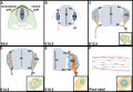
(previous page) (next page) Nasal placode diagram.jpeg 2,049 × 956; 455 KB
 Nephron development 02.jpg 640 × 409; 140 KB
 Nephron Maturation.jpg 431 × 406; 29 KB
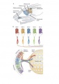
 Neural Circuit in the Cerebellum.jpg 293 × 690; 55 KB
 Neural crest cell migration in erbb3b mutants.jpg 591 × 600; 45 KB
 Neural crest migration and somite development in zebrafish.jpeg 1,280 × 472; 119 KB
 Neural Crest Migration.png 363 × 144; 107 KB
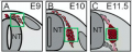
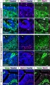 Neural crest-derived cells in the embryonic olfactory epithelium.jpg 636 × 1,075; 175 KB
 Neurogenesis and Gliogenesis Timeline.jpg 721 × 497; 90 KB
 Neuropore cell shape changes.png 1,418 × 1,940; 1.01 MB
 Neurpore cell shape changes.png 785 × 1,073; 1.11 MB
 New olfactory bulb.jpg 3,833 × 1,793; 568 KB
 Ngn1 and Ngn2 in DRG development.png 384 × 557; 212 KB
 Non-viable spermatazoa.jpg 539 × 300; 33 KB
 Normal cochlea.png 571 × 789; 141 KB
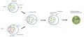
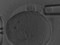
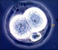 Normal Human 2-cell Embryo.jpeg 467 × 404; 107 KB
 Normal Mus musculus karyotype.jpg 371 × 450; 48 KB
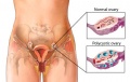
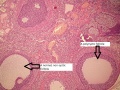
 Normal ovary compared to polycystic ovary.png 2,636 × 948; 1.36 MB
 Notch animals.png 634 × 544; 56 KB
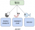
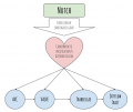
 Notch CNS simple diagram.png 579 × 490; 50 KB
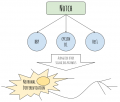
 Notch CVS simple diagram.png 652 × 548; 54 KB
 Notch Signalling - Ovulation in Drosphila.jpg 697 × 702; 68 KB
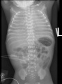 NRDS.jpg 481 × 645; 155 KB
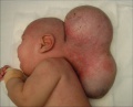 Occipital encephalocele associated with microcephaly.jpg 644 × 518; 186 KB
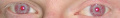 Oculocutaneous albinism eyes.jpg 755 × 113; 15 KB
 Olfaction signal transduction.JPG 365 × 514; 21 KB
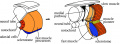
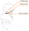 Olfactory bulb and epithelium.png 648 × 702; 95 KB
 Olfactory epithelium.jpg 3,112 × 1,848; 413 KB
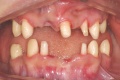 Oligodontia.jpg 773 × 517; 223 KB
 Online placement of the 1H-MRS volume of interest .jpg 600 × 569; 100 KB
 Online placement of the 1H-MRS volume of interest.tif 2,835 × 2,690; 3.1 MB
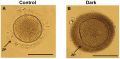 Oocytes with DZP demonstrate affect on fertility.png 1,370 × 678; 2.01 MB
 Oral cavity.png 776 × 540; 199 KB
 Otic placode embryo.jpg 500 × 461; 23 KB
 Otic vesicle.jpg 518 × 387; 24 KB
 Outflow tract anatomy.png 664 × 791; 291 KB
 Outlfow tract.png 2,828 × 1,439; 1.67 MB
 Ovarian tissue extracted after ovarian stimulation.jpeg 600 × 856; 345 KB
 Ovarian transposition.jpeg 1,280 × 942; 167 KB
 Ovary1.gif 779 × 398; 168 KB
 Overview of Notch signalling.png 1,230 × 722; 368 KB
 Oviduct Ligation .png 600 × 306; 438 KB
 Ovotestes.jpg 1,783 × 2,907; 778 KB
 P. maniculatus embryo E17.5-21.5.PNG 1,922 × 1,724; 4.8 MB
- Error creating thumbnail: File with dimensions greater than 12.5 MPPairs of conjugate sperm attached by the head.jpg 3,474 × 4,497; 1.91 MB
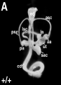
 Parathyroid position in mouse embryo.jpg 500 × 641; 100 KB
 Paternal chromatin mouse embryos.jpg 600 × 853; 102 KB
 Pathogenesis of OHSS.png 826 × 467; 462 KB
 Patholophysiology of Polycystic Ovarian Syndrome.jpg 600 × 396; 39 KB
 Patient with Marfan syndrome.jpg 502 × 1,171; 93 KB
 Patterns of ZPC Deposition in Porcine Oocytes.jpg 401 × 600; 84 KB
 PB1 transfer.jpg 2,959 × 1,311; 170 KB
 PB2 transfer.jpg 2,983 × 1,272; 169 KB
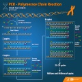 PCR.jpg 1,800 × 1,800; 1.2 MB
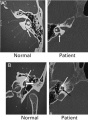 Pendred Syndrome Showing Mondini Defects.jpg 480 × 657; 53 KB
 Pericardial Development 4-6 Gestation weeks .jpg 727 × 834; 102 KB
 Pharyngeal arch one and two in mice.png 448 × 472; 326 KB
 Pharyngula.png 342 × 209; 80 KB
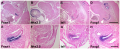
 Photomicrographs of gallbladder samples stained with hematoxylin and eosin in each group.png 1,445 × 1,097; 3.88 MB
 Pituitary Development.jpg 463 × 217; 41 KB
 PKD.jpg 664 × 252; 163 KB
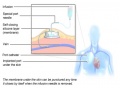
 Placenta accreta.jpg 600 × 410; 98 KB
 Placenta schematic.jpg 784 × 600; 121 KB
 Polar Body (a), Blastomere (b) and Trophectoderm (c) Biopsies.gif 567 × 128; 48 KB
 Polar Body Biopsy.jpeg 500 × 375; 74 KB
 Polar Body, Blastomere, and Trophectoderm Biopsy.jpeg 567 × 128; 29 KB
 Polycystic Ovaries.jpeg 432 × 273; 28 KB
 Polycystic rat ovary.jpg 600 × 450; 200 KB
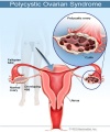 PolycysticOvarianSyndrome.jpg 450 × 540; 78 KB
 Polydactyly.jpg 230 × 188; 10 KB
 Pone.0015329.g001 z5039628.jpg 483 × 388; 135 KB
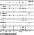 POPs and risk of hypospadias.jpg 452 × 473; 224 KB
 Port Administered Chemotherapy.jpg 475 × 350; 29 KB
 Pre-PGD workup.jpeg 722 × 1,119; 148 KB
 Preimplantation Genetic Diagnosis Procedure.jpeg 2,974 × 1,751; 395 KB
 Prepuce.jpeg 2,293 × 767; 382 KB
 Prevalence of Primary Infertility in 2010.jpeg 745 × 329; 58 KB
 Primary Brain Vesciles.pdf ; 453 KB
 Primary Brain Vesicles.jpeg 638 × 359; 64 KB
 Primary brain vesicles.jpg 2,082 × 1,221; 473 KB
 Primary Brain.png 1,005 × 655; 681 KB
 Primite Streak Cell Migration In Chick Embryo.png 320 × 214; 48 KB
 Primitive streak development in chick embryo.png 1,511 × 1,213; 1.9 MB
 Processing of Hh precurosr.jpg 1,120 × 1,280; 132 KB
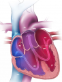
 Progressive development of the Embryonic Heart.jpeg 1,024 × 883; 154 KB
 Promising System for Selecting Healthy In Vitro Fertilized Embryos in Cattle.png 2,067 × 1,238; 1 MB
 Pronuclear transfer.jpg 2,914 × 1,270; 174 KB
 Prostate.jpeg 1,639 × 1,244; 526 KB
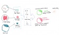
 Protein-protein interaction.jpg 600 × 776; 85 KB
 Proteomics Technologies PMID- 26471863.jpeg 966 × 1,280; 191 KB
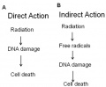 Radiation.jpeg 448 × 370; 32 KB
 Ratio of alpha & beta cells at different phases of fetal development.png 537 × 600; 609 KB
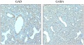 RatLungGADGABA.jpeg 1,379 × 745; 476 KB
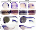 Rbm24a and rbm24b are expressed throughout somitogenesis.jpeg 773 × 664; 113 KB
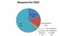 Reasons for PGD.jpg 445 × 276; 20 KB
 Reelin signalling.jpg 478 × 547; 37 KB
 Regions of varying neural cell types in ventral neural tube.jpg 600 × 537; 35 KB
 Regulation of Nodal-Activin signalling during heart formation.png 696 × 511; 238 KB
 Reproductive surgeries in Males.jpeg 1,624 × 676; 354 KB
 Respiratorysystem1.jpeg 678 × 960; 116 KB
 Respiratorysystem2.png 645 × 575; 403 KB
 Respriatory zone.png 719 × 467; 402 KB
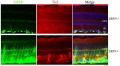 Retina-cell-clusters.JPG 600 × 331; 32 KB
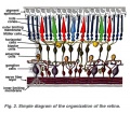 Retina-layers-diagram.jpg 570 × 500; 156 KB
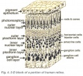 Retina-layers-diagram2.jpg 550 × 500; 96 KB
 Retinoic Acid activation pathway.png 925 × 537; 529 KB
 Rhombencephalosynapsis.jpg 600 × 619; 57 KB
 Roles and Regulation of SOX2 in Blastocyst Formation.jpeg 723 × 813; 149 KB
 Roles of LIF in embryo implantation.jpeg 600 × 422; 77 KB
 Ruffini Ending.JPG 757 × 441; 53 KB
 Rufus-eye.jpg 1,700 × 1,680; 200 KB
 Schematic diagram of heart tube looping.png 554 × 394; 117 KB
 SchizencephalicB.jpg 3,000 × 2,250; 1.37 MB
 Screen Shot 2017-10-05 at 3.29.59 pm.png 766 × 270; 243 KB
 Screen Shot 2017-10-15 at 13.31.05.png 1,664 × 864; 2.42 MB
 Screen Shot 2017-10-16 at 1.43.55 pm.png 1,183 × 535; 704 KB
 Screen Shot 2017-10-16 at 3.01.32 pm.png 615 × 645; 756 KB
 Screen Shot 2017-10-26 at 10.20.18 am.png 1,456 × 1,002; 1.02 MB
 Second Trimester Cerebellum.jpeg 475 × 385; 26 KB
 Secondary brain vesicle.jpeg 1,204 × 431; 209 KB
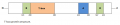
 Segmentation .png 944 × 189; 232 KB
 Semi-circular canals.jpg 1,130 × 800; 98 KB
 Semilobar holoprosencephaly.jpg 1,024 × 1,024; 54 KB
 Series 10-09.jpg 300 × 225; 19 KB
 Severe OHSS Prevention.jpeg 600 × 866; 73 KB
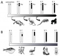 Sex Determination Across Species.jpeg 653 × 605; 71 KB
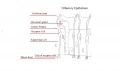
 SexualDifferentation.jpg 640 × 1,206; 90 KB
 Side effects of Chemotherapy.png 456 × 837; 170 KB
 Signalling during cerebellum development.jpeg 256 × 256; 8 KB
 Signalling factors in lung branching cartoon.png 1,201 × 657; 219 KB
 Signalling pathways in heart development.png 505 × 243; 76 KB
 Silencing of Proteins Rac1 and RhoA.jpeg 433 × 232; 81 KB
 Skin Melanocytes.jpg 704 × 278; 79 KB
 Somatosensory cortex of E20 rat.jpeg 2,003 × 2,722; 1.29 MB
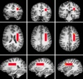
 Somatotopic Activation by corneal pain and eye blink.png 600 × 372; 220 KB
 Sox9.png 420 × 645; 368 KB
 Sperm Entry Blocked by Heparin.jpeg 478 × 344; 107 KB
 Sperm entry site and location of male proncleus.jpeg 796 × 830; 160 KB
 Stages in early development of a human zygote.png 2,059 × 348; 1.24 MB
 Stages of Atrial Septation.png 3,582 × 1,290; 5.14 MB
 Stages of development.png 912 × 597; 495 KB
 Stages of nephrogenesis.png 4,059 × 3,000; 1.8 MB
 Stages of spermatogonia.jpeg 600 × 771; 159 KB
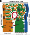 Stria Vascularis diagram 2.jpeg 291 × 336; 46 KB
 Structure of DRG in Sox10-deficient mice.jpg 969 × 2,055; 441 KB
 Structure of mouse spermatozoa.jpeg 2,293 × 900; 187 KB
 Structure of the seminiferous tubule.jpeg 600 × 847; 84 KB
 Suctionaspiration.png 800 × 600; 137 KB
 Summary Figure of Notch in Cardiac Development.jpeg 800 × 289; 61 KB
 Summary of Oncofertility.jpg 1,322 × 959; 123 KB
 Syrian Hamster In Vitro Fertilisation PMID- 24852961.jpeg 743 × 382; 70 KB
 Taste qualities.gif 421 × 440; 82 KB
 TBX 5 3D strcture.png 400 × 478; 62 KB
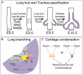 Tbx in lung and trachea development.png 1,447 × 1,303; 167 KB
 TBX1 factor figure.jpg 550 × 351; 92 KB
 Testicular cryopreservation.jpeg 600 × 330; 37 KB
 Testis and ovary.jpg 960 × 540; 37 KB
 Tetralogy of Fallot (TOF).png 364 × 477; 238 KB
 Tetralogy of fallot after surgery.png 478 × 354; 285 KB
 Tetralogy of Fallot with pulmonary atresia.jpg 600 × 753; 99 KB
 TGF-B Signalling - Formation of Receptor Hetero-Tetramers.png 2,012 × 1,696; 79 KB
 The kidney of a FA-injected mouse compared to a wildtype mouse.jpg 763 × 900; 324 KB
 The morphology of follicles after ovarian tissue vitrification.jpg 776 × 165; 76 KB
 The Process of Atrial Septation.png 3,595 × 1,110; 1.24 MB
 The Sonic Hedghehog (Shh) signalling pathway.png 572 × 586; 249 KB
 Theiler 12.JPG 733 × 395; 36 KB
 Theiler 13...JPG 1,030 × 427; 50 KB
 Theiler 13.JPG 1,047 × 578; 58 KB
 Theiler 14.JPG 815 × 406; 42 KB
 Thermoreceptor development diagram.JPG 576 × 635; 29 KB
 Thymic Epithelial Cell Development and Function.png 2,028 × 823; 3.17 MB
 ThyroidDevelopment.png 1,996 × 1,212; 49 KB
 Timeline - Fetal Tooth Development.jpg 1,156 × 220; 80 KB
 Timeline rabbit.JPG 633 × 488; 28 KB
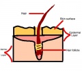 Touch Receptor- Hair Follicle.jpg 400 × 350; 35 KB
 Trachea .png 730 × 853; 125 KB
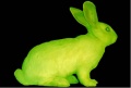 Transgenic rabbit.jpg 453 × 306; 28 KB
 Treatment of TOF surgery.png 618 × 349; 410 KB
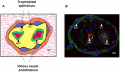 Trophoblast and villous epithelium of the placenta.png 2,058 × 1,241; 2.61 MB
 Truncus Arteriosus.png 442 × 581; 314 KB
 Typical tbx protein structure.png 773 × 164; 5 KB
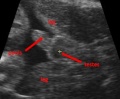 Ultrasound male.jpg 299 × 246; 29 KB
 Ultrasound of Polycystic Ovaries .jpg 600 × 718; 106 KB
 Unilateral microphthalmia patient with delection of SOX2 gene.png 439 × 205; 126 KB
 Unruptured ampullary ectopic pregnancy at laparoscopy.jpg 628 × 471; 60 KB
 Ureter.jpg 1,050 × 684; 226 KB
 Urinary Bladder Histology.jpg 581 × 399; 42 KB
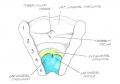
 Urorectal septum.png 400 × 407; 29 KB
 Uterus and Vagina Abnormalities.jpg 3,157 × 1,932; 1.13 MB
 Utrophin effects compared to control.jpg 452 × 449; 114 KB


.png)


_signalling_pathway.png)


,_Blastomere_(b)_and_Trophectoderm_(c)_Biopsies.gif)