Category:Spermatozoa: Difference between revisions
From Embryology
No edit summary |
mNo edit summary |
||
| Line 1: | Line 1: | ||
This page lists | This page lists Embryology pages and media related to spermatozoa development. | ||
Revision as of 14:27, 23 January 2014
This page lists Embryology pages and media related to spermatozoa development.
Subcategories
This category has the following 2 subcategories, out of 2 total.
Pages in category 'Spermatozoa'
The following 68 pages are in this category, out of 68 total.
A
B
F
H
M
P
- Paper - A study of the function of the epididymis 1 (1929)
- Paper - Cytology of the human spermatozoon
- Paper - Electron microscopy of the sperm tail - results obtained with a new fixative
- Paper - Studies in the physiology of spermatozoa
- Paper - The duration of life of the spermatozoa in the human uterine tube
- Paper - The mammalian spermatozoon
- Template:Primary spermatocyte
- Template:Primordial germ cell
- Template:Pronuclei
- Template:Pronucleus
R
S
- Sertoli cell
- Template:Sperm
- Template:Spermatogenesis
- Template:Spermatogonia
- Template:Spermatogonial stem cell
- Template:Spermatogonium
- Template:Spermatozoa
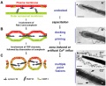
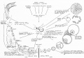
- Spermatozoa Chemotaxis
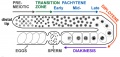
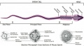
- Spermatozoa Development
- Spermatozoa Meiosis Movie 1
- Spermatozoa Structure Movie
- Template:Spermatozoa Terms
- Template:Spermatozoa Terms collapse table
- Template:Spermatozoon
- Template:Spermiogenesis
- Template:SSC
Media in category 'Spermatozoa'
The following 150 files are in this category, out of 150 total.
 Adult hermaphrodite gonad arm.jpg 800 × 377; 66 KB
 Azoospermia.jpg 768 × 554; 77 KB
 Bailey004.jpg 364 × 1,013; 40 KB
 Bailey005.jpg 747 × 1,050; 138 KB
 Bailey006.jpg 354 × 1,113; 131 KB
 Bailey007.jpg 772 × 803; 138 KB
 Bailey008.jpg 838 × 815; 89 KB
 Bailey009.jpg 774 × 766; 68 KB
 Bailey012.jpg 946 × 530; 73 KB
 Bailey013.jpg 866 × 896; 197 KB
 BurgosFawcett1955 fig11.jpg 1,453 × 2,015; 528 KB
 BurgosFawcett1955 fig13.jpg 1,460 × 2,049; 501 KB
 BurgosFawcett1955 fig14.jpg 1,456 × 1,965; 381 KB
 BurgosFawcett1955 text-fig01.jpg 1,280 × 1,137; 143 KB
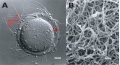
 Cat spermatozoa bound to oocyte zona pellucida.jpg 1,000 × 917; 161 KB
 Cytomegalovirus infected spermatozoa EM01.jpg 990 × 991; 204 KB
 Cytomegalovirus infected spermatozoa.jpg 1,000 × 1,260; 324 KB
 Cytomegalovirus virions EM.jpg 911 × 987; 212 KB
 Detection and Localisation of HPV in Sperms.png 600 × 238; 288 KB
 Disomic XY spermatozoa.jpg 393 × 440; 7 KB
 Dog- spermatozoa NANOG expression.jpg 800 × 691; 109 KB
 Fawcett1975 fig31.jpg 1,280 × 403; 128 KB
 Fawcett1975 fig34.jpg 1,280 × 1,746; 506 KB
 Frazer002 bw600.jpg 600 × 501; 45 KB
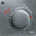
 Hamster fused oocyte and spermatozoa.jpg 888 × 405; 98 KB
 Hamster oocyte and spermatozoa.jpg 883 × 836; 266 KB
 Hilfer1990 Fig01.jpg 461 × 1,000; 28 KB
 Human fertilization movie 1 frame 01.jpg 600 × 409; 27 KB
 Human fertilization movie 1 frame 02.jpg 600 × 409; 27 KB
 Human fertilization movie 1 frame 03.jpg 600 × 409; 26 KB
 Human fertilization movie 1 frame 04.jpg 600 × 409; 24 KB
 Human fertilization movie 1 frame 05.jpg 600 × 409; 25 KB
 Human fertilization movie 1 frame 06.jpg 600 × 409; 25 KB
 Human fertilization movie 1 frame 07.jpg 600 × 409; 24 KB
 Human fertilization movie 1 frame 08.jpg 600 × 409; 25 KB
 Human fertilization movie 1 frame 09.jpg 600 × 409; 24 KB
 Human fertilization movie 1 frame 10.jpg 600 × 409; 25 KB
 Human sperm pathologies EM01.jpg 761 × 759; 148 KB
 Human sperm pathology EM02.jpg 800 × 256; 22 KB
 Human spermatid electron micrograph.jpg 619 × 918; 206 KB
 Human spermatid EM01.jpg 1,000 × 762; 162 KB
 Human spermatid EM02.jpg 1,000 × 762; 186 KB
 Human spermatozoa acrosomal protein SP-10.jpg 1,100 × 1,189; 239 KB
 Human spermatozoa chemotaxis labeled model.jpg 1,260 × 699; 158 KB
 Human spermatozoa chemotaxis model.jpg 1,260 × 699; 132 KB
 Human spermatozoa nucleus EM01.jpg 600 × 476; 27 KB
 Human spermatozoa nucleus EM02.jpg 597 × 476; 52 KB
 Human spermatozoa nucleus EM03.jpg 600 × 475; 44 KB
 Human spermatozoa phospholipase C zeta.jpg 1,000 × 571; 119 KB
 Human testis NANOG expression.jpg 1,000 × 328; 77 KB
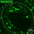 Human- spermatozoa NANOG expression 01.jpg 798 × 797; 79 KB
 Human- spermatozoa NANOG expression.jpg 1,000 × 333; 77 KB
 Human- vacuolated spermatozoa.jpg 1,000 × 801; 77 KB
 Human-spermatozoa 01.jpg 1,000 × 805; 90 KB
 Human-spermatozoa 01a.jpg 800 × 644; 66 KB
 Human-spermatozoa 01b.jpg 600 × 483; 43 KB
 Human-spermatozoa 01c.jpg 400 × 322; 22 KB
 Human-spermatozoa EM01.jpg 1,000 × 204; 26 KB
 Human-spermatozoa.jpg 600 × 581; 19 KB
 Keibel Mall 004.jpg 195 × 1,080; 42 KB
 Keith1921 fig011.jpg 1,200 × 614; 76 KB
 Kollmann006.jpg 494 × 680; 21 KB
 Kollmann007.jpg 956 × 582; 45 KB
 Kollmann008.jpg 641 × 577; 61 KB
 Kollmann009.jpg 572 × 552; 24 KB
 Kollmann010.jpg 550 × 541; 19 KB
 Kollmann011.jpg 550 × 541; 28 KB
 Kollmann013.jpg 732 × 626; 95 KB
 Kollmann458.jpg 1,000 × 520; 120 KB
 Kollmann459.jpg 1,000 × 385; 50 KB
 Kollmann460.jpg 551 × 569; 40 KB
 Meiotic prophase I stages.jpg 1,000 × 341; 66 KB
 Minot1897 fig001.jpg 293 × 644; 14 KB
 Minot1897 fig002.jpg 828 × 746; 162 KB
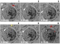
 Model capacitation-induced acrosome docking to sperm membrane.jpg 600 × 489; 73 KB
 Mouse oocyte fertilization 01.jpg 675 × 494; 58 KB
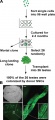
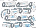 Mouse spermatogenesis stage cartoon.jpg 1,020 × 800; 298 KB
 Mouse spermatogonial self-renewal.jpg 500 × 794; 49 KB
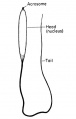
 Mouse spermatozoa cartoon.jpg 231 × 729; 26 KB
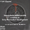
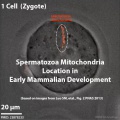 Mouse spermatozoa mito movie icon.jpg 495 × 495; 43 KB
 Mouse spermatozoa mitochondria 01.jpg 831 × 1,280; 141 KB
 Mouse spermiogenesis 01.jpg 1,200 × 299; 48 KB
 Mouse spermiogenesis model.png 600 × 571; 448 KB
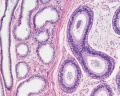
 Mouse- epididymis histology.jpg 751 × 383; 82 KB
 Mouse- gonadal supporting cell development.jpg 1,000 × 588; 74 KB
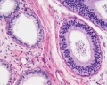
 Mouse- seminiferous tubule histology.jpg 715 × 427; 76 KB
 Mouse- spermatozoa EM and diagram.jpg 729 × 407; 49 KB
 Mouse- spermatozoa NANOG expression.jpg 800 × 512; 111 KB
 Mouse- zona pellucida 01.jpg 800 × 430; 83 KB
 Mouse- zona pellucida 02.jpg 700 × 688; 95 KB
 Mouse- zona pellucida 03.jpg 1,000 × 345; 64 KB
 Mouse-fertilization 01.jpg 600 × 593; 30 KB
 Mouse-fertilization 02.jpg 1,342 × 691; 118 KB
 Mouse-spermatozoa SLY protein.jpg 637 × 767; 352 KB
 Nelsen1953 fig022.jpg 1,200 × 839; 207 KB
 Pig sperm capacitation 01.jpg 1,000 × 840; 204 KB
 Pig sperm capacitation 02.jpg 600 × 504; 82 KB
 Rat- immortal germ cells are spermatogonial stem cells.jpg 459 × 1,000; 72 KB
 Rugh 019.jpg 383 × 600; 15 KB
 Seminiferous tubule cartoon.jpg 800 × 544; 92 KB
 Single human spermatozoa.jpg 1,000 × 780; 53 KB
 Spermatocyte prophase 1 stages 01.jpg 1,280 × 266; 81 KB
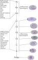
 Spermatogenesis androgen action cartoon.jpg 1,000 × 659; 125 KB
 Spermatogenesis cartoon 01.jpg 1,064 × 759; 142 KB
 Spermatozoa animation icon.jpg 300 × 200; 6 KB
 Spermatozoa animation.gif 300 × 200; 123 KB
 Spermatozoa animation.mov ; 113 KB
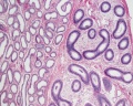
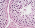
 Spermatozoa histology 001.jpg 1,280 × 1,024; 366 KB
 Spermatozoa histology 002.jpg 1,280 × 1,024; 246 KB
 Spermatozoa histology 003.jpg 1,280 × 1,024; 166 KB
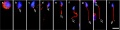
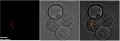
 Spermatozoa mitochondria 1cell.jpg 906 × 306; 33 KB
 Spermatozoa mitochondria 2cell.jpg 906 × 306; 34 KB
 Spermatozoa mitochondria 4cell.jpg 906 × 306; 42 KB
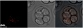 Spermatozoa mitochondria 8cell.jpg 906 × 306; 37 KB
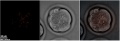 Spermatozoa mitochondria morula.jpg 906 × 306; 38 KB
 Spermatozoa mitochondria PMID23878233.gif 495 × 495; 974 KB
- Spermatozoa motility 01.mov ; 486 KB
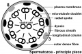 Spermatozoa principal piece.jpg 800 × 532; 74 KB
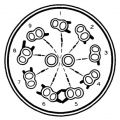 Spermatozoa tail cross-section cartoon.jpg 429 × 429; 43 KB
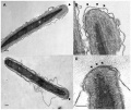
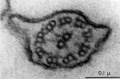 Spermatozoa tail EM01.jpg 932 × 613; 75 KB
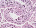
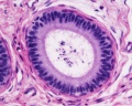
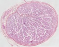 Testis histology 001.jpg 1,280 × 1,024; 574 KB
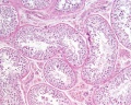
 Testis histology 002.jpg 1,280 × 1,024; 599 KB
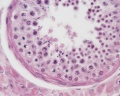
 Testis histology 003.jpg 1,280 × 1,024; 183 KB
 Testis histology 004.jpg 1,280 × 1,024; 396 KB
 Testis histology 005.jpg 1,280 × 1,024; 266 KB
 Testis histology 006.jpg 1,280 × 1,024; 251 KB
 Testis histology 007.jpg 1,280 × 1,024; 256 KB
 Testis histology 008.jpg 1,280 × 1,024; 454 KB
 Testis histology 009.jpg 1,280 × 1,024; 339 KB
 Testis histology 010.jpg 1,280 × 1,024; 422 KB
 Testis histology 011.jpg 1,280 × 1,024; 245 KB
 Testis histology 012.jpg 1,280 × 1,024; 266 KB
 Testis histology 013.jpg 1,280 × 1,024; 418 KB
 Testis histology 014.jpg 1,280 × 1,024; 352 KB
 Testis histology 015.jpg 1,280 × 1,024; 281 KB
 Testis histology 016.jpg 1,280 × 1,024; 322 KB
 Testis histology 017.jpg 1,280 × 1,024; 283 KB
 Testis histology 018.jpg 1,280 × 1,024; 350 KB
 Testis histology 019.jpg 1,280 × 1,024; 239 KB
 Testis histology 02.jpg 246 × 481; 49 KB
 Testis histology 023.jpg 600 × 375; 35 KB
 Testis histology 1.jpg 400 × 500; 113 KB
 Testis histology 2.jpg 400 × 500; 32 KB
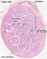 Testis histology.jpg 400 × 500; 54 KB
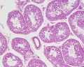 Testis, young H&E reproductive system, male, convoluted seminiferous tubules x10.jpg 1,280 × 1,024; 396 KB
 Y chromosome haplogroup distribution.jpg 800 × 522; 42 KB


