Category:Limb: Difference between revisions
From Embryology
mNo edit summary |
mNo edit summary |
||
| (2 intermediate revisions by the same user not shown) | |||
| Line 1: | Line 1: | ||
This page lists {{Embryology}} content related to limb development and abnormalities. The limb is also an excellent model for how tissue pattern is established. | This page lists {{Embryology}} content related to limb development and limb abnormalities. The limb is also an excellent model for how tissue pattern is established. | ||
:'''Links:''' [[Musculoskeletal System - Limb Development|Limb Development]] | [[Book_-_Manual_of_Human_Embryology_11D| | |||
:'''Links:''' [[Musculoskeletal System - Limb Development|Limb Development]] | [[Musculoskeletal System - Limb Abnormalities|Limb Abnormalities]] | [[Musculoskeletal System - Appendicular Skeleton Development|Appendicular Skeleton]] | [[Book_-_Manual_of_Human_Embryology_11D|1910 - Limb Development]] | |||
<br> | |||
{{Musculoskeletal Links}} | |||
Revision as of 14:05, 3 October 2017
This page lists Embryology content related to limb development and limb abnormalities. The limb is also an excellent model for how tissue pattern is established.
Pages in category 'Limb'
The following 148 pages are in this category, out of 148 total.
B
- Template:Bardeen1906 figures
- BGDA Practical 7 - Week 6
- Book - An Atlas of Topographical Anatomy 19
- Book - An Atlas of Topographical Anatomy 20
- Book - An Atlas of Topographical Anatomy 21
- Book - An Atlas of Topographical Anatomy 22
- Book - An Atlas of Topographical Anatomy 23
- Book - An Atlas of Topographical Anatomy 24
- Book - An Atlas of Topographical Anatomy 26
- Book - An Atlas of Topographical Anatomy 27
- Book - An Atlas of Topographical Anatomy 28
- Book - Human Embryology and Morphology 20
- Book - Manual of Human Embryology 11D
- Book - Manual of Human Embryology 12
C
D
L
- Lecture - Limb Development
- Template:Limb
- Template:Limb abnormalities
- Template:Limb axis
- Template:Limb Links
- Template:Limb Rotation table
- Template:Limb skeleton mesenchyme-cartilage-bone table
- Template:Lower limb ossification collapse table
- Template:Lower limb ossification collapsible table
- Template:Lower limb ossification table
- Template:Lower limb ossification timeline
M
P
- Paper - A contribution to the embryology of the fore-limb
- Paper - A study of the development of the mammalian pelvis
- Paper - An unusual form of brachyphalangy and syndactyly with double proximal phalanx in the middle fingers (1932)
- Paper - Anatomy of a seven months foetus exhibiting bilateral absence of the ulna accompanied by monodactyly
- Paper - Chondrification in the hands and feet of staged human embryos
- Paper - Development and variation of the nerves and the musculature of the inferior extremity and of the neighboring regions of the trunk in man
- Paper - Development of the innervation pattern in the upper limb of staged human embryos (1990)
- Paper - Extra digits and digital reductions
- Paper - Familial abnormalities of the middle phalanges of each hand (1923)
- Paper - On an instance of two subclavian arteries of the early arm bud of man and its fundamental significance
- Paper - Rare congenital malformation of hands and feet (1924)
- Paper - Studies of the development of the human skeleton (1905)
- Paper - The arrangement of the bursae in the superior extremities of the full-time foetus
- Paper - The development and evolution of the "papillary" ridges and patterns on the volar surface of the hand (1906)
- Paper - The development of joints
- Paper - The development of the arm in man (1902)
- Paper - The development of the arteries of the human lower extremity
- Paper - The development of the limbs, body-wall and back
- Paper - The development of the limbs, body-wall and back (1901)
- Paper - The development of the patella
- Paper - The development of the principal arterial stems in the forelimb of the pig (1922)
- Paper - The development of the veins in the limbs of rabbit embryos
- Paper - The early development of the knee joint in staged human embryos
- Paper - The embryology of the human hip joint (1943)
- Paper - The epiphysis of the head of the femur (1915)
- Paper - The growth of the human foot
- Paper - The proximo-distal sequence of origin of the parts of the chick wing and the role of the ectoderm
- Paper - The sexual differences of the fetal pelvis
- Paper - The sexual differences of the fetal pelvis (1899)
- Template:Pelvis
- Template:Pelvis Timeline table
R
- Template:Ref-Bardeen1907
- Template:Ref-BardeenLewis1901
- Template:Ref-Brailsford1943
- Template:Ref-Carey1922
- Template:Ref-Cockayne1932
- Template:Ref-Cummins1929
- Template:Ref-Davenport1932
- Template:Ref-deLima1928
- Template:Ref-Evans1908
- Template:Ref-Evatt1906
- Template:Ref-GardnerO'Rahilly1968
- Template:Ref-Geddes1912b
- Template:Ref-GrayGardener1950
- Template:Ref-Haines1947
- Template:Ref-Haines1953
- Template:Ref-Hann1923
- Template:Ref-Harrison1915
- Template:Ref-Johnson1899
- Template:Ref-Keith1910
- Template:Ref-Lewis1902
- Template:Ref-Lewis1905b
- Template:Ref-Lewis1910a
- Template:Ref-Manson1924
- Template:Ref-O'Rahilly1957
- Template:Ref-PMID55906
- Template:Ref-Prentiss1906
- Template:Ref-Rutherford1914
- Template:Ref-Saunders1947a
- Template:Ref-Saunders1948
- Template:Ref-ShinoharaTanaka1990
- Template:Ref-Stacey1938
- Template:Ref-Walmsley1915
- Template:Ref-Walmsley1940
- Template:Ref-Watt1917
- Template:Ref-Whittaker1910
- Template:Ref-Wigoder1928
- Template:Ref-Woollard1922
S
U
V
Media in category 'Limb'
The following 200 files are in this category, out of 274 total.
(previous page) (next page)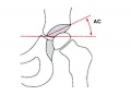 Acetabular angle.jpg 600 × 433; 22 KB
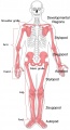 Appendicular skeleton developmental regions.jpg 1,000 × 1,849; 143 KB
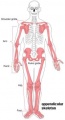 Appendicular skeleton small.jpg 220 × 407; 12 KB
 Appendicular skeleton.jpg 1,000 × 1,849; 125 KB
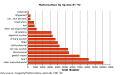 Australian abnormalities graph allsystem.png 509 × 320; 7 KB
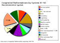 Australian abnormalities pie skmus.png 481 × 344; 9 KB
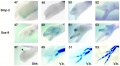 Axolotl developing limb Bmp2 and Sox9.jpg 1,159 × 638; 105 KB
 Bailey143.jpg 911 × 673; 114 KB
 Bailey144.jpg 491 × 398; 39 KB
 Bailey145.jpg 777 × 654; 80 KB
 Bailey149.jpg 576 × 520; 38 KB
 Bailey150.jpg 406 × 596; 42 KB
 Bailey189.jpg 810 × 632; 63 KB
 Bailey190.jpg 801 × 584; 69 KB
 Bailey204.jpg 534 × 653; 62 KB
 Bailey205.jpg 534 × 653; 68 KB
 Bardeen1905 plate13.jpg 1,000 × 1,337; 85 KB
 Bardeen1906-fig02.jpg 1,598 × 1,183; 228 KB
 Bardeen1906-fig03.jpg 1,598 × 1,166; 231 KB
 Bardeen1906-plate01.jpg 1,565 × 2,322; 238 KB
 Bardeen1906-plate02.jpg 1,719 × 2,302; 512 KB
 Bardeen1906-plate06.jpg 1,568 × 2,299; 379 KB
 Bardeen1906-plate31.jpg 1,571 × 2,330; 257 KB
 Bardeen1906-plate32.jpg 1,588 × 2,341; 292 KB
 Bardeen1906-plate41.jpg 1,555 × 2,323; 261 KB
 Bardeen1906-plate42.jpg 1,570 × 2,331; 240 KB
 Bardeen1906-plate51.jpg 1,570 × 2,330; 392 KB
 Bardeen1906-plate52.jpg 1,596 × 2,350; 404 KB
 Bat - adult and fetal limbs.jpg 600 × 612; 80 KB
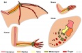
 Bat and mouse limb comparison.jpg 1,000 × 591; 77 KB
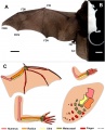
 Bat limb 01.jpg 1,688 × 699; 149 KB
 Bat limb 02.jpg 1,200 × 430; 59 KB
 Bone-femur.jpg 798 × 1,000; 150 KB
 Cat 6 toes.jpg 420 × 280; 13 KB
 Chicken limb BMP modulator expression 01.jpg 1,200 × 1,224; 211 KB
 Chicken limb gene expression 01.jpg 1,200 × 758; 85 KB
 Chicken limb gene expression 02.jpg 1,000 × 478; 42 KB
 Chicken limb gene expression 03.jpg 1,200 × 711; 108 KB
 Chicken- limb bud chondrogenesis.jpg 600 × 1,032; 163 KB
 Chicken- wing cartilage.jpg 1,200 × 1,022; 218 KB
 Chicken-limb sox9 wnt6.jpg 1,173 × 373; 68 KB
 Clefthand-apical-defect.jpg 500 × 568; 19 KB
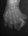
 Congenital limb reduction xray.jpg 793 × 600; 18 KB
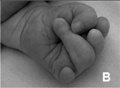
 Congenital limb reduction.jpg 400 × 289; 11 KB
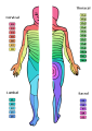 Dermatomes.png 424 × 600; 82 KB
 Ectrodactyly 01.jpg 1,200 × 619; 113 KB
 Ectrodactyly.jpg 500 × 244; 9 KB
 Embryonic upper limb - brachial and superficial brachial artery.jpg 1,280 × 314; 122 KB
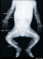
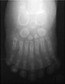
 Fetal limb abnormalities X-ray-02.jpg 685 × 931; 62 KB
 Fetal limb abnormalities X-ray-03.jpg 685 × 931; 57 KB
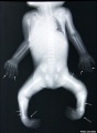
 Fetal limb X-ray-01.jpg 685 × 931; 57 KB
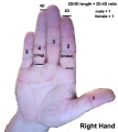 Finger length ratio - 2D4D.jpg 428 × 480; 26 KB
 Gray0595.jpg 600 × 491; 94 KB
 Gray0606.jpg 686 × 800; 93 KB
 Gray0608.jpg 400 × 623; 74 KB
 Gray0609.jpg 527 × 500; 69 KB
 Gray0610.jpg 303 × 1,000; 81 KB
 Gray0807.gif 587 × 500; 44 KB
 Gray0807.jpg 704 × 600; 97 KB
 Gray0822.jpg 599 × 600; 69 KB
 Gray1235.jpg 800 × 389; 41 KB
 Gray1236.jpg 800 × 281; 29 KB
 Gray1237.jpg 371 × 600; 57 KB
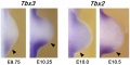
 Hindlimb Tbx2 model.jpg 1,000 × 607; 84 KB
 Hoxa gene expression in limb bud 01.jpg 1,376 × 533; 85 KB
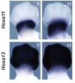 Hoxa gene expression in limb bud 02.jpg 546 × 600; 40 KB
 Human embryo femur CS18 to CS23.png 1,200 × 1,624; 1.42 MB
 Human embryonic shoulder girdle 01.jpg 1,000 × 726; 81 KB
 Human embryonic shoulder girdle 02.jpg 1,025 × 713; 109 KB
 Human embryonic shoulder girdle 04.jpg 1,000 × 755; 71 KB
 Human upper limb mri-01.jpg 1,409 × 936; 108 KB
 Keibel Mall 077.jpg 424 × 700; 33 KB
 Keibel Mall 078.jpg 278 × 236; 18 KB
 Keibel Mall 2 451.jpg 1,000 × 714; 73 KB
 Keibel Mall 215.jpg 727 × 379; 53 KB
 Keibel Mall 216.jpg 366 × 356; 30 KB
 Keibel Mall 274-278.jpg 717 × 1,072; 123 KB
 Keibel Mall 279-284.jpg 709 × 707; 84 KB
 Keibel Mall 285-288.jpg 703 × 674; 72 KB
 Keibel Mall 289.jpg 682 × 645; 85 KB
 Keibel Mall 290.jpg 800 × 452; 59 KB
 Keibel Mall 291.jpg 800 × 421; 54 KB
 Keibel Mall 292.jpg 400 × 439; 30 KB
 Keibel Mall 293.jpg 687 × 857; 137 KB
 Keibel Mall 301.jpg 734 × 945; 94 KB
 Keibel Mall 302.jpg 300 × 233; 18 KB
 Keibel Mall 307.jpg 800 × 803; 112 KB
 Keibel Mall 346.jpg 685 × 670; 71 KB
 Keibel Mall 347.jpg 700 × 706; 68 KB
 Keibel Mall 348.jpg 635 × 642; 72 KB
 Keibel Mall 349.jpg 661 × 692; 69 KB
 Keibel Mall 352.jpg 611 × 558; 52 KB
 Keibel Mall 353.jpg 649 × 489; 44 KB
 Keibel Mall 354.jpg 800 × 459; 55 KB
 Keibel Mall 355.jpg 767 × 800; 66 KB
 Keibel Mall 356.jpg 775 × 516; 50 KB
 Keibel Mall 357.jpg 870 × 535; 60 KB
 Keibel Mall 358.jpg 700 × 693; 49 KB
 Keibel Mall 359.jpg 654 × 460; 46 KB
 Keibel Mall 360.jpg 765 × 490; 55 KB
 Keibel Mall 361.jpg 673 × 468; 37 KB
 Keibel Mall 362.jpg 801 × 516; 57 KB
 Keibel Mall 363.jpg 605 × 407; 34 KB
 Keibel Mall 364.jpg 594 × 378; 35 KB
 Keibel Mall 365.jpg 747 × 550; 59 KB
 Keibel Mall 366.jpg 780 × 535; 60 KB
 Keith1902 fig233.jpg 933 × 800; 122 KB
 Keith1902 fig234.jpg 1,200 × 387; 84 KB
 Keith1902 fig235.jpg 1,200 × 329; 74 KB
 Keith1902 fig236.jpg 1,200 × 652; 109 KB
 Keith1902 fig237.jpg 896 × 800; 111 KB
 Keith1902 fig238.jpg 1,000 × 752; 166 KB
 Keith1902 fig239.jpg 595 × 800; 58 KB
 Keith1902 fig240.jpg 1,000 × 491; 55 KB
 Keith1902 fig241.jpg 458 × 800; 50 KB
 Keith1902 fig242.jpg 1,000 × 458; 75 KB
 Keith1902 fig243.jpg 1,000 × 631; 98 KB
 Keith1902 fig244.jpg 1,000 × 503; 100 KB
 Keith1902 fig245.jpg 1,000 × 614; 88 KB
 Keith1902 fig246.jpg 943 × 600; 98 KB
 Keith1902 fig247.jpg 1,000 × 488; 68 KB
 Keith1902 fig248.jpg 800 × 480; 41 KB
 Keith1902 fig249.jpg 1,000 × 713; 108 KB
 Keith1902 fig250.jpg 1,000 × 457; 81 KB
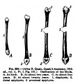
 Keith1902 fig251.jpg 600 × 446; 31 KB
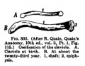
 Keith1902 fig252.jpg 600 × 510; 39 KB
 Left foot polydactyly 01.jpg 481 × 600; 27 KB
 Left foot polydactyly 02.jpg 307 × 396; 10 KB
 Left hand reduction defect 01.jpg 400 × 293; 14 KB
 Left hand reduction defect 02.jpg 396 × 509; 12 KB
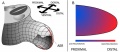
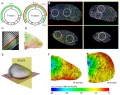 Limb 3D map of cell proliferation rates.jpg 1,000 × 794; 242 KB
 Limb bud geometry and patterning.jpg 583 × 765; 76 KB
 Limb bud growth model 01.jpg 600 × 261; 30 KB
 Limb bud growth model 02.jpg 600 × 637; 80 KB
 Limb changing 3D geometry.jpg 1,000 × 1,062; 196 KB
 Limb comparison cartoon 01.jpg 1,780 × 2,203; 379 KB
 Limb comparison cartoon 02.jpg 1,200 × 778; 119 KB
 Limb hairy2 expression model.jpg 1,280 × 523; 76 KB
 Limb induction-initiation signal 01.jpg 664 × 664; 204 KB
 Limb induction-initiation signal 02.jpg 1,280 × 1,009; 124 KB
 Limb mesenchymal cell shape.jpg 775 × 1,000; 180 KB
 Limb patterning factors 01.jpg 800 × 794; 76 KB
 Limb patterning factors 02.jpg 800 × 794; 73 KB
 Limb patterning factors 03.jpg 800 × 794; 36 KB
 Limb patterning factors 04.jpg 800 × 794; 67 KB
 Limb patterning factors 05.jpg 800 × 794; 37 KB
 Limb patterning factors 06.jpg 800 × 794; 41 KB
 Limb patterning factors 07.jpg 800 × 794; 44 KB
 Limb patterning factors 08.jpg 1,200 × 576; 79 KB
 Limb patterning factors 09.jpg 1,200 × 601; 82 KB
 Limb patterning factors 11.jpg 1,200 × 655; 78 KB
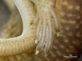 Lizard embryo 08.jpg 1,200 × 900; 177 KB
 Malformation1.jpg 354 × 570; 40 KB
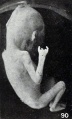 Mall Meyer1921 fig90.jpg 479 × 791; 64 KB
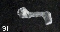 Mall Meyer1921 fig91.jpg 352 × 191; 14 KB
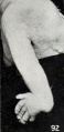 Mall Meyer1921 fig92.jpg 359 × 734; 46 KB
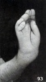 Mall Meyer1921 fig93.jpg 413 × 734; 42 KB
 Mall1906 fig05.jpg 561 × 1,055; 45 KB
 Mall1906 fig06.jpg 522 × 966; 41 KB
 Mall1906 table06.jpg 2,117 × 1,074; 469 KB
 Mall1906 table07.jpg 2,118 × 1,034; 432 KB
 McMurrich1930 fig88.jpg 1,280 × 1,509; 218 KB
 McMurrich1930 fig89.jpg 1,280 × 681; 98 KB
 Model digit origin.png 599 × 505; 128 KB
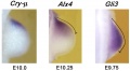
 Model GATA6 hindlimb.jpg 737 × 600; 60 KB
 Morgan 1925 fig65.jpg 1,175 × 766; 189 KB
 Morgan 1925 fig66.jpg 649 × 969; 108 KB
 Morgan 1925 fig68.jpg 1,000 × 744; 188 KB
 Mouse Bmp4 expression limb and face 01.jpg 1,200 × 513; 91 KB
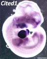 Mouse Cited1.jpg 446 × 550; 37 KB
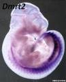 Mouse Dmrt2.jpg 446 × 550; 35 KB
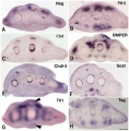
 Mouse E10.5 gene expression.jpg 1,747 × 1,650; 404 KB
 Mouse E10.5 hindlimb gene expression.jpg 1,000 × 336; 49 KB
 Mouse E11.5 gene expression.jpg 1,760 × 1,650; 396 KB
 Mouse E12.5 gene expression.jpg 1,823 × 1,100; 272 KB
 Mouse E13.5 gene expression.jpg 1,764 × 1,100; 266 KB
 Mouse E9.5 gene expression.jpg 1,757 × 1,100; 269 KB
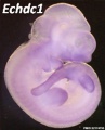 Mouse Echdc1.jpg 446 × 550; 29 KB
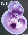 Mouse Egr1.jpg 446 × 550; 34 KB
 Mouse embryo cortex radial expansion.jpg 1,200 × 624; 249 KB
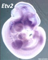 Mouse Etv2.jpg 446 × 550; 33 KB
 Mouse Fbxo41.jpg 446 × 550; 24 KB
 Mouse Figf.jpg 446 × 550; 25 KB
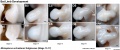
 Mouse forelimb cartilage and bone E14.5 E18.5.jpg 1,000 × 1,300; 156 KB
 Mouse forelimb cartilage and bone E18.5.jpg 774 × 600; 70 KB
 Mouse forelimb E10.5 to E11.5.jpg 1,001 × 2,000; 331 KB
 Mouse forelimb gene expression.jpg 2,205 × 1,650; 456 KB
 Mouse forelimb.jpg 600 × 812; 71 KB
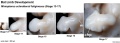
 Mouse hindlimb cartilage and bone E18.5.jpg 774 × 600; 75 KB
 Mouse hindlimb gene expression.jpg 2,237 × 550; 147 KB
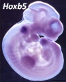 Mouse Hoxb5.jpg 446 × 550; 36 KB
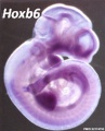 Mouse Hoxb6.jpg 446 × 550; 35 KB
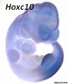 Mouse Hoxc10.jpg 446 × 550; 23 KB
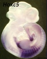 Mouse Hoxc5.jpg 446 × 550; 30 KB
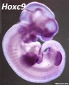 Mouse Hoxc9.jpg 446 × 550; 34 KB
 Mouse interdigit apoptosis 01.jpg 800 × 800; 81 KB
 Mouse interdigit apoptosis 02.jpg 764 × 764; 61 KB
 Mouse limb bone development timeline.jpg 1,256 × 469; 107 KB
 Mouse limb cartilage and bone E14.5.jpg 800 × 526; 42 KB
 Mouse limb cartilage and bone E14.5L.jpg 1,000 × 658; 73 KB


