Book - Text-Book of Embryology (1921) - Figures: Difference between revisions
mNo edit summary |
|||
| (48 intermediate revisions by 3 users not shown) | |||
| Line 1: | Line 1: | ||
{{Template:Bailey 1921}} | {{Template:Bailey 1921}} | ||
[[File:Bailey_and_Miller_1921.jpg|right|250px]] | |||
==The germ cells== | ==The germ cells== | ||
| Line 40: | Line 40: | ||
File:Bailey017.jpg|Fig. 17. Polyspermy in sea-urchin eggs treated with 0.005 per cent, nicotine solution | File:Bailey017.jpg|Fig. 17. Polyspermy in sea-urchin eggs treated with 0.005 per cent, nicotine solution | ||
</gallery> | </gallery> | ||
:'''Links:''' [[Fertilization]] | [[:Category:Fertilization|Category:Fertilization]] | |||
==Early development of amphioxus== | ==Early development of amphioxus== | ||
| Line 61: | Line 64: | ||
<gallery> | <gallery> | ||
File:Bailey026.jpg | File:Bailey026.jpg|Fig. 26. Section through the fully formed ovarian egg of a frog. | ||
File:Bailey027.jpg | File:Bailey027.jpg|Fig. 27. A frog's egg before and after fertilization, showing the formation of the gray crescent. | ||
File:Bailey028.jpg | File:Bailey028.jpg|Fig. 28. Cleavage of the frog's egg. | ||
File:Bailey029.jpg | File:Bailey029.jpg|Fig. 29. From a sagittal section through blastula of frog. | ||
File:Bailey030.jpg | File:Bailey030.jpg|Fig. 30. Diagrams showing the position of the blastopore at successive stages of gastrulation in the frog's egg. | ||
File:Bailey031.jpg | File:Bailey031.jpg|Fig. 31. Median sagittal sections showing successive stages of gastrulation in the frog's egg | ||
File:Bailey032.jpg | File:Bailey032.jpg|Fig. 32. Transverse section of embryo of frog (Rana fusca). | ||
File:Bailey033.jpg | File:Bailey033.jpg|Fig. 33. Transverse section through embryo of frog (Rana fusca). | ||
File:Bailey034.jpg | File:Bailey034.jpg|Fig. 34. Portion of a transverse section still continuous at the lower lateral angles of the larva of a frog (Rana fusca) | ||
File:Bailey035.jpg | File:Bailey035.jpg|Fig. 35. Diagrams of median sagittal sections through an eight-cell stage and four stages during gastrulation of the frog's egg. | ||
File:Bailey036.jpg | File:Bailey036.jpg|Fig. 36. Postero-lateral views of successive stages following gastrulation in the frog. | ||
File:Bailey037.jpg|Fig. 37. Median sagittal sections of frog larvae. | File:Bailey037.jpg|Fig. 37. Median sagittal sections of frog larvae. | ||
</gallery> | </gallery> | ||
| Line 80: | Line 83: | ||
<gallery> | <gallery> | ||
File:Bailey038.jpg|Fig. 38 | File:Bailey038.jpg|Fig. 38. Cleavage in hen's egg | ||
File:Bailey039.jpg|Fig. 39 | File:Bailey039.jpg|Fig. 39. Vertical section germ disk of a fresh-laid hen's egg. | ||
File:Bailey040.jpg|Fig. 40 | File:Bailey040.jpg|Fig. 40. Cross section blastoderm of a pigeon 14.5 hours after fertilization. | ||
File:Bailey041.jpg|Fig. 41 | File:Bailey041.jpg|Fig. 41. Median longitudinal section blastoderm of a pigeon 31 hours after fertilization. | ||
File:Bailey042.jpg|Fig. 42 | File:Bailey042.jpg|Fig. 42. Median longitudinal section blastoderm of a pigeon 36 hours after fertilization. | ||
File:Bailey043.jpg|Fig. 43 | File:Bailey043.jpg|Fig. 43. Surface views of blastoderms of the pigeon. | ||
File:Bailey044.jpg|Fig. 44 | File:Bailey044.jpg|Fig. 44. Surface views of blastoderms of Haliplana primitive streak. | ||
File:Bailey045.jpg|Fig. | File:Bailey045.jpg|Fig. 45. Surface view of embryonic disk of chick. | ||
File:Bailey046.jpg|Fig. 46 | File:Bailey046.jpg|Fig. 46. Surface view of chick blastoderm. | ||
File:Bailey047.jpg|Fig. 47 | File:Bailey047.jpg|Fig. 47. Transverse sections of blastoderm of chick 21 hours. | ||
File:Bailey048.jpg|Fig. 48 | File:Bailey048.jpg|Fig. 48. Transverse section of blastoderm of chick 21 hours. | ||
File:Bailey049.jpg|Fig. | File:Bailey049.jpg|Fig. 49. Median longitudinal section blastoderm of chick after the primitive axis | ||
File:Bailey050.jpg|Fig. 50 | File:Bailey050.jpg|Fig. 50. Transverse section of blastoderm of chick 40 hours. | ||
File:Bailey051.jpg|Fig. 51 | File:Bailey051.jpg|Fig. 51. Dorsal view of chick embryo with ten pairs of mesodermal somites. | ||
File:Bailey052.jpg|Fig. 52 | File:Bailey052.jpg|Fig. 52. Transverse section of chick embryo 2 days incubation. | ||
</gallery> | </gallery> | ||
:'''Links:''' [[Chicken Development]] | |||
==Early mammalian development== | ==Early mammalian development== | ||
| Line 102: | Line 108: | ||
<gallery> | <gallery> | ||
File:Bailey053.jpg|Fig. 53 | File:Bailey053.jpg|Fig. 53. Four stages in the cleavage of the ovum of the white rat. | ||
File:Bailey054.jpg|Fig. 54 | File:Bailey054.jpg|Fig. 54. Four stages in cleavage of the ovum of the mouse. | ||
File:Bailey055.jpg|Fig. 55 | File:Bailey055.jpg|Fig. 55. Four stages in the development of the bat. | ||
File:Bailey056.jpg|Fig. 56 | File:Bailey056.jpg|Fig. 56. Sections of blastocysts of the white rat 5 days. | ||
File:Bailey057.jpg|Fig. 57 | File:Bailey057.jpg|Fig. 57. Section of a 16-cell stage of an ovum of the opossum. | ||
File:Bailey058.jpg|Fig. 58 | File:Bailey058.jpg|Fig. 58. Section of the blastocyst of the lemur Tarsius spectrum. | ||
File:Bailey059.jpg|Fig. 59 | File:Bailey059.jpg|Fig. 59. Sections of blastodermic vesicle of bat. | ||
File:Bailey060.jpg|Fig. 60 | File:Bailey060.jpg|Fig. 60. Three stages in the formation of the germ layers in the lemur Tarsius spectrum. | ||
File:Bailey061.jpg|Fig. 61 | File:Bailey061.jpg|Fig. 61 | ||
File:Bailey062.jpg|Fig. 62 | File:Bailey062.jpg|Fig. 62 | ||
| Line 139: | Line 145: | ||
<gallery> | <gallery> | ||
File:Bailey083.jpg|Fig. 83 | File:Bailey083.jpg|Fig. 83. Human embryo with 8 pairs of mesodermal somites | ||
File:Bailey084.jpg|Fig. 84. Human embryo with 14 pairs of mesodermal somites. | |||
File:Bailey084.jpg|Fig. 84 | File:Bailey085.jpg|Fig. 85. Human embryo of 2.6 mm. | ||
File:Bailey085.jpg|Fig. 85 | File:Bailey086.jpg|Fig. 86. Human embryo of 4 mm. | ||
File:Bailey086.jpg|Fig. 86 | File:Bailey087.jpg|Fig. 87. Human embryo 27 primitive segments. | ||
File:Bailey087.jpg|Fig. 87 | File:Bailey088.jpg|Fig. 88. Human embryo with 28 primitive segments. | ||
File:Bailey088.jpg|Fig. 88 | File:Bailey089.jpg|Fig. 89. Human embryo 11 mm. | ||
File:Bailey089.jpg|Fig. 89 | File:Bailey090.jpg|Fig. 90. Human embryo of 15.5 mm. | ||
File:Bailey090.jpg|Fig. 90 | File:Bailey091.jpg|Fig. 91. Human embryo of 17.5 mm (47-51 days). | ||
File:Bailey091.jpg|Fig. 91 | File:Bailey092.jpg|Fig. 92. Human embryo of 18.5 mm (52-54 days). | ||
File:Bailey092.jpg|Fig. 92 | File:Bailey093.jpg| Fig. 93. Human embryo of 23 mm (2 months). | ||
File:Bailey093.jpg|Fig. 93 | File:Bailey094.jpg|Fig. 94. Human embryo of 78 mm (3 months). | ||
File:Bailey094.jpg|Fig. 94 | File:Bailey095.jpg|Fig. 95. Human embryo of 4 months. | ||
File:Bailey095.jpg|Fig. 95 | File:Bailey096.jpg|Fig. 96. Ventral view of head of 8 mm human embryo. | ||
File:Bailey096.jpg|Fig. 96 | File:Bailey097.jpg|Fig. 97. Ventral view of head of 11.3 mm human embryo. | ||
File:Bailey097.jpg|Fig. 97 | File:Bailey098.jpg|Fig. 98. Ventral view of head of 13.7 mm human embryo. | ||
File:Bailey098.jpg|Fig. 98 | File:Bailey099.jpg|Fig. 99. Ventral view of head of human embryo of 8 weeks. | ||
File:Bailey099.jpg|Fig. 99 | |||
</gallery> | </gallery> | ||
| Line 276: | Line 281: | ||
File:Bailey202.jpg|Fig. 202 | File:Bailey202.jpg|Fig. 202 | ||
File:Bailey203.jpg|Fig. 203 | File:Bailey203.jpg|Fig. 203 | ||
File:Bailey204.jpg|Fig. 204 | File:Bailey204.jpg|Fig. 204 Fig. 205 | ||
File:Bailey206.jpg|Fig. 206 | File:Bailey206.jpg|Fig. 206 | ||
File:Bailey207.jpg|Fig. 207 | File:Bailey207.jpg|Fig. 207 | ||
| Line 298: | Line 302: | ||
File:Baileytable02.jpg|Table 2 | File:Baileytable02.jpg|Table 2 | ||
</gallery> | </gallery> | ||
:'''Links:''' [[Cardiovascular System Development]] | |||
==The muscular system== | ==The muscular system== | ||
| Line 316: | Line 323: | ||
<gallery> | <gallery> | ||
File:Bailey228.jpg|Fig. 228 | |||
File:Bailey229.jpg|Fig. 229 | |||
File:Bailey230.jpg|Fig. 230 | |||
File:Bailey231.jpg|Fig. 231 | |||
File:Bailey232.jpg|Fig. 232 | |||
File:Bailey233.jpg|Fig. 233 | |||
File:Bailey234.jpg|Fig. 234 | |||
File:Bailey235.jpg|Fig. 235 | |||
File:Bailey236.jpg|Fig. 236 | |||
File:Bailey237.jpg|Fig. 237 | |||
File:Bailey238.jpg|Fig. 238 | |||
File:Bailey239.jpg|Fig. 239 | |||
File:Bailey240.jpg|Fig. 240 | |||
File:Bailey241.jpg|Fig. 241 | |||
File:Bailey242.jpg|Fig. 242 | |||
File:Bailey243.jpg|Fig. 243 | |||
File:Bailey244.jpg|Fig. 244 | |||
File:Bailey245.jpg|Fig. 245 | |||
File:Bailey246.jpg|Fig. 246 | |||
File:Bailey247.jpg|Fig. 247 | |||
File:Bailey248.jpg|Fig. 248 | |||
File:Bailey249.jpg|Fig. 249 | |||
File:Bailey250.jpg|Fig. 250 | |||
File:Bailey251.jpg|Fig. 251 | |||
File:Bailey252.jpg|Fig. 252 | |||
File:Bailey253.jpg|Fig. 253 | |||
File:Bailey254.jpg|Fig. 254 | |||
File:Bailey255.jpg|Fig. 255 | |||
File:Bailey256.jpg|Fig. 256 | |||
File:Bailey257.jpg|Fig. 257 | |||
File:Bailey258.jpg|Fig. 258 | |||
File:Bailey259.jpg|Fig. 259 | |||
File:Bailey260.jpg|Fig. 260 | |||
File:Bailey261.jpg|Fig. 261 | |||
File:Bailey262.jpg|Fig. 262 | |||
File:Bailey263.jpg|Fig. 263 | |||
File:Bailey264.jpg|Fig. 264 | |||
File:Bailey265.jpg|Fig. 265 | |||
File:Bailey266.jpg|Fig. 266 | |||
File:Bailey267.jpg|Fig. 267 | |||
File:Bailey268.jpg|Fig. 268 | |||
File:Bailey269.jpg|Fig. 269 | |||
File:Bailey270.jpg|Fig. 270 | |||
File:Bailey271.jpg|Fig. 271 | |||
File:Bailey272.jpg|Fig. 272 | |||
File:Bailey273.jpg|Fig. 273 | |||
File:Bailey274.jpg|Fig. 274 | |||
File:Bailey275.jpg|Fig. 275 | |||
File:Bailey276.jpg|Fig. 276 | |||
File:Bailey277.jpg|Fig. 277 | File:Bailey277.jpg|Fig. 277 | ||
File:Bailey278_279.jpg|Fig. 278 279 | File:Bailey278_279.jpg|Fig. 278 279 | ||
| Line 325: | Line 381: | ||
[[Book_-_Text-Book_of_Embryology_13|The development of the respiratory system]] | [[Book_-_Text-Book_of_Embryology_13|The development of the respiratory system]] | ||
<gallery> | |||
File:Bailey282.jpg|Fig. 282 | |||
File:Bailey283.jpg|Fig. 283 | |||
File:Bailey284.jpg|Fig. 284 | |||
File:Bailey285.jpg|Fig. 285 | |||
File:Bailey286.jpg|Fig. 286. Transverse section of a 14 mm. pig embryo, at the level of the upper limb buds, showing especially the two bronchi | |||
File:Bailey287.jpg|Fig. 287. Anlage of lungs of a human embryo of 4.3 mm. His. | |||
File:Bailey288.jpg|Fig. 288. Anlage of lungs of a human embryo of 8.5 mm. His. | |||
File:Bailey289.jpg|Fig. 289. Anlage of lungs of a human embryo of 10.5 mm. His. | |||
File:Bailey290.jpg|Fig. 290. Transverse section of a pig embryo of 35 mm, showing the developing lungs (bronchial rami surrounded by mesoderm). | |||
File:Bailey291.jpg|Fig. 291. Transverse sections of a rabbit embryo Showing how the omphalomesenteric veins (vom) push outward across the ccelom and fuse with the lateral body wall. | |||
File:Bailey292.jpg|Fig. 292 | |||
File:Bailey293.jpg|Fig. 293 | |||
File:Bailey294.jpg|Fig. 294 | |||
File:Bailey295.jpg|Fig. 295 | |||
File:Bailey296_297.jpg|Fig. 296-297 | |||
File:Bailey298.jpg|Fig. 298 | |||
</gallery> | |||
==The coelom, pericardium, pleuroperitoneum, diaphragm and mesenteries== | ==The coelom, pericardium, pleuroperitoneum, diaphragm and mesenteries== | ||
[[Book_-_Text-Book_of_Embryology_14|The development of the coelom, the pericardium, pleuroperitoneum, diaphragm and mesenteries]] | [[Book_-_Text-Book_of_Embryology_14|The development of the coelom, the pericardium, pleuroperitoneum, diaphragm and mesenteries]] | ||
<gallery> | |||
File:Bailey299_300.jpg|Fig. 299-300 | |||
File:Bailey301-303.jpg|Fig. 301-303 | |||
File:Bailey304.jpg|Fig. 304 | |||
</gallery> | |||
==The urogenital system== | ==The urogenital system== | ||
| Line 348: | Line 429: | ||
File:Bailey316.jpg|Fig. 316 | File:Bailey316.jpg|Fig. 316 | ||
File:Bailey317-319.jpg|Fig. 317-319 | File:Bailey317-319.jpg|Fig. 317-319 | ||
File:Bailey320.jpg|Fig. 320 | |||
File:Bailey321.jpg|Fig. 321 | |||
File:Bailey322.jpg|Fig. 322 | |||
File:Bailey323.jpg|Fig. 323 | |||
File:Bailey324.jpg|Fig. 324 | |||
File:Bailey325.jpg|Fig. 325 | |||
File:Bailey326.jpg|Fig. 326 | |||
File:Bailey327.jpg|Fig. 327 | |||
File:Bailey328.jpg|Fig. 328 | |||
File:Bailey329.jpg|Fig. 329 | |||
File:Bailey330.jpg|Fig. 330 | |||
File:Bailey331.jpg|Fig. 331 | |||
File:Bailey332.jpg|Fig. 332 | |||
File:Bailey333.jpg|Fig. 333 | |||
File:Bailey334.jpg|Fig. 334 | |||
File:Bailey335.jpg|Fig. 335 | |||
File:Bailey336.jpg|Fig. 336 | |||
File:Bailey337.jpg|Fig. 337 | |||
File:Bailey338.jpg|Fig. 338 | |||
File:Bailey339.jpg|Fig. 339 | |||
File:Bailey340.jpg|Fig. 340 | |||
File:Bailey341.jpg|Fig. 341 | |||
File:Bailey342.jpg|Fig. 342 | |||
File:Bailey343-344.jpg|Fig. 343-344 | |||
File:Bailey345-346.jpg|Fig. 345-346 | |||
File:Bailey347-348.jpg|Fig. 347-348 | |||
File:Bailey349.jpg|Fig. 349 | |||
File:Bailey350.jpg|Fig. 350 | |||
File:Bailey351.jpg|Fig. 351 | |||
File:Bailey352.jpg|Fig. 352 | |||
</gallery> | </gallery> | ||
| Line 353: | Line 464: | ||
[[Book_-_Text-Book_of_Embryology_16|The development of the integumentary system]] | [[Book_-_Text-Book_of_Embryology_16|The development of the integumentary system]] | ||
<gallery> | |||
File:Bailey353.jpg|Fig. 353 | |||
File:Bailey354.jpg|Fig. 354 | |||
</gallery> | |||
==The nervous system== | ==The nervous system== | ||
[[Book_-_Text-Book_of_Embryology_17|The nervous system]] | [[Book_-_Text-Book_of_Embryology_17|The nervous system]] | ||
<gallery> | |||
File:Bailey358.jpg|Fig. 358 | |||
File:Bailey359.jpg|Fig. 359 | |||
File:Bailey360.jpg|Fig. 360 | |||
File:Bailey361.jpg|Fig. 361 | |||
File:Bailey362.jpg|Fig. 362 | |||
File:Bailey363.jpg|Fig. 363 | |||
File:Bailey364.jpg|Fig. 364 | |||
File:Bailey365.jpg|Fig. 365 | |||
File:Bailey366.jpg|Fig. 366 | |||
File:Bailey367.jpg|Fig. 367 | |||
File:Bailey368.jpg|Fig. 368 | |||
File:Bailey369.jpg|Fig. 369 | |||
File:Bailey370.jpg|Fig. 370 | |||
File:Bailey371.jpg|Fig. 371 | |||
File:Bailey372.jpg|Fig. 372 | |||
File:Bailey373.jpg|Fig. 373 | |||
File:Bailey374.jpg|Fig. 374 | |||
File:Bailey375.jpg|Fig. 375 | |||
File:Bailey376.jpg|Fig. 376 | |||
File:Bailey377.jpg|Fig. 377 | |||
File:Bailey378.jpg|Fig. 378 | |||
File:Bailey379-382.jpg|Fig. 379-382 | |||
File:Bailey383.jpg|Fig. 383 | |||
File:Bailey384.jpg|Fig. 384 | |||
File:Bailey385.jpg|Fig. 385 | |||
File:Bailey386.jpg|Fig. 386 | |||
File:Bailey387.jpg|Fig. 387 | |||
File:Bailey388.jpg|Fig. 388 | |||
File:Bailey389.jpg|Fig. 389 | |||
File:Bailey390.jpg|Fig. 390 | |||
File:Bailey391.jpg|Fig. 391 | |||
File:Bailey392.jpg|Fig. 392 | |||
File:Bailey393.jpg|Fig. 393 | |||
File:Bailey394.jpg|Fig. 394 | |||
File:Bailey395.jpg|Fig. 395 | |||
File:Bailey396.jpg|Fig. 396 | |||
File:Bailey397.jpg|Fig. 397 | |||
File:Bailey398.jpg|Fig. 398 | |||
File:Bailey399.jpg|Fig. 399 | |||
File:Bailey400.jpg|Fig. 400 | |||
File:Bailey401.jpg|Fig. 401 | |||
File:Bailey402.jpg|Fig. 402 | |||
File:Bailey403.jpg|Fig. 403 | |||
File:Bailey404.jpg|Fig. 404 | |||
File:Bailey405.jpg|Fig. 405 | |||
File:Bailey406.jpg|Fig. 406 | |||
File:Bailey407.jpg|Fig. 407 | |||
File:Bailey408.jpg|Fig. 408 | |||
File:Bailey409.jpg|Fig. 409 | |||
File:Bailey410.jpg|Fig. 410 | |||
File:Bailey411.jpg|Fig. 411 | |||
File:Bailey412.jpg|Fig. 412 | |||
File:Bailey413.jpg|Fig. 413 | |||
File:Bailey414.jpg|Fig. 414 | |||
File:Bailey415.jpg|Fig. 415 | |||
File:Bailey416.jpg|Fig. 416 | |||
File:Bailey417.jpg|Fig. 417 | |||
File:Bailey418.jpg|Fig. 418 | |||
File:Bailey419.jpg|Fig. 419 | |||
File:Bailey420.jpg|Fig. 420 | |||
File:Bailey421.jpg|Fig. 421 | |||
File:Bailey422.jpg|Fig. 422 | |||
File:Bailey423.jpg|Fig. 423 | |||
File:Bailey424.jpg|Fig. 424 | |||
File:Bailey425.jpg|Fig. 425 | |||
File:Bailey426.jpg|Fig. 426 | |||
File:Bailey427.jpg|Fig. 427 | |||
File:Bailey428.jpg|Fig. 428 | |||
File:Bailey429.jpg|Fig. 429 | |||
File:Bailey430.jpg|Fig. 430 | |||
File:Bailey431.jpg|Fig. 431 | |||
File:Bailey432.jpg|Fig. 432 | |||
File:Bailey433.jpg|Fig. 433 | |||
File:Bailey434.jpg|Fig. 434 | |||
File:Bailey435.jpg|Fig. 435 | |||
File:Bailey436.jpg|Fig. 436 | |||
File:Bailey437.jpg|Fig. 437 | |||
File:Bailey438.jpg|Fig. 438 | |||
File:Bailey439.jpg|Fig. 439 | |||
File:Bailey440.jpg|Fig. 440 | |||
File:Bailey441.jpg|Fig. 441 | |||
File:Bailey442.jpg|Fig. 442 | |||
File:Bailey443.jpg|Fig. 443 | |||
File:Bailey444.jpg|Fig. 444 | |||
File:Bailey445.jpg|Fig. 445 | |||
File:Bailey446.jpg|Fig. 446 | |||
File:Bailey447.jpg|Fig. 447 | |||
File:Bailey448.jpg|Fig. 448 | |||
File:Bailey449.jpg|Fig. 449 | |||
File:Bailey450.jpg|Fig. 450 | |||
File:Bailey451-452.jpg|Fig. 451 452 | |||
File:Bailey453.jpg|Fig. 453 | |||
File:Bailey454.jpg|Fig. 454 | |||
File:Bailey455.jpg|Fig. 455 | |||
</gallery> | |||
==The organs of special sense== | ==The organs of special sense== | ||
[[Book_-_Text-Book_of_Embryology_18|The organs of special sense]] | [[Book_-_Text-Book_of_Embryology_18|The organs of special sense]] | ||
<gallery> | |||
File:Bailey456.jpg|Fig. 456 | |||
File:Bailey457.jpg|Fig. 457 | |||
File:Bailey458-459.jpg|Fig. 458-459 | |||
File:Bailey460.jpg|Fig. 460 | |||
File:Bailey461.jpg|Fig. 461 | |||
File:Bailey462.jpg|Fig. 462 | |||
File:Bailey463.jpg|Fig. 463 | |||
File:Bailey464.jpg|Fig. 464 | |||
File:Bailey465.jpg|Fig. 465 | |||
File:Bailey466.jpg|Fig. 466 | |||
File:Bailey467.jpg|Fig. 467 | |||
File:Bailey468.jpg|Fig. 468 | |||
File:Bailey469.jpg|Fig. 469 | |||
File:Bailey470.jpg|Fig. 470 | |||
File:Bailey471.jpg|Fig. 471 | |||
File:Bailey472.jpg|Fig. 472 | |||
File:Bailey473.jpg|Fig. 473 | |||
File:Bailey474.jpg|Fig. 474 | |||
File:Bailey475.jpg|Fig. 475 | |||
File:Bailey476.jpg|Fig. 476 | |||
File:Bailey477.jpg|Fig. 477 | |||
</gallery> | |||
==Foetal membranes== | ==Foetal membranes== | ||
[[Book_-_Text-Book_of_Embryology_19|Foetal membranes]] | [[Book_-_Text-Book_of_Embryology_19|Foetal membranes]] | ||
<gallery> | |||
File:Bailey478.jpg|Fig. 478 | |||
File:Bailey479.jpg|Fig. 479 | |||
File:Bailey480.jpg|Fig. 480 | |||
File:Bailey481.jpg|Fig. 481 | |||
File:Bailey482.jpg|Fig. 482 | |||
File:Bailey483.jpg|Fig. 483 | |||
File:Bailey484.jpg|Fig. 484 | |||
File:Bailey485.jpg|Fig. 485 | |||
File:Bailey486.jpg|Fig. 486 | |||
File:Bailey487.jpg|Fig. 487 | |||
File:Bailey488.jpg|Fig. 488 | |||
File:Bailey489.jpg|Fig. 489 | |||
File:Bailey490.jpg|Fig. 490 | |||
File:Bailey491.jpg|Fig. 491 | |||
File:Bailey492.jpg|Fig. 492 | |||
File:Bailey493.jpg|Fig. 493 | |||
File:Bailey494.jpg|Fig. 494 | |||
File:Bailey495.jpg|Fig. 495 | |||
File:Bailey496.jpg|Fig. 496 | |||
File:Bailey497.jpg|Fig. 497 | |||
File:Bailey498.jpg|Fig. 498 | |||
File:Bailey499.jpg|Fig. 499 | |||
File:Bailey500.jpg|Fig. 500 | |||
File:Bailey501.jpg|Fig. 501 | |||
File:Bailey502.jpg|Fig. 502 | |||
File:Bailey503.jpg|Fig. 503 | |||
</gallery> | |||
==Teratogenesis== | ==Teratogenesis== | ||
Latest revision as of 10:12, 13 January 2015
| Embryology - 26 Apr 2024 |
|---|
| Google Translate - select your language from the list shown below (this will open a new external page) |
|
العربية | català | 中文 | 中國傳統的 | français | Deutsche | עִברִית | हिंदी | bahasa Indonesia | italiano | 日本語 | 한국어 | မြန်မာ | Pilipino | Polskie | português | ਪੰਜਾਬੀ ਦੇ | Română | русский | Español | Swahili | Svensk | ไทย | Türkçe | اردو | ייִדיש | Tiếng Việt These external translations are automated and may not be accurate. (More? About Translations) |
Bailey FR. and Miller AM. Text-Book of Embryology (1921) New York: William Wood and Co.
- Contents: Germ cells | Maturation | Fertilization | Amphioxus | Frog | Chick | Mammalian | External body form | Connective tissues and skeletal | Vascular | Muscular | Alimentary tube and organs | Respiratory | Coelom, Diaphragm and Mesenteries | Urogenital | Integumentary | Nervous System | Special Sense | Foetal Membranes | Teratogenesis | Figures
| Historic Disclaimer - information about historic embryology pages |
|---|
 |

The germ cells
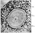
Fig. 1. From a section of the ovary of a 12-year old girl
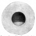
Fig. 2. Ovum of frog (Rana sylvatica).
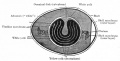
Fig. 3. Diagram of a vertical section through an unfertilized hen's egg
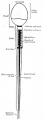
Fig. 4. Diagram of a human spermatozoon.
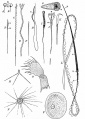
Fig. 5. Various types of spermatozoa


Maturation
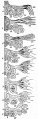
Fig. 6. Schematic outline of spermatogenesis in the rat.
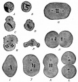
Fig. 7. Reduction of chromosomes in spermatogenesis in Ascaris megalocephala (bivalens).
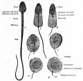
Fig. 8. Transformation of a spermatid into a spermatozoon (human).
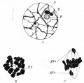
Fig. 9. Three stages in spermatogenesis in man (negro).
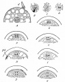
Fig. 10. Maturation of the ovum of Ascaris megalocephala (bivalens)
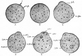
Fig. 11. From sections of ova of the mouse, showing stages in the maturation process
Fig. 12. Diagram representing the histogenesis of (a) the female sex cells and (6) the male sex cells
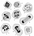
Fig. 13. Stages in the spermatogenesis of a grasshopper (Stenobothrus viridulus)
Fig. 14. From section of human ovary, showing mature Graafian follicle ready to rupture


Fertilization
Fig. 15. Diagram of fertilization of the ovum
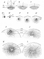
Fig. 16. Fertilization of the eggs of the star-fish and sea-urchin
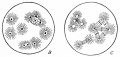
Fig. 17. Polyspermy in sea-urchin eggs treated with 0.005 per cent, nicotine solution


- Links: Fertilization | Category:Fertilization
Early development of amphioxus
Early development of amphioxus
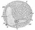
Fig. 18. Diagram of a median sagittal section through an Amphioxus ovum
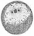
Fig. 19. Prophase of first cleavage the sagittal plane of the embryo
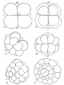
Fig. 20. Cleavage in Amphioxus
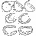
Fig. 21. Gastrulation in Amphioxus
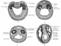
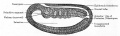
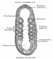
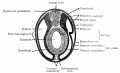
Fig. 25. Diagram to show differentiation of primitive segment into muscle plate (myotome) and cutis plate and relation of myocoel and splanchnocoel


Early development of the frog
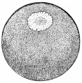
Fig. 26. Section through the fully formed ovarian egg of a frog.
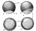
Fig. 27. A frog's egg before and after fertilization, showing the formation of the gray crescent.
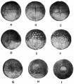
Fig. 28. Cleavage of the frog's egg.
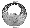
Fig. 29. From a sagittal section through blastula of frog.
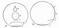
Fig. 30. Diagrams showing the position of the blastopore at successive stages of gastrulation in the frog's egg.
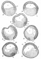
Fig. 31. Median sagittal sections showing successive stages of gastrulation in the frog's egg
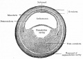
Fig. 32. Transverse section of embryo of frog (Rana fusca).
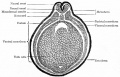
Fig. 33. Transverse section through embryo of frog (Rana fusca).
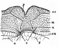
Fig. 34. Portion of a transverse section still continuous at the lower lateral angles of the larva of a frog (Rana fusca)
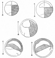
Fig. 35. Diagrams of median sagittal sections through an eight-cell stage and four stages during gastrulation of the frog's egg.
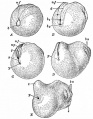
Fig. 36. Postero-lateral views of successive stages following gastrulation in the frog.
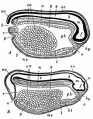
Fig. 37. Median sagittal sections of frog larvae.


Early development of the chick
Early development of the chick
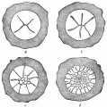
Fig. 38. Cleavage in hen's egg
Fig. 39. Vertical section germ disk of a fresh-laid hen's egg.
Fig. 40. Cross section blastoderm of a pigeon 14.5 hours after fertilization.
Fig. 41. Median longitudinal section blastoderm of a pigeon 31 hours after fertilization.
Fig. 42. Median longitudinal section blastoderm of a pigeon 36 hours after fertilization.
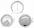
Fig. 43. Surface views of blastoderms of the pigeon.
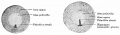
Fig. 44. Surface views of blastoderms of Haliplana primitive streak.
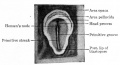
Fig. 45. Surface view of embryonic disk of chick.
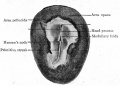
Fig. 46. Surface view of chick blastoderm.
Fig. 47. Transverse sections of blastoderm of chick 21 hours.
Fig. 48. Transverse section of blastoderm of chick 21 hours.
Fig. 49. Median longitudinal section blastoderm of chick after the primitive axis
Fig. 50. Transverse section of blastoderm of chick 40 hours.
Fig. 51. Dorsal view of chick embryo with ten pairs of mesodermal somites.
Fig. 52. Transverse section of chick embryo 2 days incubation.


- Links: Chicken Development
Early mammalian development
Fig. 53. Four stages in the cleavage of the ovum of the white rat.
Fig. 54. Four stages in cleavage of the ovum of the mouse.
Fig. 55. Four stages in the development of the bat.
Fig. 56. Sections of blastocysts of the white rat 5 days.
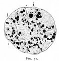
Fig. 57. Section of a 16-cell stage of an ovum of the opossum.
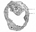
Fig. 58. Section of the blastocyst of the lemur Tarsius spectrum.
Fig. 59. Sections of blastodermic vesicle of bat.
Fig. 60. Three stages in the formation of the germ layers in the lemur Tarsius spectrum.
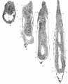
Fig. 61
Fig. 62
Fig. 63
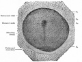
Fig. 64
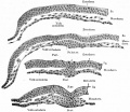
Fig. 65
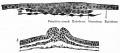
Fig. 66
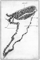
Fig. 67
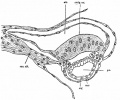
Fig. 68
Fig. 69
Fig. 70
Fig. 71
Fig. 72
Fig. 73
Fig. 74
Fig. 75
Fig. 76
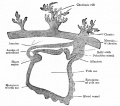
Fig. 77
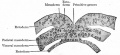
Fig. 78
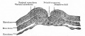
Fig. 79
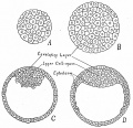
Fig. 80
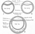
Fig. 81
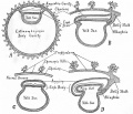
Fig. 82


Development of the external form of the body
Development of the external form of the body
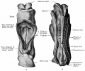
Fig. 83. Human embryo with 8 pairs of mesodermal somites
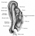
Fig. 84. Human embryo with 14 pairs of mesodermal somites.
Fig. 85. Human embryo of 2.6 mm.
Fig. 86. Human embryo of 4 mm.
Fig. 87. Human embryo 27 primitive segments.
Fig. 88. Human embryo with 28 primitive segments.
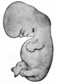
Fig. 89. Human embryo 11 mm.
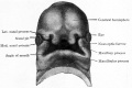
Fig. 90. Human embryo of 15.5 mm.
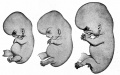
Fig. 91. Human embryo of 17.5 mm (47-51 days).
- Bailey092.jpg
Fig. 92. Human embryo of 18.5 mm (52-54 days).
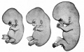
Fig. 93. Human embryo of 23 mm (2 months).
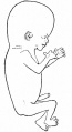
Fig. 94. Human embryo of 78 mm (3 months).
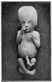
Fig. 95. Human embryo of 4 months.
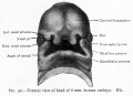
Fig. 96. Ventral view of head of 8 mm human embryo.
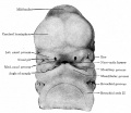
Fig. 97. Ventral view of head of 11.3 mm human embryo.
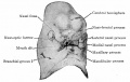
Fig. 98. Ventral view of head of 13.7 mm human embryo.
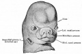
Fig. 99. Ventral view of head of human embryo of 8 weeks.


The development of connective tissues and the skeletal system
The development of connective tissues and the skeletal system
Fig. 100
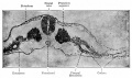
Fig. 101
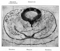
Fig. 102
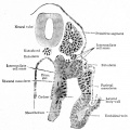
Fig. 103
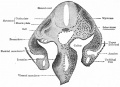
Fig. 104
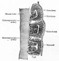
Fig. 105
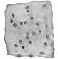
Fig. 106
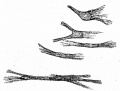
Fig. 107
- Bailey108.jpg
Fig. 108
Fig. 109
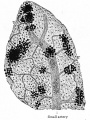
Fig. 110
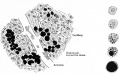
Fig. 111
Fig. 112
Fig. 113
Fig. 114
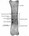
Fig. 115
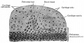
Fig. 116
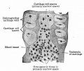
Fig. 117
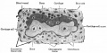
Fig. 118
Fig. 119
Fig. 120
Fig. 121
Fig. 122
Fig. 123
Fig. 124
Fig. 125
Fig. 126
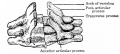
Fig. 127
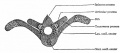
Fig. 128
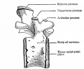
Fig. 129
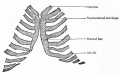
Fig. 130
Fig. 131
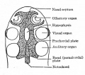
Fig. 132
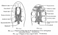
Fig. 132+133
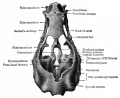
Fig. 133
- Bailey134.jpg
Fig. 134
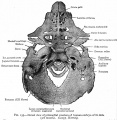
Fig. 135
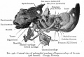
Fig. 136
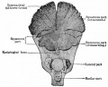
Fig. 137
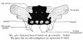
Fig. 138
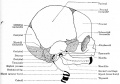
Fig. 139
Fig. 140
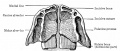
Fig. 141
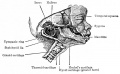
Fig. 142
Fig. 143
Fig. 144
Fig. 145
Fig. 146
Fig. 147
Fig. 148
Fig. 149
Fig. 150

Fig. 151
Fig. 152
Fig. 153
Fig. 154
Fig. 155


The development of the vascular system
The development of the vascular system
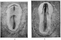
Fig. 156
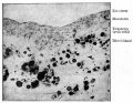
Fig. 157
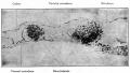
Fig. 158
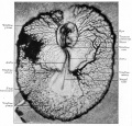
Fig. 159
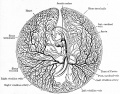
Fig. 160
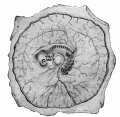
Fig. 161
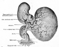
Fig. 162
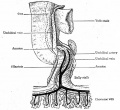
Fig. 163
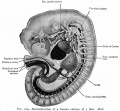
Fig. 164
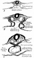
Fig. 165
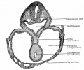
Fig. 166
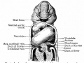
Fig. 167
Fig. 168
Fig. 169
Fig. 170
Fig. 171
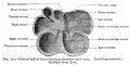
Fig. 172
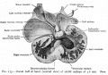
Fig. 173
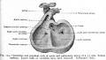
Fig. 174

Fig. 175
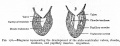
Fig. 176
Fig. 177
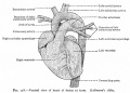
Fig. 178
Fig. 179
Fig. 180
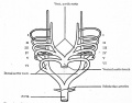
Fig. 181
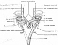
Fig. 182
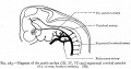
Fig. 183
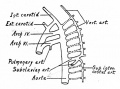
Fig. 184
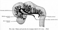
Fig. 185
- Bailey186.jpg
Fig. 186
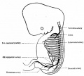
Fig. 187
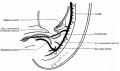
Fig. 188
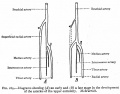
Fig. 189

Fig. 180
Fig. 191
Fig. 192
Fig. 193
Fig. 194
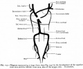
Fig. 195
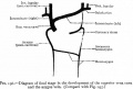
Fig. 196
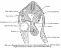
Fig. 197
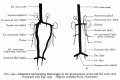
Fig. 198
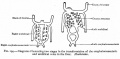
Fig. 199
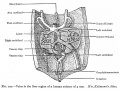
Fig. 200
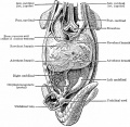
Fig. 201
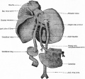
Fig. 202
Fig. 203
Fig. 204 Fig. 205
Fig. 206
Fig. 207
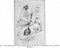
Fig. 208
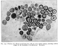
Fig. 209
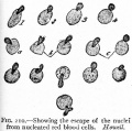
Fig. 210
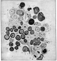
Fig. 211
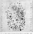
Fig. 212
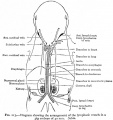
Fig. 213
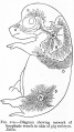
Fig. 214
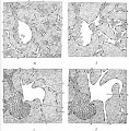
Fig. 215
Fig. 216
Fig. 217
Fig. 218
Fig. 219
Fig. 220
Fig. 221
Fig. 222
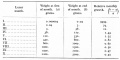
Table 1
Table 2


The muscular system
The development of the muscular system
Fig. 223
Fig. 224
Fig. 225
Fig. 226
Fig. 227


The alimentary and organs
The development of the alimentary tube and appended organs
Fig. 228
Fig. 229
Fig. 230
Fig. 231
Fig. 232
Fig. 233
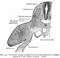
Fig. 234
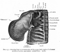
Fig. 235
Fig. 236
Fig. 237
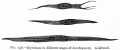
Fig. 238
- Bailey239.jpg
Fig. 239
- Bailey240.jpg
Fig. 240
Fig. 241
Fig. 242
Fig. 243
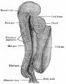
Fig. 244
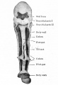
Fig. 245
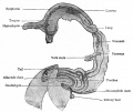
Fig. 246
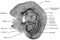
Fig. 247
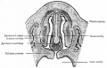
Fig. 248
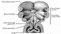
Fig. 249
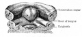
Fig. 250
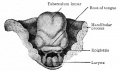
Fig. 251
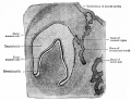
Fig. 252
Fig. 253
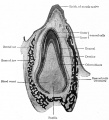
Fig. 254
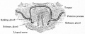
Fig. 255
Fig. 256
Fig. 257
Fig. 258
Fig. 259
Fig. 260
Fig. 261
Fig. 262
Fig. 263
Fig. 264
Fig. 265
Fig. 266
Fig. 267
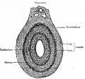
Fig. 268
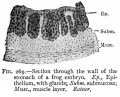
Fig. 269

Fig. 270
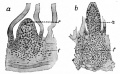
Fig. 271
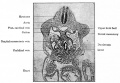
Fig. 272
Fig. 273
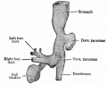
Fig. 274
Fig. 275
Fig. 276
Fig. 277
Fig. 278 279
Fig. 280
Fig. 281


The respiratory system
The development of the respiratory system
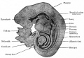
Fig. 282
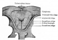
Fig. 283
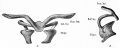
Fig. 284
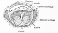
Fig. 285
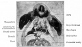
Fig. 286. Transverse section of a 14 mm. pig embryo, at the level of the upper limb buds, showing especially the two bronchi
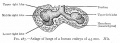
Fig. 287. Anlage of lungs of a human embryo of 4.3 mm. His.
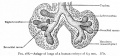
Fig. 288. Anlage of lungs of a human embryo of 8.5 mm. His.
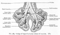
Fig. 289. Anlage of lungs of a human embryo of 10.5 mm. His.
Fig. 290. Transverse section of a pig embryo of 35 mm, showing the developing lungs (bronchial rami surrounded by mesoderm).
Fig. 291. Transverse sections of a rabbit embryo Showing how the omphalomesenteric veins (vom) push outward across the ccelom and fuse with the lateral body wall.
Fig. 292
Fig. 293
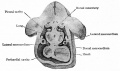
Fig. 294
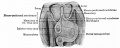
Fig. 295
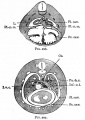
Fig. 296-297
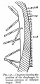
Fig. 298


The coelom, pericardium, pleuroperitoneum, diaphragm and mesenteries
The development of the coelom, the pericardium, pleuroperitoneum, diaphragm and mesenteries
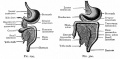
Fig. 299-300
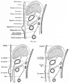
Fig. 301-303
Fig. 304


The urogenital system
The development of the urogenital system
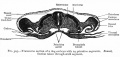
Fig. 305
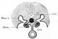
Fig. 306
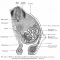
Fig. 307
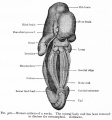
Fig. 308
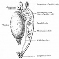
Fig. 309
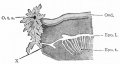
Fig. 310
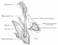
Fig. 311
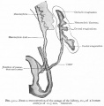
Fig. 312
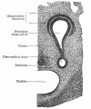
Fig. 313
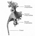
Fig. 314

Fig. 315
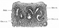
Fig. 316
Fig. 317-319
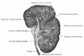
Fig. 320
Fig. 321
Fig. 322
Fig. 323
Fig. 324
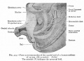
Fig. 325
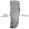
Fig. 326
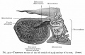
Fig. 327
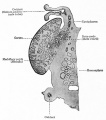
Fig. 328
Fig. 329
Fig. 330
Fig. 331
Fig. 332
Fig. 333
Fig. 334
Fig. 335
Fig. 336
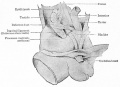
Fig. 337
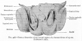
Fig. 338
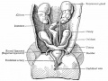
Fig. 339
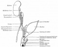
Fig. 340
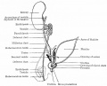
Fig. 341
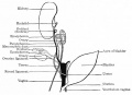
Fig. 342
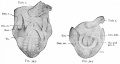
Fig. 343-344
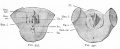
Fig. 345-346
Fig. 347-348
Fig. 349
Fig. 350
Fig. 351
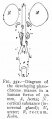
Fig. 352


The integumentary system
The development of the integumentary system
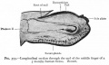
Fig. 353
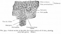
Fig. 354


The nervous system
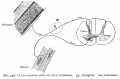
Fig. 358
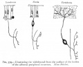
Fig. 359
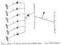
Fig. 360

Fig. 361
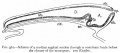
Fig. 362
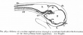
Fig. 363
Fig. 364
Fig. 365
Fig. 366
Fig. 367
Fig. 368
Fig. 369
Fig. 370
Fig. 371
Fig. 372
Fig. 373
Fig. 374
Fig. 375
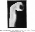
Fig. 376
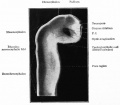
Fig. 377
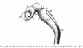
Fig. 378
Fig. 379-382
Fig. 383
Fig. 384
Fig. 385
Fig. 386
Fig. 387
Fig. 388
Fig. 389
Fig. 390
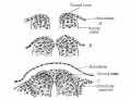
Fig. 391
Fig. 392
Fig. 393
Fig. 394
Fig. 395
Fig. 396
Fig. 397
Fig. 398
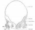
Fig. 399
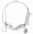
Fig. 400
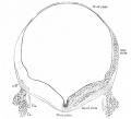
Fig. 401
Fig. 402
Fig. 403
Fig. 404
Fig. 405

Fig. 406
Fig. 407
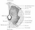
Fig. 408
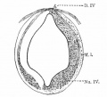
Fig. 409
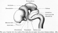
Fig. 410
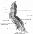
Fig. 411
Fig. 412
Fig. 413
Fig. 414
Fig. 415
Fig. 416
Fig. 417
Fig. 418
Fig. 419
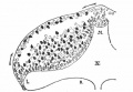
Fig. 420
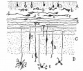
Fig. 421
Fig. 422
Fig. 423
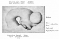
Fig. 424
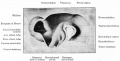
Fig. 425
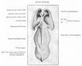
Fig. 426
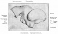
Fig. 427
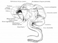
Fig. 428
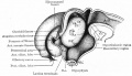
Fig. 429
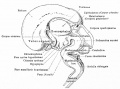
Fig. 430
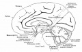
Fig. 431
Fig. 432
Fig. 433
Fig. 434
Fig. 435
Fig. 436
Fig. 437
Fig. 438
Fig. 439
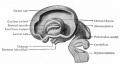
Fig. 440
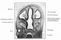
Fig. 441
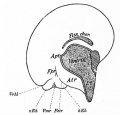
Fig. 442
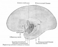
Fig. 443
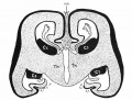
Fig. 444
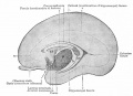
Fig. 445
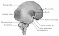
Fig. 446
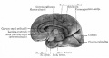
Fig. 447
Fig. 448
Fig. 449
Fig. 450
Fig. 451 452
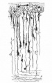
Fig. 453
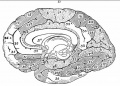
Fig. 454
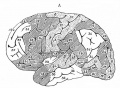
Fig. 455


The organs of special sense
Fig. 456
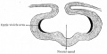
Fig. 457
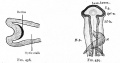
Fig. 458-459
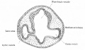
Fig. 460
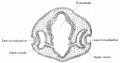
Fig. 461
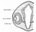
Fig. 462
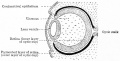
Fig. 463
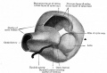
Fig. 464

Fig. 465
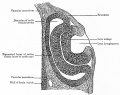
Fig. 466
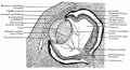
Fig. 467
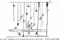
Fig. 468
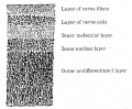
Fig. 469
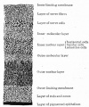
Fig. 470
Fig. 471
Fig. 472
Fig. 473
Fig. 474
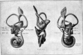
Fig. 475
Fig. 476
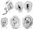
Fig. 477


Foetal membranes
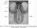
Fig. 478
Fig. 479
Fig. 480
Fig. 481
Fig. 482
Fig. 483
Fig. 484
Fig. 485
Fig. 486
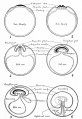
Fig. 487
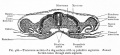
Fig. 488
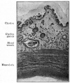
Fig. 489
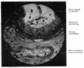
Fig. 490
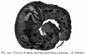
Fig. 491
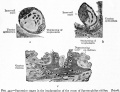
Fig. 492
Fig. 493
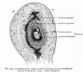
Fig. 494
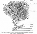
Fig. 495
Fig. 496
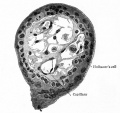
Fig. 497
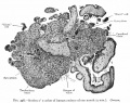
Fig. 498
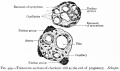
Fig. 499
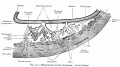
Fig. 500
Fig. 501
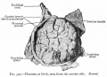
Fig. 502
Fig. 503


Teratogenesis
No images in this chapter.
| Historic Disclaimer - information about historic embryology pages |
|---|
|
Text-Book of Embryology: Germ cells | Maturation | Fertilization | Amphioxus | Frog | Chick | Mammalian | External body form | Connective tissues and skeletal | Vascular | Muscular | Alimentary tube and organs | Respiratory | Coelom, Diaphragm and Mesenteries | Urogenital | Integumentary | Nervous System | Special Sense | Foetal Membranes | Teratogenesis | Figures
Glossary Links
- Glossary: A | B | C | D | E | F | G | H | I | J | K | L | M | N | O | P | Q | R | S | T | U | V | W | X | Y | Z | Numbers | Symbols | Term Link
Cite this page: Hill, M.A. (2024, April 26) Embryology Book - Text-Book of Embryology (1921) - Figures. Retrieved from https://embryology.med.unsw.edu.au/embryology/index.php/Book_-_Text-Book_of_Embryology_(1921)_-_Figures
- © Dr Mark Hill 2024, UNSW Embryology ISBN: 978 0 7334 2609 4 - UNSW CRICOS Provider Code No. 00098G