BGDA Practical 12 - Embryo to Fetus: Difference between revisions
mNo edit summary |
mNo edit summary |
||
| Line 61: | Line 61: | ||
Limbs: hands and feet turned inward | Limbs: hands and feet turned inward | ||
|} | |} | ||
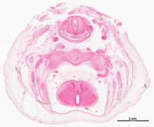
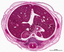
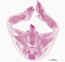
===Virtual Slides=== | |||
=== | {{Stage 22 virtual slides table}} | ||
{{ | |||
==Week 10 - Fetus (40mm)== | ==Week 10 - Fetus (40mm)== | ||
Revision as of 18:00, 25 May 2016
Introduction
Identify the development and form of the week 8 embryo (Stage 22 and 23). This 3D reconstruction animations showing specific systems and the histological sections from which these were prepared. There are also virtual slides and selected cross-sections showing specific features.
Then look at the week 10 early fetus and observe developmental changes. The selected mid-sagittal section shows the overall fetal anatomy and the gallery of excerpts have further detailed descriptions of these regions.
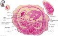
Week 8 - Stage 22
 |
stage 22, Week 8, 54 - 56 days, 23 - 28 mm
Mesoderm: heart prominence, ossification continues Head: nose, eye, external acoustic meatus Body:straightening of trunk, heart, liver, umbilicus: placental cord, midgut herniation, allantois, vitelline duct Limb: upper limbs longer and bent at elbow, foot plate with webbed digits, wrist, hand plate with separated digits Straightening of trunk, pigmented eye, eyelid, nose, external acoustic meatus, ear auricle, scalp vascular plexus, separated digits (fingers), thigh, ankle, umbilical cord |
Embryo Anatomy
Now examine selected regions of the Stage 22 embryo and their overall development in the 3D animations.
Head Region
Shoulder Region
Chest Region
Abdomen Region
Hip Region


|
|
|
|
|
|
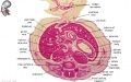
Week 8 - Stage 23
 |
Week 8, 56 - 60 days, 27 - 31 mm
scalp vascular plexus, eylid, eye, nose, auricle of external ear, mouth, sholder, arm, elbow, wrist, toes separated, sole of foot, umbilical cord Mesoderm: ossification continues Head: eyelids, external ears, rounded head Body: straightening of trunk, intestines herniated at umbilicus Limbs: hands and feet turned inward |
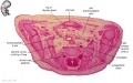
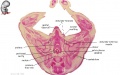
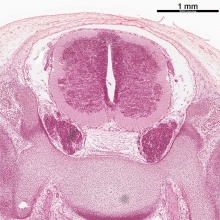
Virtual Slides
|
|
|
| ||||||||||||
|
|
| |||||||||||||
|
|
|


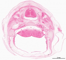
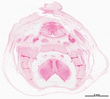

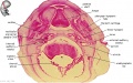
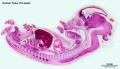
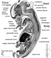
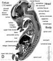
Week 10 - Fetus (40mm)

- Human Female Fetus (week 10)
Sagittal Section (plane D)

Pituitary and Lamina Terminalis

Olfactory Nerve

Atlas and Axis

Sacrum

Oral Cavity

Epiglottis

Heart

Spleen

Midgut Herniation

Midgut Herniation (label)

Pelvic Region

Pelvic Region (label)


|
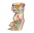
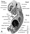
This is an image of the actual size comparison shown in the introduction of the Embryonic stage 13, 23 and Fetal stage 10 week 40mm. This stage of development is after the embryonic period (up to week 8), but only 2 weeks into early fetal development. |

|
The fetal period is a time of extensive growth in size and mass as well as differentiation of organ systems established in the embryonic period. In particular, the brain continues to grow and develop, the respiratory system differentiates, the urogenital system further differentiates between male/female, endocrine and gastrointestinal tract begins to function.
Compare this 10 week fetus with the earlier Carnegie stage embryos: size, head/body proportions, brain, head, skeletal development. Note that in the early fetus the midgut remains herniated and will only be taken into the peritoneal cavity on further body wall growth. |
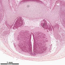
There are 4 sections taken in the sagittal plane (moving from the right at Plane A towards the midline at Plane D). Click on the small images (or the text below) to open the linked large image pages.
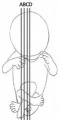
Planes
Plane A

Plane B
Plane C
Plane D


Midgut Herniation
Related Images
Fetus (week 10) Planes A (most lateral), B (lateral), C (medial) and D (midline) from lateral towards the midline.
- Human Fetus - most lateral | lateral | medial | midline
- Head - most lateral | lateral | medial | midline
- Cerebellum - most lateral | lateral | medial | midline
- Urogenital Unlabelled - most lateral | lateral | medial | midline
- Urogenital Labelled - most lateral | lateral | medial | midline
- Large Images - midline
- Image Source: UNSW Embryology, no reproduction without permission.
Additional Information
Links: Fetal Development - 10 Weeks | Stage 22 embryo slices
BGDA: Lecture 1 | Lecture 2 | Practical 3 | Practical 6 | Practical 12 | Lecture Neural | Practical 14 | Histology Support - Female | Male | Tutorial
Glossary Links
- Glossary: A | B | C | D | E | F | G | H | I | J | K | L | M | N | O | P | Q | R | S | T | U | V | W | X | Y | Z | Numbers | Symbols | Term Link
Cite this page: Hill, M.A. (2024, June 1) Embryology BGDA Practical 12 - Embryo to Fetus. Retrieved from https://embryology.med.unsw.edu.au/embryology/index.php/BGDA_Practical_12_-_Embryo_to_Fetus
- © Dr Mark Hill 2024, UNSW Embryology ISBN: 978 0 7334 2609 4 - UNSW CRICOS Provider Code No. 00098G