AACP Meeting 2013 - Face Embryology: Difference between revisions
mNo edit summary |
mNo edit summary |
||
| Line 59: | Line 59: | ||
:'''Links:''' [[Media:Stage15to22 head 01.mp4|MP4 version]] | [[Face Development Movie]] | [[Lecture - Head Development]] | [[Movies]] | :'''Links:''' [[Media:Stage15to22 head 01.mp4|MP4 version]] | [[Face Development Movie]] | [[Lecture - Head Development]] | [[Movies]] | ||
|} | |||
==Secondary Palate Development== | |||
{| border='0px' | |||
|- | |||
| width=220px|<mediaplayer width='200' height='220' image="http://embryology.med.unsw.edu.au/embryology/images/4/4f/Palate_001_icon.jpg">File:Palate_001.mp4</mediaplayer> | |||
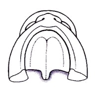
| valign="top" |[[File:Palate_001_icon.jpg|200px|right]] Animation shows an inferior view of the developmental sequence of secondary palate formation. | |||
* Animation shows the left and right maxillary processes of the first pharyngeal arch. | |||
* Primary palate formation is the fusion of these maxillary processes with the frontonasal prominence (FNP) in the midline. | |||
** Frontonasal prominence (FNP) contributing midline component of the upper jaw as well as the the philtrum of the upper lip. | |||
* Secondary palate formation is the growth of the palatal shelves towards the midline. | |||
|- | |||
| width=220px|<mediaplayer width='200' height='240' image="http://embryology.med.unsw.edu.au/embryology/images/a/a3/Palate_002_icon.jpg">File:Palate_002.mp4</mediaplayer> | |||
| valign="top" |[[File:Palate_002_icon.jpg|200px|right]] Animation shows an anterior view of the developmental sequence of secondary palate formation. | |||
* Animation shows the left and right maxillary processes of the first pharyngeal arch. | |||
* Primary palate formation is the fusion of these maxillary processes with the frontonasal prominence (FNP) in the midline. | |||
** Frontonasal prominence (FNP) contributing midline component of the upper jaw as well as the the philtrum of the upper lip. | |||
* Secondary palate formation is the growth of the palatal shelves towards the midline. | |||
{{Palate movie links}} | |||
|- | |||
|} | |} | ||
==Movies== | ==Movies== | ||
Revision as of 11:31, 15 May 2013

Face Embryology
2013 Australian Chapter, American Academy of Craniofacial Pain (AACP) Meeting
- May 18 May 2013
- Links: Embryology
Draft Page - notice removed when complete.
Introduction
This page will be updated and contain the final conference presentation.
| <mediaplayer width='420' height='500' image="http://embryology.med.unsw.edu.au/embryology/images/3/33/Face_001_icon.jpg">file:Face_001.mp4</mediaplayer> |
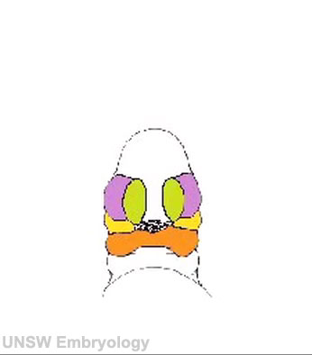
Development of the Face This animation shows a ventral view of development of the human face from approximately week 5 through to neonate. The separate embryonic components that contribute to the face have been colour coded.
The stomodeum is the primordial mouth region and a surface central depression lying between the forebrain bulge and the heart bulge. At the floor of the stomodeum indentation is the buccopharyngeal membrane (oral membrane). Note the complex origin of the maxillary region (upper jaw) requiring the fusion of several embryonic elements, abnormalities of this process lead to cleft lip and cleft palate.
|
Week 6-7 (GA 8-9)
| <mediaplayer width='320' height='420' image="http://embryology.med.unsw.edu.au/embryology/images/9/9c/Stage16-18_face_02.jpg">File:Stage16to18 face 01.mp4</mediaplayer> |  Movie shows a quick animation of the ventral views of the human embryo face, between Carnegie stage 16 to stage 18 (Week 6 to Week 7). Animation based on Kyoto embryos.
|
Week 5-8 (GA 7-10)
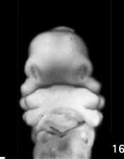
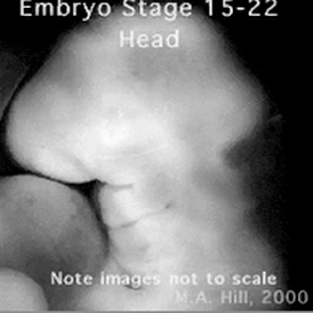
| <mediaplayer width='380' height='400' image="http://embryology.med.unsw.edu.au/embryology/images/9/92/Stage15to22_head_icon.jpg">File:Stage15to22 head 01.mp4</mediaplayer> |
Movie shows a quick animation of the lateral view of the human embryo head, between Carnegie stage 15 to stage 22 (Week 5 to Week 8). Note that these stage images are not to scale.
|
Secondary Palate Development
| <mediaplayer width='200' height='220' image="http://embryology.med.unsw.edu.au/embryology/images/4/4f/Palate_001_icon.jpg">File:Palate_001.mp4</mediaplayer> |
|
| <mediaplayer width='200' height='240' image="http://embryology.med.unsw.edu.au/embryology/images/a/a3/Palate_002_icon.jpg">File:Palate_002.mp4</mediaplayer> |
|
Movies
|
|
|
|
|
Histology

|

|
| Medial view | Lateral view |

Related Pages
Glossary Links
- Glossary: A | B | C | D | E | F | G | H | I | J | K | L | M | N | O | P | Q | R | S | T | U | V | W | X | Y | Z | Numbers | Symbols | Term Link
Cite this page: Hill, M.A. (2024, May 1) Embryology AACP Meeting 2013 - Face Embryology. Retrieved from https://embryology.med.unsw.edu.au/embryology/index.php/AACP_Meeting_2013_-_Face_Embryology
- © Dr Mark Hill 2024, UNSW Embryology ISBN: 978 0 7334 2609 4 - UNSW CRICOS Provider Code No. 00098G