Lecture - Mesoderm Development
Objectives

- Understanding of events during the third week of development
- Understanding the process of early somite development
- Understanding the process of body cavity formation
- Brief understanding of the future fate of mesoderm components
- Brief understanding of early heart formation
Notochord (Axial mesoderm)
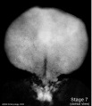
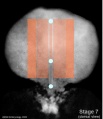
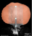
Stage 7 embryonic disc
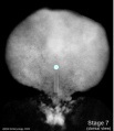
Stage 7 primitive-streak-node
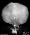
Stage 7 cloacal-oral-membranes
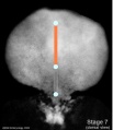
Stage 7 notochord


Mesoderm

- generated from epiblast cells migrating through the primitive streak
- epiblast cells expressing fibroblast growth factor (FGF2)
- forms a layer between ectoderm and endoderm with notochord down midline
- present before neural tube formation
- divides initially into 3 components

Stage 7 paraxial mesoderm
Stage 7 intermediate mesoderm
Stage 7 lateral plate


- Paraxial mesoderm - somites - musculoskeletal structures
- Intermediate mesoderm - kidney
- Lateral plate mesoderm - body wall structures

Mesoderm Development

The four images below beginning at week 3 show cross-sections of the trilaminar embryo and the sequence of mesoderm development.


Mesenchyme
- Embryonic connective tissue, describes the cell morphology (Histology is not epithelial organization)
- epithelial to mesenchymal transitions
- mesenchymal to epithelial transitions
Paraxial Mesoderm
- lies adjacent to notochord
- Forms 2 components
- Head - unsegmented paraxial mesoderm
- Body - segmented paraxial mesoderm
- Generates trunk muscles, skeleton, dermis of skin, blood vessels, connective tissue
- Segmented Paraxial Mesoderm
- segments called somites
- first pair of somites (day 20)
- segmentation imposes a pattern on
- nerves, vasculature, vertebra....
- somites appear in ordered sequence cranial to caudal
- appearance so regular used to stage the embryo
- Hamburger & Hamilton 1951- chicken
- thought to be generated by a "clock" (1 pair every 90 minutes)
- neural tube begins to close at 4th somite level
- 44 pairs of somites
Co-ordinator Note

Dr Mark Hill |
ANAT2341 Embryology S2 2011
|
Course Content 2011
2011 Timetable: | Embryology Introduction | Fertilization | Cell Division/Fertilization | Week 1 and 2 Development | Week 3 Development | Week 1 to 3 | Mesoderm Development | Ectoderm, Early Neural, Neural Crest | Trilaminar Embryo to Early Embryo | Early Vascular Development | Placenta | Vascular and Placenta | Endoderm, Early Gastrointestinal | Respiratory Development | Endoderm and Respiratory | Head Development | Neural Crest Development | Head and Neural Crest | Musculoskeletal Development | Limb Development | Musculoskeletal | Renal Development | Genital | Kidney and Genital | Sensory | Stem Cells | Stem Cells | Endocrine Development | Endocrine | Heart | Integumentary Development | Heart and Integumentary | Fetal | Birth and Revision | Fetal
Glossary Links
- Glossary: A | B | C | D | E | F | G | H | I | J | K | L | M | N | O | P | Q | R | S | T | U | V | W | X | Y | Z | Numbers | Symbols | Term Link
Cite this page: Hill, M.A. (2024, June 10) Embryology Lecture - Mesoderm Development. Retrieved from https://embryology.med.unsw.edu.au/embryology/index.php/Lecture_-_Mesoderm_Development
- © Dr Mark Hill 2024, UNSW Embryology ISBN: 978 0 7334 2609 4 - UNSW CRICOS Provider Code No. 00098G