File:Keibel Mall 2 447.jpg
From Embryology
Size of this preview: 451 × 599 pixels. Other resolution: 1,280 × 1,701 pixels.
Original file (1,280 × 1,701 pixels, file size: 250 KB, MIME type: image/jpeg)
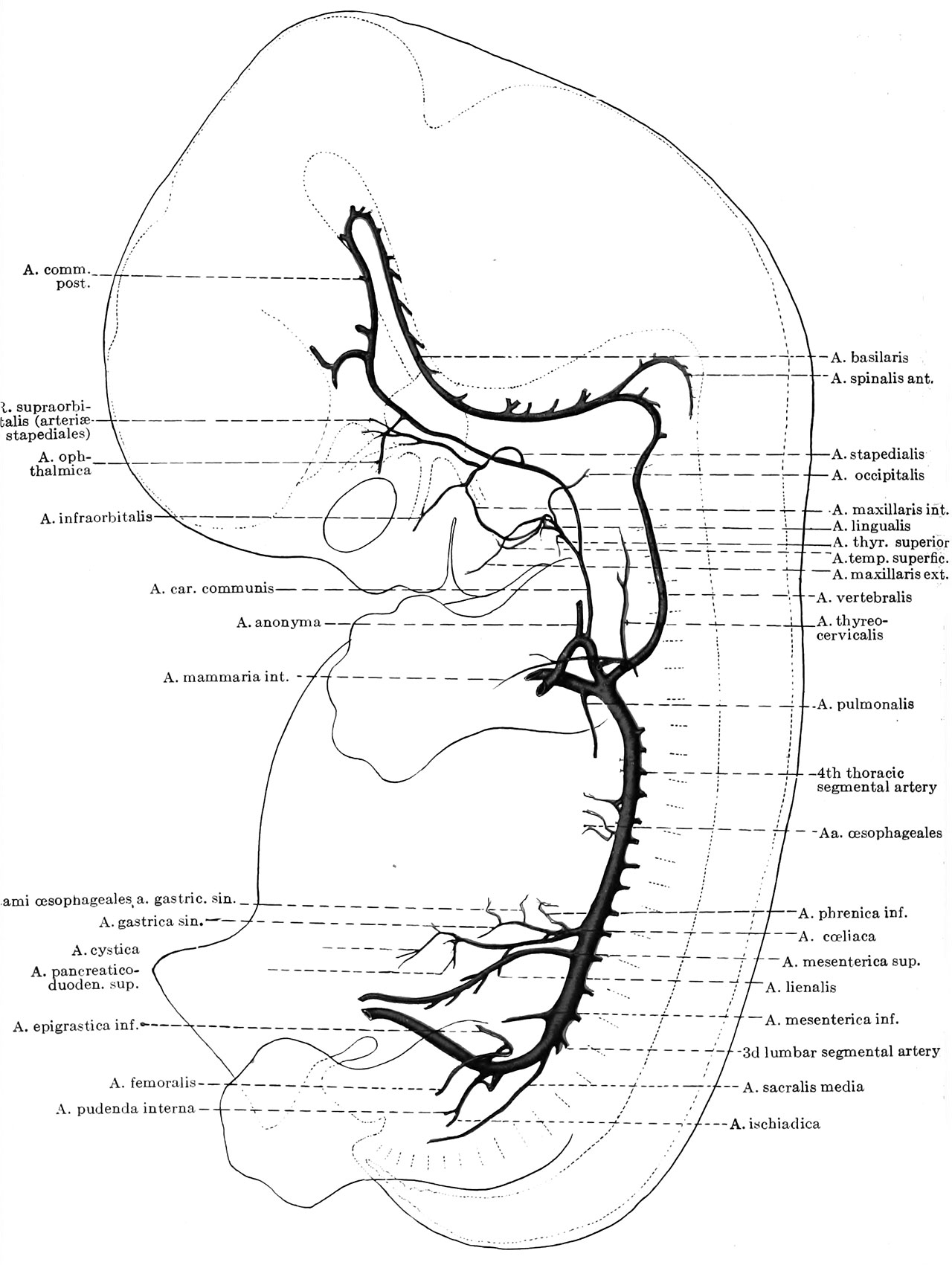
Fig. 447. Graphic reconstruction of the arterial system of a human embryo 15.5 mm. long
Which had been injected while the heart was still beating by Mr. Broedel. The embryo was subsequently cut into a series of sagittal sections. (No. 390, Mall collection.)
Online Editor - Carnegie Embryo No. 390 in classified as Carnegie stage 19 occurring in Week 7 GA week 9. See also Circulation Development and Blood Vessel Development
- XVIII. Development of Blood, Vascular System and Spleen: Introduction | Origin of the Angioblast and Development of the Blood | Development of the Heart | The Development of the Vascular System | General | Special Development of the Blood-vessels | Origin of the Blood-vascular System | Blood-vascular System in Series of Human Embryos | Arteries | Veins | Development of the Lymphatic System | Development of the Spleen
Reference
Keibel F. and Mall FP. Manual of Human Embryology II. (1912) J. B. Lippincott Company, Philadelphia.
File history
Click on a date/time to view the file as it appeared at that time.
| Date/Time | Thumbnail | Dimensions | User | Comment | |
|---|---|---|---|---|---|
| current | 13:23, 14 January 2017 | | 1,280 × 1,701 (250 KB) | Z8600021 (talk | contribs) | |
| 13:22, 14 January 2017 | 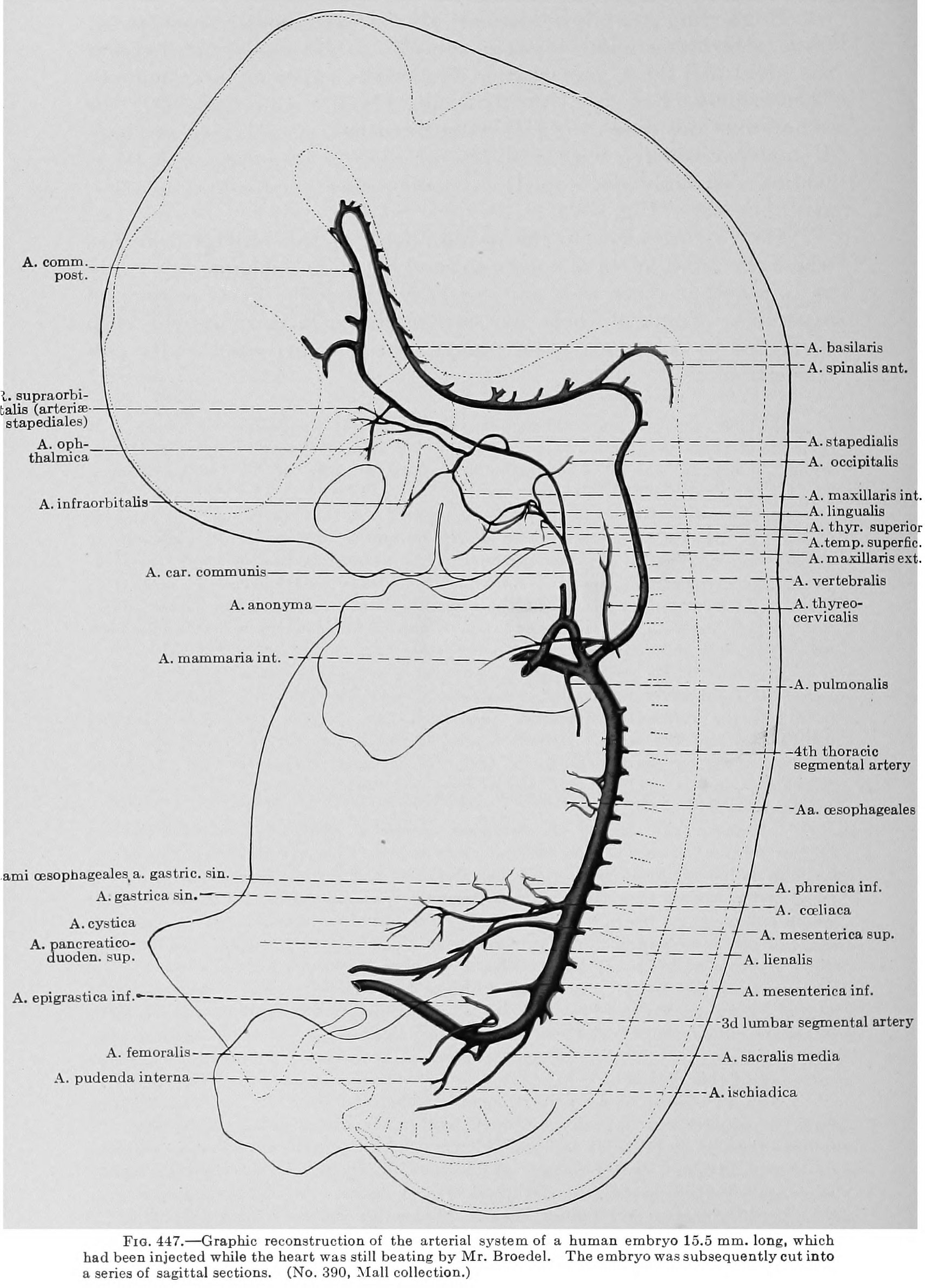 | 1,711 × 2,382 (519 KB) | Z8600021 (talk | contribs) |
You cannot overwrite this file.
File usage
The following 2 pages use this file: